Review
. 2024 Aug 7;227(4):iyae088.
doi: 10.1093/genetics/iyae088.

Extracellular vesicles

Affiliations
Review

Extracellular vesicles

Juan Wang et al. Genetics. .

Abstract

Extracellular vesicles (EVs) encompass a diverse array of membrane-bound organelles released outside cells in response to developmental and physiological cell needs. EVs play important roles in remodeling the shape and content of differentiating cells and can rescue damaged cells from toxic or dysfunctional content. EVs can send signals and transfer metabolites between tissues and organisms to regulate development, respond to stress or tissue damage, or alter mating behaviors. While many EV functions have been uncovered by characterizing ex vivo EVs isolated from body fluids and cultured cells, research using the nematode Caenorhabditis elegans has provided insights into the in vivo functions, biogenesis, and uptake pathways. The C. elegans EV field has also developed methods to analyze endogenous EVs within the organismal context of development and adult physiology in free-living, behaving animals. In this review, we summarize major themes that have emerged for C. elegans EVs and their relevance to human health and disease. We also highlight the diversity of biogenesis mechanisms, locations, and functions of worm EVs and discuss open questions and unexplored topics tenable in C. elegans, given the nematode model is ideal for light and electron microscopy, genetic screens, genome engineering, and high-throughput omics.

Keywords: Caenorhabditis elegans; cilia; exosome; extracellular vesicle; microvesicle; midbody remnant; spermatogenesis.


Conflict of interest statement

Conflicts of interest The author(s) declare no conflicts of interest.

Figures

Fig. 1.
Fig. 1.
Caenorhabditis elegans release diverse EVs from various tissues. Distinct EV subtypes are released from diverse tissues during different developmental states in male and hermaphrodite worms, such as epidermal cells, neurons, muscle, and germ cells, as well as undifferentiated embryonic blastomeres. Each EV subtype has a characteristic size range from tens of nanometers to several microns.
Fig. 2.
Fig. 2.
Exosome biogenesis pathways. Cargo proteins can be endocytosed and sorted into the late endosome to localize to the surface of ILVs within the MVB. Cargo proteins can also be incorporated into the lumen of ILVs by budding of the limiting endosomal membrane. RAL-1, actin crosslinkers, and the ESCRT machinery are involved in ILV formation in C. elegans. The MVB is then transported proximally to the plasma membrane, where the MVB docks and releases its contents into the extracellular space. MVBs can also mature into lysosomes for degradation of ILV contents.
Fig. 3.
Fig. 3.
Biogenesis of MV EVs in embryos. MVs are formed by ectocytosis, the progressive budding, and scission of the plasma membrane. TAT-5 is a PE flippase that transports PE lipids to the inner leaflet of the plasma membrane to maintain lipid asymmetry. TAT-5 is activated by PAD-1 to maintain membrane homeostasis. During ectocytosis, other proteins cause the exposure of cone-shaped PE, which may contribute to the curvature necessary for budding. The ESCRT machinery is recruited to the plasma membrane to create the bud and release the MV.
Fig. 4.
Fig. 4.
EV biogenesis from cilia. a) Sensory cilia release EVs from both the ciliary tip and base. The ciliary transport system promotes EV release at the tip while inhibiting EV release at the base. EVs released from the ciliary base can be taken up by neighboring amphid glia. b) Male-specific EVNs utilize cell type-specific ciliary kinesin (KLP-6) to traffic cargos to the tip for sorting into EVs. KLP-6 and the tubulin TBA-6, along with the post-translational modification enzyme polyglutamylase TTLL-11 and its counteracting enzyme, CCPP-1, specialize the axoneme to facilitate abundant EV release.
Fig. 5.
Fig. 5.
EV biogenesis from microvilli. Glial phagocytosis regulates the sensitivity of AFD neurons by engulfing the ends of AFD microvilli as EVs. This process involves the glial sheath recognizing “eat-me”? signals on microvilli, such as PS lipids (red PS) after their exposure by lipid scramblases, including SCRM-1. The PS flippase TAT-1 maintains the normal asymmetry of PS lipids in the rest of the cell. PS exposure is recognized by phagocytic pathways for the selective pruning of microvilli to become EVs.
Fig. 6.
Fig. 6.
Biogenesis of double membrane EVs in sperm. The main cytoskeletal protein MSP (star) drives the formation of membrane protrusions in spermatids and spermatozoa. Protrusions can fold back to form double-membrane vesicles (adapted from Kosinski et al. 2005).
Fig. 7.
Fig. 7.
Large EV biogenesis involving contractile rings. a) During cell division, an actomyosin ring contracts to pull the plasma membrane between the two cells toward the spindle midbody. Cytokinesis forms an intercellular bridge, which undergoes active constriction and membrane remodeling to release the midbody remnant as an EV. b) During meiosis II, spermatocytes use two types of myosin motors to polarize their contents and bud off from a shared cytoplasm, resulting in the release of a large EV known as a residual body. c) Mid-embryogenesis, primordial germ cells reduce their cell volume in half using an actomyosin ring to form a large lobe. The lobe is engulfed by the neighboring endodermal cell to non-autonomously form a large EV.
Fig. 8.
Fig. 8.
Caenorhabditis elegans extrudes exophers from stressed neurons and body wall muscle cells. a) Adult hermaphrodites release exophers from stressed neurons, with ALMR neurons releasing the most. Under starved conditions, body wall muscles release exophers to feed oocytes. b) Stressed neurons accumulate protein aggregates in aggresomes near the nucleus and dysfunctional organelles, including mitochondria. Exopher formation begins with membrane bud formation, followed by enlargement and constriction. Aggresomes caged by intermediate filaments promote exopher formation. The exopher remains connected to the cell body by a nanotube, which facilitates exopher growth. Exophers are released as EVs after phagocytosis by a neighboring epidermal cell. Mito, mitochondria; Agg, aggresome.
Fig. 9.
Fig. 9.
Functional roles of EVs for EV-releasing cells. a) At the end of cell division, cells release the intercellular bridge full of cytoskeletal and membrane trafficking regulators as a large EV known as the midbody remnant (MBR). The bridge also uses the ESCRT machinery to bud off tubules that can be taken up by neighboring cells as small EVs. b) Sensory cilia release EVs from the ciliary tip and base to maintain the balance of ciliary proteins. c) Protease-activated spermatids bud mitochondria in EVs called mitophers to decrease their mitochondrial content as they differentiate into spermatozoa. d) Neurons concentrate toxic protein aggregates and dysfunctional organelles in exophers to remove them from the cell body and improve neuronal function.
Fig. 10.
Fig. 10.
Functional roles of EVs for EV-receiving cells. a) In L1 larvae and adults, two lateral rows of cuticle fold into three ridges, known as alae, formed by the deposition of structural extracellular matrix. The underlying epidermal cells release exosomes carrying morphogens, such as hedgehog-like proteins WRT-2 and WRT-8, to instruct alae formation. b) PVD neurons form extensive dendritic arbors to cover the body area beneath the cuticle and sense harsh touch. Dendritic damage stimulates seam cells to release EVs containing fusogens like AFF-1 that repair dendritic connections. c) Embryos emit signals inducing the body wall muscle to convert muscle mass into yolk nutrition, released in large exophers to feed oocytes for embryogenesis and larval growth. d) Apoptotic cells expose PS on the outer leaflet of the plasma membrane and release small PS-positive EVs that facilitate PS exposure on neighboring cells and promote phagocytic clearance.
Fig. 11.
Fig. 11.
EVs mediate inter-organismal communication. a) Isolated environmental EVs spotted on a plate induce males to switch from normal sinusoidal movement to backward tail-chasing behavior. b) Males transfer multiple types of EVs to hermaphrodites during mating, including ciliary EVs, seminal fluid containing ENPP-1, and sperm-derived EVs carrying MSP that induce ovulation.
Fig. 12.
Fig. 12.
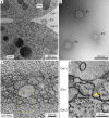
Visualization of EVs using electron microscopy. a) TEM of the cell contract from a wild-type two-cell embryo after high-pressure freezing. Data were collected for Wehman et al. (2011). b) Negative staining of environmental EVs isolated from C. elegans culture. Data were collected for Wang et al. (2014). c), d) Computed section from a 3D electron tomogram of a 200-nm section of the cell–cell contact (C) from a two-cell embryo that overproduces EVs by ectocytosis. The magnified region in d shows the cross section of a plasma membrane bud. Data were collected for Wehman et al. (2011).
Fig. 13.
Fig. 13.
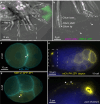
Visualization, characterization, and tracking of EVs using fluorescence microscopy. a), b) RnB neurons (1–5, 7–9) in the male tail release many EVs into the environment from their cilia. The transmembrane cargos, LOV-1 (magenta) and PKD-2 (green), are conserved markers for ciliary EVs (arrows and arrowheads). Data were collected for Walsh et al. (2022). Midbody remnants (arrowheads) are labeled with a plasma membrane reporter (PH, cyan) and a contractile ring reporter (NMY-2, yellow) before (c) and after (d) phagocytosis, enabling their tracking. Data were collected for Fazeli et al. (2016). e), f) A plasma membrane-localized degron reporter (PH::ZF1, yellow) allows the specific labeling of MVs in vivo after degradation is initiated in somatic cells (left side of embryo in e). More MVs (arrowheads) are observed between the eggshell and cell surface in partial loss-of-function pad-1 mutant embryos. PB, polar body. Data were collected for Fazeli et al. (2020).
Fig. 14.
Fig. 14.
Methods for EV isolation from c. elegans cultures. a) EVs are harvested by washing culture plates, which contain worms, bacteria, different EV subtypes, and other secreted macromolecules. EVs can also be collected from liquid culture. b) Differential centrifugation sequentially pellets worms and bacteria from the mixed sample. c) EVs can be isolated from the cleaned supernatant using ultracentrifugation on a cushion. Different speeds can be used to pellet large and small EVs. d) For increased purity, EVs can be pelleted from the cleaned supernatant by ultracentrifugation on a cushion and then loaded onto a gradient solution. Ultracentrifugation allows gradient fractionation to separate dense and light EVs. e) EVs can be isolated from cleaned supernatants using size exclusion chromatography, which allows the separation of EVs based on size.
Fig. 15.
Fig. 15.
MyEVome, a tool for identifying EV cargo candidates for c. elegans cell types. MyEVome combines single-cell transcriptomic data with environmental EV proteomics to plot likely cell-specific EV cargos (Cao et al. 2017; Nikonorova et al. 2022). MyEVome is currently available at https://myevome.shinyapps.io/evome-app.

References

    1. Aaronson S, Behrens U, Orner R, Haines TH. 1971. Ultrastructure of intracellular and extracellular vesicles, membranes, and myelin figures produced by Ochromonas danica. J Ultrastruct Res. 35(5–6):418–430. doi:10.1016/S0022-5320(71)80003-5. - DOI - PubMed
    1. Abdu Y, Maniscalco C, Heddleston JM, Chew T-L, Nance J. 2016. Developmentally programmed germ cell remodelling by endodermal cell cannibalism. Nat Cell Biol. 18(12):1302–1310. doi:10.1038/ncb3439. - DOI - PMC - PubMed
    1. Akella JS, Barr MM. 2021. The tubulin code specializes neuronal cilia for extracellular vesicle release. Dev Neurobiol. 81(3):231–252. doi:10.1002/dneu.22787. - DOI - PMC - PubMed
    1. Akella JS, Carter SP, Nguyen K, Tsiropoulou S, Moran AL, Silva M, Rizvi F, Kennedy BN, Hall DH, Barr MM, et al. 2020. Ciliary Rab28 and the BBSome negatively regulate extracellular vesicle shedding. Elife. 9:e50580. doi:10.7554/eLife.50580. - DOI - PMC - PubMed
    1. Al-Jawabreh R, Lastik D, McKenzie D, Reynolds K, Suleiman M, Mousley A, Atkinson L, Hunt V. 2024. Advancing Strongyloides omics data: bridging the gap with Caenorhabditis elegans. Philos Trans R Soc Lond B Biol Sci. 379(1894):20220437. doi:10.1098/rstb.2022.0437. - DOI - PMC - PubMed

LinkOut - more resources