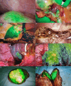
Application of indocyanine green fluorescence imaging in hepatobiliary surgery
- PMID: 38884267
- PMCID: PMC11634118
- DOI: 10.1097/JS9.0000000000001802
Application of indocyanine green fluorescence imaging in hepatobiliary surgery
Abstract
Indocyanine green (ICG) is a fluorescent dye with an emission wavelength of about 840 nm, which is selectively absorbed by the liver after intravenous or bile duct injection, and then it is excreted into the intestines through the biliary system. With the rapid development of fluorescence laparoscopy, ICG fluorescence imaging is safe, feasible, and widely used in hepatobiliary surgery. ICG fluorescence imaging is of great significance in precise preoperative and intraoperative localization of liver lesions, real-time visualization of hepatic segmental anatomy, intrahepatic and extrahepatic biliary tract visualization, and liver transplantation. ICG fluorescence imaging facilitates efficient intraoperative hepatobiliary decision-making and improves the safety of minimally invasive hepatobiliary surgery. Advances in imaging systems will increase the use of fluorescence imaging as an intraoperative navigation tool, improving the safety and accuracy of open and laparoscopic/robotic hepatobiliary surgery. Herin, we have reviewed the status of ICG applications in hepatobiliary surgery, aiming to provide new insights for the development of hepatobiliary surgery.
Trial registration: ClinicalTrials.gov NCT05546619.
Copyright © 2024 The Author(s). Published by Wolters Kluwer Health, Inc.
Conflict of interest statement
All authors have no conflicts of interest related to this publication. No benefits in any form have been received or will be received from a commercial party related directly or indirectly to the subject of this article.
Sponsorships or competing interests that may be relevant to content are disclosed at the end of this article.
Figures


References
-
- Hernot S, van Manen L, Debie P, et al. . Latest developments in molecular tracers for fluorescence image-guided cancer surgery. Lancet Oncol 2019;20:e354–e367. - PubMed
-
- Liu Y, Wang Q, Du B, et al. . A meta-analysis of the three-dimensional reconstruction visualization technology for hepatectomy. Asian J Surg 2023;46:669–676. - PubMed
-
- Clarke MP, Kane RA, Steele G, et al. . Prospective comparison of preoperative imaging and intraoperative ultrasonography in the detection of liver tumors. Surgery 1989;106:849–855. - PubMed
-
- Makuuchi M, Hasegawa H, Yamazaki S. Intraoperative ultrasonic examination for hepatectomy. Ultrasound Med Biol 1983;(Suppl2):493–497. - PubMed
Publication types
MeSH terms
Substances
Associated data
LinkOut - more resources
Full Text Sources
Medical

