Review
. 2024 Jun 26;52(3):1131-1148.
doi: 10.1042/BST20230724.

Proofreading mechanisms of the innate immune receptor RIG-I: distinguishing self and viral RNA

Affiliations
Review

Proofreading mechanisms of the innate immune receptor RIG-I: distinguishing self and viral RNA

Mihai Solotchi et al. Biochem Soc Trans. .

Abstract

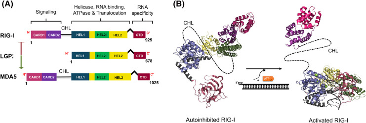
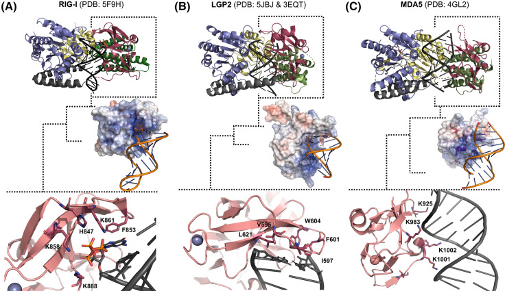
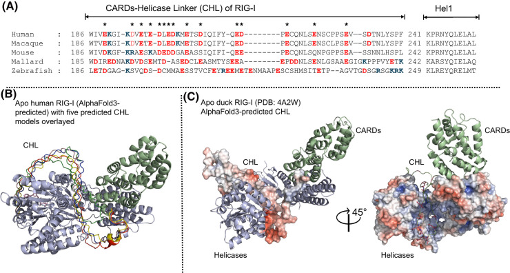
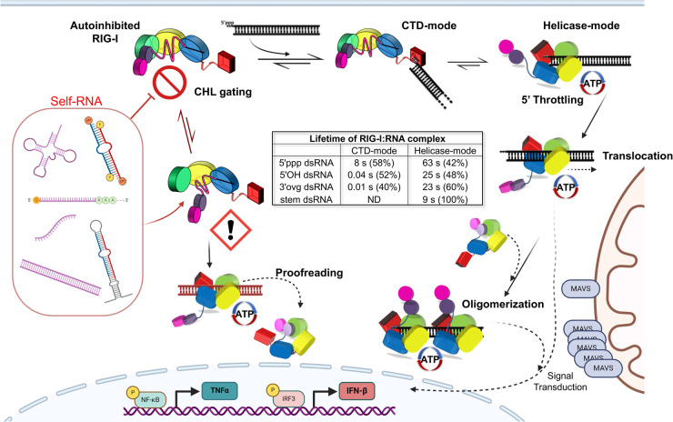
The RIG-I-like receptors (RLRs), comprising retinoic acid-inducible gene I (RIG-I), melanoma differentiation-associated gene 5 (MDA5), and laboratory of genetics and physiology 2 (LGP2), are pattern recognition receptors belonging to the DExD/H-box RNA helicase family of proteins. RLRs detect viral RNAs in the cytoplasm and respond by initiating a robust antiviral response that up-regulates interferon and cytokine production. RIG-I and MDA5 complement each other by recognizing different RNA features, and LGP2 regulates their activation. RIG-I's multilayered RNA recognition and proofreading mechanisms ensure accurate viral RNA detection while averting harmful responses to host RNAs. RIG-I's C-terminal domain targets 5'-triphosphate double-stranded RNA (dsRNA) blunt ends, while an intrinsic gating mechanism prevents the helicase domains from non-specifically engaging with host RNAs. The ATPase and RNA translocation activity of RIG-I adds another layer of selectivity by minimizing the lifetime of RIG-I on non-specific RNAs, preventing off-target activation. The versatility of RIG-I's ATPase function also amplifies downstream signaling by enhancing the signaling domain (CARDs) exposure on 5'-triphosphate dsRNA and promoting oligomerization. In this review, we offer an in-depth understanding of the mechanisms RIG-I uses to facilitate viral RNA sensing and regulate downstream activation of the immune system.

Keywords: RIG-I like receptors; helicase; nucleic acid receptors.


Conflict of interest statement

The authors declare that there are no competing interests associated with the manuscript.

Figures

Figure 1.
Figure 1.. Structural architecture of the RIG-I-like receptors (RLRs).
(A) Comparison of domain architecture of the RIG-I-like receptor family. The green arrow indicates activation of MDA5, and the red indicates inhibition of RIG-I. (B) Activation model of RIG-I. 5′ppp dsRNA and ATP binding induce a conformational change from autoinhibited (PDB: 4A2W) (CTD modeled from PDB: 2YKG) to the activated state (PDB: 5F9H). The exposure of N-terminal CARDs marks RIG-I activation.
Figure 2.
Figure 2.. The CTD of RLRs determines their RNA binding specificities.
The RNA-bound states of RIG-I, MDA5, and LGP2 show similar structure and RNA footprint within the helicase domains’ channel. The specificity of the RLRs arises from their C-terminal domain (brick red), distinguished by a conserved positively charged patch, as depicted by the blue regions in the electrostatic surface. (A) RIG-I (PDB: 5F9H) uses positively charged residues in the capping loop to form strong interactions with the 5′-triphosphate of dsRNA, while base-stacking interactions with aromatic F residues form a preference for blunt-ended RNA. (B) LGP2 (chicken full-length PDB: 5JBJ) and (human CTD PDB: 3EQT) has a hydrophobic capping loop that facilitates blunt end RNA binding. (C) MDA5 (PDB: 4GL2) does not have RNA end-binding preference. Residues from the positively charged patch on MDA5's CTD interact with the backbone of the RNA and within the minor groove.
Figure 3.
Figure 3.. RIG-I's intrinsically disordered, charge-conserved CHL provides an RNA-gating mechanism.
(A) RIG-I sequences spanning residues ∼180–280 are aligned from various vertebrates. While sequence conservation in the CHL is not as pronounced as in the Helicase domains, the prevalence and conservation of negatively charged residues suggest their significant functional role in RIG-I regulation. Glutamate and aspartate residues are colored red, and lysines are blue. Asterisks (*) indicate positions where a charge is conserved in the sequence across species. (B) The AlphaFold3 predictions of full-length human apo RIG-I render the CHL within the Helicase RNA-binding channel. The top 5 ranked predictions of the CHL are overlayed on the top rank prediction of the full-length protein to emphasize the confidence of spatial fitting for the CHL, rather than the weak confidence score of the region's folding propensity. The CTD of RIG-I has been hidden to reveal the CHL predictions, but it is predicted to sit between Hel1 and the CARDs at the end of the V-linker. (C) The electrostatic surface of the autoinhibited duck RIG-I Helicase domains (PDB: 4A2W) displays a positively charged RNA-binding channel. AlphaFold and biochemical studies predict that the negatively charged CHL will reside in this channel when RNA is absent. The electrostatic surface of the predicted CHL displays a high degree of charge complementation to the Helicase electrostatics.
Figure 4.
Figure 4.. RIG-I's activation pathway with ordered RNA binding and ATPase proofreading.
Top left, RIG-I initiates its cycle in the autoinhibited state, characterized by CARD-Hel2i sequestration and CHL (pink) gating the Helicase's RNA binding site to prevent nonspecific RNA binding. The CTD (red) is free to interact with RNA and forms high-affinity interactions with 5′ppp dsRNA ends, a characteristic feature of viral RNA replication intermediates, resulting in the CTD-mode conformation. The left side of the figure shows binding to non-specific RNA and the right side shows binding to viral RNA. The viral RNA with a 5′ppp RNA end has a longer lifetime on the CTD compared with non-ppp RNAs, as indicated in the center table that tabulates the kinetic lifetime measurements of RIG-I CTD- and Helicase-mode. This prolonged lifetime facilitates the activated Helicase-mode conformation, where the RNA is bound to the Helicase, and CARDs are free. The ATPase activity induces translocation-mediated oligomerization, clustering multiple RIG-I molecules on one RNA, thereby enhancing the likelihood of CARD oligomerization. Strong CTD interactions with 5′ppp prolong the activated state's duration by inhibiting RIG-I's translocation. The CARDs oligomers transmit their signal by interacting with downstream partner MAVS, establishing an antiviral and pro-inflammatory state through the up-regulation of TNF-α and IFN-β. Ultimately, RIG-I's ATPase activity dissociates and inactivates the complex. If RIG-I binds to a nonspecific RNA end, weak association with CTD results in rapid dissociation. If RIG-I's gating mechanism is compromised, self-RNA regions can bind to the Helicase directly. In the absence of a 5′ppp-mediated throttling effect, however, RIG-I's ATPase and translocation processes rapidly dissociate such self-RNAs, minimizing aberrant RIG-I activation.
Figure 5.
Figure 5.. An unstructured Hel2 loop is a signature of RIG-I translocation throttling.
(A) The Hel2 translocation loop is unresolved in the structure of RIG-I bound to 5′ppp dsRNA (PDB: 5F9H), signifying the long-lived translocation throttling state. (B) Resolution of the Hel2 translocation loop (orange) in the structure of RIG-I bound to 5′-OH dsRNA (PDB: 5F9F) indicates the propensity for this complex to rapidly dissociate via translocation.
Figure 6.
Figure 6.. Host mRNA capping and ribose methylation (Cap-1) are essential for evading recognition by RIG-I.
RIG-I must avoid engagement with host mRNAs present in the cytoplasm. (A) The distinguishing features of mRNA capping include an m7G moiety (Cap-0) and a 2′-O-methylation of the end-nucleotide ribose (Cap-1). (B) RIG-I crystal structure (PDB: 5F98) bound to 5′ cap-0 dsRNA shows the CTD surface electrostatics accommodating the bulky m7Gppp moiety. (C) 5′ triphosphate and base-stacking interactions are maintained by the CTD capping loop with the terminal base pair of the Cap-0 dsRNA. (D) H830 interacts with the 2′OH of the ribose of the 5′-end nucleotide in dsRNA. The H830 is expected to clash with the 2′O-Me of the ribose, allowing RIG-I to discriminate against Cap-1 mRNA. Mutation to H830A abolishes this checkpoint entirely.
Figure 7.
Figure 7.. RIG-I discriminates against 5′ monophosphate dsRNA.
There is an abundance of 5′ monophosphate dsRNA in the cell that RIG-I must actively discriminate against, including microRNA, tRNA, and leaky mitochondrial RNA transcripts. (A) The 5′-p dsRNA bound to the CTD of WT RIG-I shows potential interactions of 5′-p with a subset of lysines in the CTD capping loop (PDB: 7TNZ), yet RIG-I is not activated by 5′-p dsRNA. (B) The 5′-p dsRNA bound to the CTD of I875A RIG-I (PDB: 7BAI). No structural differences between panels A and B are apparent, although I875A mutation rescues RIG-I activation by 5′-p dsRNA.
Figure 8.
Figure 8.. RIG-I signal transduction is mediated by CARDs tetramerization.
The crystal structure of RIG-I CARDs reveals a tetrameric lock-washer formation, stabilized by K63-linked di-ubiquitin chains (PDB: 4NQK). Experimental results and an extended structure (PDB: 4P4H) of this formation suggest this scaffold is necessary to nucleate MAVS filamentation and activate downstream signaling events. RIG-I either clusters on long dsRNA (PDB: 7JL3) or intermolecularly across RNA ligands (PDB: 5F9H) to facilitate CARDs tetramer formation. Localized CARDs can interact with each other, and ubiquitination events stabilize these interactions.

Similar articles

Cited by

References

    1. Yoneyama, M., Kikuchi, M., Natsukawa, T., Shinobu, N., Imaizumi, T., Miyagishi, M.et al. (2004) The RNA helicase RIG-I has an essential function in double-stranded RNA-induced innate antiviral responses. Nat. Immunol. 5, 730–737 10.1038/ni1087 - DOI - PubMed
    1. Yoneyama, M., Kikuchi, M., Matsumoto, K., Imaizumi, T., Miyagishi, M., Taira, K.et al. (2005) Shared and unique functions of the DExD/H-box helicases RIG-I, MDA5, and LGP2 in antiviral innate immunity. J. Immunol. 175, 2851–2858 10.4049/jimmunol.175.5.2851 - DOI - PubMed
    1. Onomoto, K., Onoguchi, K. and Yoneyama, M. (2021) Regulation of RIG-I-like receptor-mediated signaling: interaction between host and viral factors. Cell Mol. Immunol. 18, 539–555 10.1038/s41423-020-00602-7 - DOI - PMC - PubMed
    1. Thoresen, D., Wang, W., Galls, D., Guo, R., Xu, L. and Pyle, A.M. (2021) The molecular mechanism of RIG-I activation and signaling. Immunol. Rev. 304, 154–168 10.1111/imr.13022 - DOI - PMC - PubMed
    1. Rodriguez, K.R., Bruns, A.M. and Horvath, C.M. (2014) MDA5 and LGP2: accomplices and antagonists of antiviral signal transduction. J. Virol. 88, 8194–8200 10.1128/JVI.00640-14 - DOI - PMC - PubMed

Publication types

MeSH terms

LinkOut - more resources