

[Erythema nodosum]
- PMID: 38884811
- PMCID: PMC11485184
- DOI: 10.1007/s00393-024-01529-4
[Erythema nodosum]
Abstract
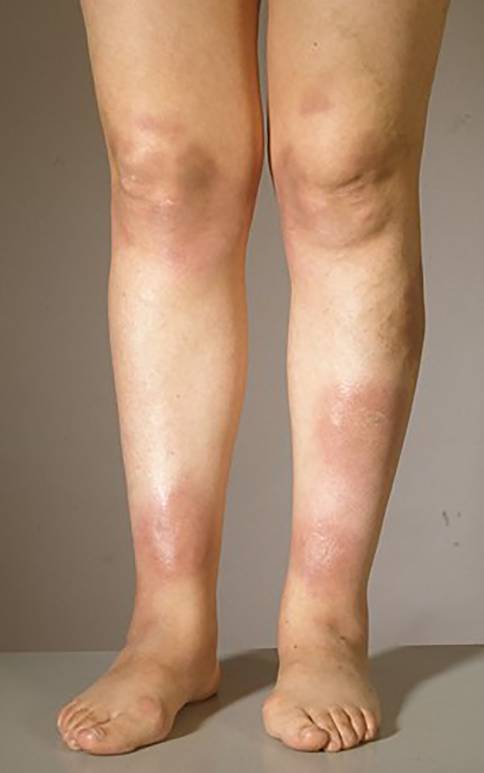
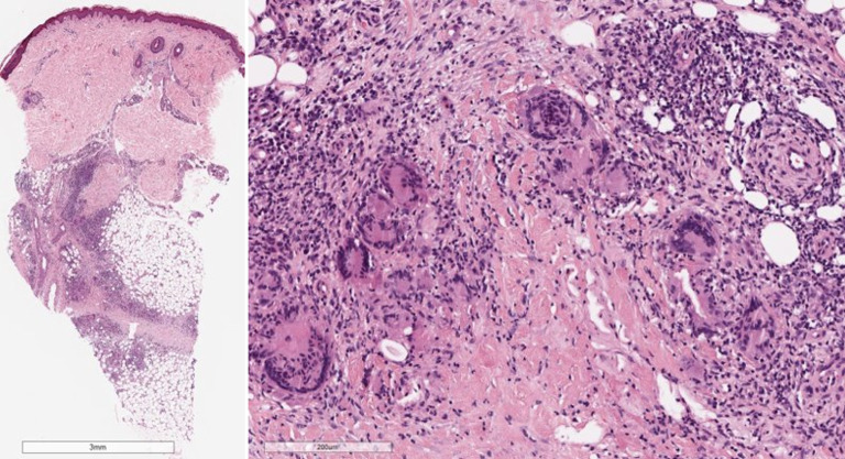
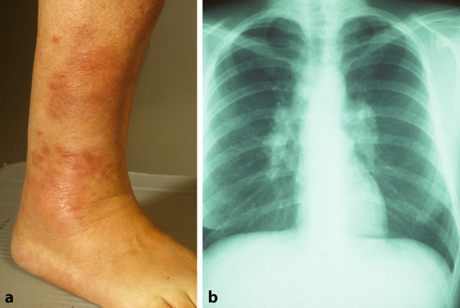
Erythema nodosum (EN) is the most frequently occurring form of acute panniculitis. It is characterized by painful red to livid raised nodules or bumps that typically occur symmetrically in the shin area. The cause of EN is often a reaction of the immune system to various triggers including infections, inflammatory diseases or medications. In approximately half of the cases no trigger can be identified. After treatment of the underlying pathology EN is typically self-limiting.
Das Erythema nodosum (EN) ist die am häufigsten auftretende Form einer akuten Pannikulitis. Es wird charakterisiert durch schmerzhafte, rote bis livide, erhabene Knötchen oder Beulen, die typischerweise symmetrisch im Bereich der Schienbeine auftreten. Häufig handelt es sich beim EN um eine Reaktion des Immunsystems auf Infektionen, entzündliche Erkrankungen oder Medikamente. In etwa der Hälfte der Fälle ist kein Auslöser zu eruieren. Nach Behandlung der zugrunde liegenden Ursache ist das EN in der Regel selbstlimitierend.
Keywords: Drug reaction; Inflammatory nodules; Sarcoidosis; Septal panniculitis; Streptococci.
© 2024. The Author(s).
Figures
References
-
- Schwartz RA, Nervi SJ (2007) Erythema nodosum: a sign of systemic disease. Am Fam Physician 75(5):695–700 - PubMed
-
- Mert A et al (2004) Erythema nodosum: an experience of 10 years. Scand J Infect Dis 36(6–7):424–427 - PubMed
-
- Requena L, Sanchez Yus E (2007) Erythema nodosum. Semin Cutan Med Surg 26(2):114–125 - PubMed
-
- Garcia-Porrua C et al (2000) Erythema nodosum: etiologic and predictive factors in a defined population. Arthritis Rheum 43(3):584–592 - PubMed
Publication types
MeSH terms
LinkOut - more resources
Full Text Sources
Research Materials
