

Suppression of HIV-TAT and cocaine-induced neurotoxicity and inflammation by cell penetrable itaconate esters
- PMID: 38884890
- PMCID: PMC11512888
- DOI: 10.1007/s13365-024-01216-9
Suppression of HIV-TAT and cocaine-induced neurotoxicity and inflammation by cell penetrable itaconate esters
Abstract
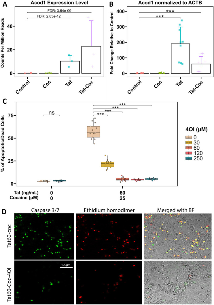
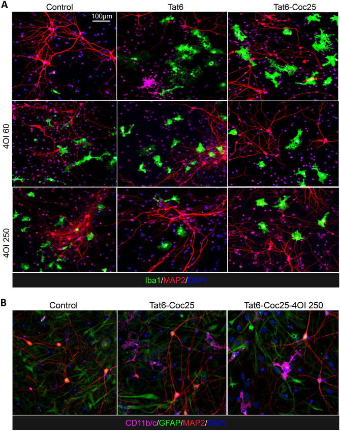
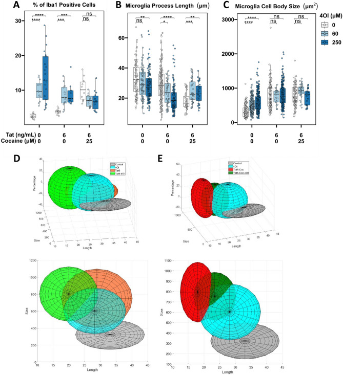
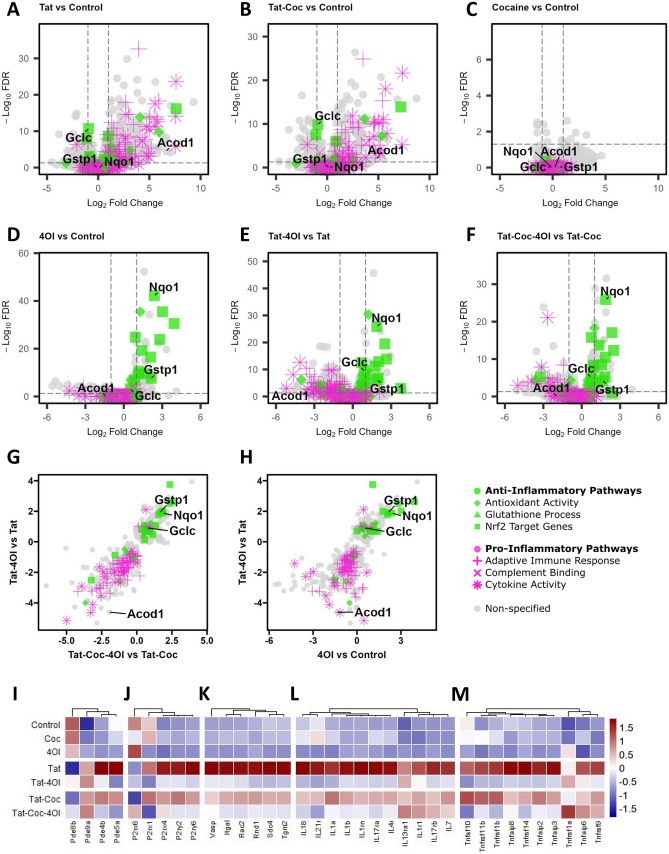
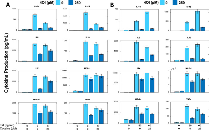
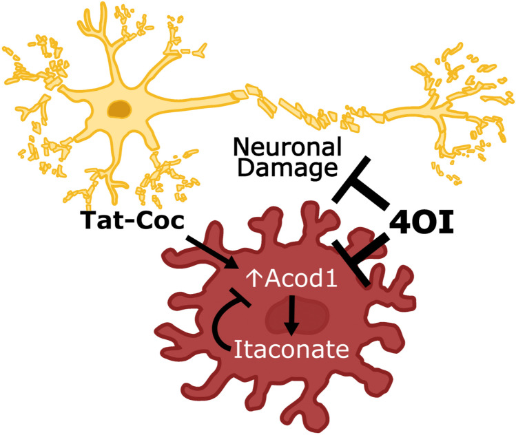
HIV-associated neurological disorder (HAND) is a serious complication of HIV infection marked by neurotoxicity induced by viral proteins like Tat. Substance abuse exacerbates neurocognitive impairment in people living with HIV. There is an urgent need for therapeutic strategies to combat HAND comorbid with Cocaine Use Disorder (CUD). Our analysis of HIV and cocaine-induced transcriptomes in primary cortical cultures revealed significant overexpression of the macrophage-specific gene aconitate decarboxylase 1 (Acod1). The ACOD1 protein converts the tricarboxylic acid intermediate cis-aconitate into itaconate during the activation of inflammation. Itaconate then facilitates cytokine production and activates anti-inflammatory transcription factors, shielding macrophages from infection-induced cell death. However, the immunometabolic function of itaconate was unexplored in HIV and cocaine-exposed microglia. We assessed the potential of 4-octyl-itaconate (4OI), a cell-penetrable ester form of itaconate known for its anti-inflammatory properties. When primary cortical cultures exposed to Tat and cocaine were treated with 4OI, microglial cell number increased and the morphological altercations induced by Tat and cocaine were reversed. Microglial cells also appeared more ramified, resembling the quiescent microglia. 4OI treatment inhibited secretion of the proinflammatory cytokines IL-1α, IL-1β, IL-6, and MIP1-α induced by Tat and cocaine. Transcriptome profiling determined that Nrf2 target genes were significantly activated in Tat and 4OI treated cultures relative to Tat alone. Further, genes associated with cytoskeleton dynamics in inflammatory microglia were downregulated by 4OI treatment. Together, the results strongly suggest 4-octyl-itaconate holds promise as a potential candidate for therapeutic development to treat HAND coupled with CUD comorbidities.
Keywords: Acod 1; HAND; HIV; Itaconate; Microglia; Neuroinflammation; Tat.
© 2024. The Author(s).
Conflict of interest statement
None of the authors have any conflict of interest.
Figures
Update of
-
Suppression of HIV and cocaine-induced neurotoxicity and inflammation by cell penetrable itaconate esters.bioRxiv [Preprint]. 2023 Sep 26:2023.09.25.559154. doi: 10.1101/2023.09.25.559154. bioRxiv. 2023. Update in: J Neurovirol. 2024 Aug;30(4):337-352. doi: 10.1007/s13365-024-01216-9. PMID: 37808776 Free PMC article. Updated. Preprint.
Similar articles
-
Suppression of HIV and cocaine-induced neurotoxicity and inflammation by cell penetrable itaconate esters.bioRxiv [Preprint]. 2023 Sep 26:2023.09.25.559154. doi: 10.1101/2023.09.25.559154. bioRxiv. 2023. Update in: J Neurovirol. 2024 Aug;30(4):337-352. doi: 10.1007/s13365-024-01216-9. PMID: 37808776 Free PMC article. Updated. Preprint.
-
Itaconate suppresses neonatal intestinal inflammation via metabolic reprogramming of M1 macrophage.Clin Transl Med. 2025 Jul;15(7):e70419. doi: 10.1002/ctm2.70419. Clin Transl Med. 2025. PMID: 40673634 Free PMC article.
-
Itaconate and obesity-related hormones promote tumor progression - new insights on metabolic dysfunction in early-onset colon cancer.Front Immunol. 2025 Jun 9;16:1572985. doi: 10.3389/fimmu.2025.1572985. eCollection 2025. Front Immunol. 2025. PMID: 40552282 Free PMC article.
-
Population-based interventions for reducing sexually transmitted infections, including HIV infection.Cochrane Database Syst Rev. 2004;(2):CD001220. doi: 10.1002/14651858.CD001220.pub2. Cochrane Database Syst Rev. 2004. Update in: Cochrane Database Syst Rev. 2011 Mar 16;(3):CD001220. doi: 10.1002/14651858.CD001220.pub3. PMID: 15106156 Updated.
-
Systemic pharmacological treatments for chronic plaque psoriasis: a network meta-analysis.Cochrane Database Syst Rev. 2021 Apr 19;4(4):CD011535. doi: 10.1002/14651858.CD011535.pub4. Cochrane Database Syst Rev. 2021. Update in: Cochrane Database Syst Rev. 2022 May 23;5:CD011535. doi: 10.1002/14651858.CD011535.pub5. PMID: 33871055 Free PMC article. Updated.
Cited by
-
Exposure to angiotensin-converting enzyme inhibitors that cross the blood-brain barrier and the risk of dementia among patients with human immunodeficiency virus.medRxiv [Preprint]. 2024 Jan 17:2024.01.16.24301275. doi: 10.1101/2024.01.16.24301275. medRxiv. 2024. Update in: AIDS. 2024 Dec 1;38(15):1999-2009. doi: 10.1097/QAD.0000000000004009. PMID: 38293017 Free PMC article. Updated. Preprint.
-
Activation of Nrf2 pathway by 4-Octyl itaconate enhances donor lung function in cold preservation settings.Respir Res. 2025 Feb 27;26(1):69. doi: 10.1186/s12931-025-03151-7. Respir Res. 2025. PMID: 40016745 Free PMC article.
-
Exposure to angiotensin-converting enzyme inhibitors that cross the blood-brain barrier and the risk of dementia among people with HIV.AIDS. 2024 Dec 1;38(15):1999-2009. doi: 10.1097/QAD.0000000000004009. Epub 2024 Sep 9. AIDS. 2024. PMID: 39250700
References
-
- Aksenov MY, Aksenova MV, Nath A, Ray PD, Mactutus CF, Booze RM (2006) Cocaine-mediated enhancement of Tat toxicity in rat hippocampal cell cultures: the role of oxidative stress and D1 dopamine receptor. Neurotoxicology 27:217–228 - PubMed
-
- Aksenova M, Sybrandt J, Cui B, Sikirzhytski V, Ji H, Odhiambo D, Lucius MD, Turner JR, Broude E, Pena E, Lizarraga S, Zhu J, Safro I, Wyatt MD, Shtutman M (2019) Inhibition of the dead box RNA helicase 3 prevents HIV-1 Tat and cocaine-induced neurotoxicity by targeting microglia activation. J Neuroimmune Pharmacol 15(2):209–223 - PMC - PubMed
Publication types
MeSH terms
Substances
Grants and funding
LinkOut - more resources
Full Text Sources
Molecular Biology Databases
