. 2024 Jun 17;19(6):e0305741.
doi: 10.1371/journal.pone.0305741. eCollection 2024.

Exploring laser-induced acute and chronic retinal vein occlusion mouse models: Development, temporal in vivo imaging, and application perspectives

Affiliations

Exploring laser-induced acute and chronic retinal vein occlusion mouse models: Development, temporal in vivo imaging, and application perspectives

Xiaowei Xu et al. PLoS One. .

Abstract

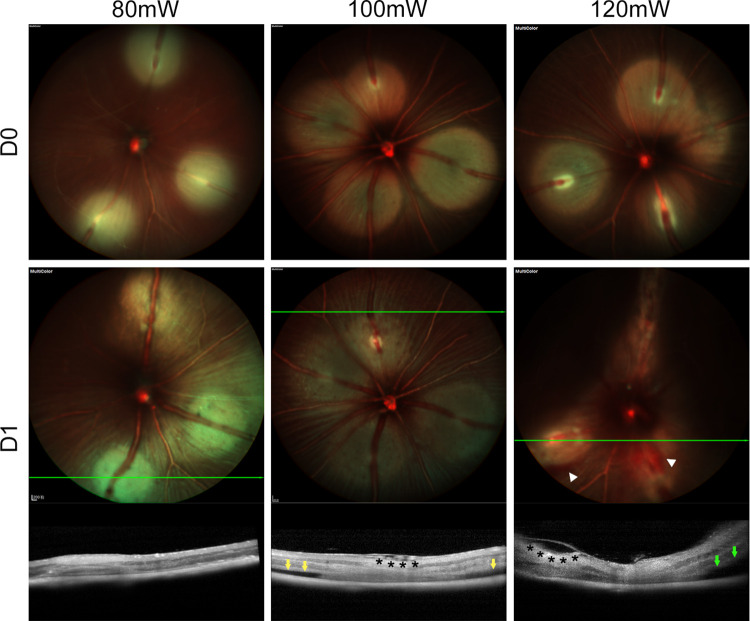
Photodynamic venous occlusion is a commonly accepted method for establishing mouse models of retinal vein occlusion (RVO). However, existing model parameters do not distinguish between acute and chronic RVO subtypes. Large variations in laser energy seem to correlate with fluctuating retinopathy severity and high rates of venous recanalization during the acute phase, along with the variable levels of retinal perfusion during the chronic phase. After optimizing the modeling procedure and defining success and exclusion criteria, laser energy groups of 80mW, 100mW, and 120mW were established. Multimodal imaging confirmed that higher energy levels increased the incidence of retinal cystoid edema and intraretinal hemorrhage, exacerbated the severity of exudative retinal detachment, and reduced the venous recanalization rate. For the acute model, 100mW was considered an appropriate parameter for balancing moderate retinopathy and venous recanalization. Continuous imaging follow-up revealed that day 1 after RVO was the optimal observation point for peaking of retinal thickness and intensive occurrence of retinal cystic edema and intraretinal hemorrhage. After excluding the influence of venous recanalization on retinal thickness, acute retinal edema demonstrated a positive response to standard anti-vascular endothelial growth factor therapy, validating the clinical relevance of the acute RVO model for further study in pathogenic mechanisms and therapeutic efficacy. For the chronic model, the 120mW parameter with the lowest venous recanalization rate was applied, accompanied by an increase in both photocoagulation shots and range to ensure sustained vein occlusion. Imaging follow-up clarified non-ischemic retinopathy characterized by tortuosity and dilation of the distal end, branches, and adjacent veins of the occluded vein. These morphological changes are quantifiable and could be combined with electrophysiological functional assessment for treatment effectiveness evaluation. Moreover, the stable state of venous occlusion may facilitate investigations into response and compensation mechanisms under conditions of chronic retinal hypoperfusion.


Conflict of interest statement

The authors have declared that no competing interests exist.

Figures

Fig 1
Fig 1. Major vein identification and laser focus adjustment for photocoagulation.
(A) Distinction of the major retina arteries and veins at the posterior pole of the fundus. Red and blue marks represented retinal major arteries and veins, respectively. Five to six arteries and veins respectively emanated from the optic disc and were arranged in an alternating spoke-like pattern. Retinal arteries were thin and bright red, with obvious reflective walls, and might send out two to three branches at the posterior pole of the fundus. Retinal veins were thick and dark red, and did not give off branches at the posterior pole of the fundus. (B) Focus adjustment procedure during the photocoagulation. The visual axis was aligned with the laser path for centering the optic disc and clear visibility of major blood vessels. Partial fundus structure blurring caused by misalignment should be avoided. The laser focus was adjusted to target a small segment of the vein at the laser indication point until the wall reflection appeared, while simultaneously blurring the surrounding retinal structure to achieve an optimal photocoagulation condition.
Fig 2
Fig 2. Success and exclusion criteria for the RVO mouse model.
Success criteria for RVO after photocoagulation: (A) extreme vein stenosis on SLO, (B) photothrombosis on SLO and the corresponding hyperreflective lump within the venous lumen on OCT, (C) venous filling defect on FFA. Exclusion criteria during the laser procedure and follow-up phase: (D) unoccluded veins on SLO, (E) irregularity of the laser spot on SLO, (F) hemorrhage of the target vein on SLO and OCT, (G) outer retina damage as the formation of evaporative bubbles on SLO and OCT during the laser procedure; (H) artery involvement manifested as retinal arteriovenous occlusion on FFA in the follow-up phase. Abbreviations: RVO, retinal vein occlusion. SLO, scanning laser ophthalmoscopy. OCT, optical coherence tomography. FFA, fundus fluorescein angiography.
Fig 3
Fig 3. Typical fundus changes of 80mW, 100mW, and 120mW groups after RVO.
Post-photocoagulation RVO was all achieved in 80mW, 100mW and 120mW groups by SLO. 1 day after RVO, 3 groups exhibited typical retinopathies on SLO and OCT. The 80mW group mainly developed venous recanalization on SLO. The 100mW group displayed cystoid edema (black asterisk) in NFL-GCL and superficial RD (yellow arrow) on SLO. The 120mW group exhibited intraretinal hemorrhage on SLO (white arrowhead), severe RD on both SLO (dark areas) and OCT (green arrow), as well as cystoid edema on OCT (black asterisk). Abbreviations: RVO, retinal vein occlusion. SLO, scanning laser ophthalmoscopy. OCT, optical coherence tomography. NFL-GCL, nerve fiber layer-ganglion cell layer. RD, retinal detachment.
Fig 4
Fig 4. Retinopathy progression of acute RVO in SLO.
(A) The progression of intraretinal hemorrhage in the 100mW acute RVO model (Laser group) compared with its control (Sham group) after photocoagulation (Day 0) and at days 1, 3, 5, and 7 after RVO. In the Laser group, intraretinal hemorrhage (white arrowhead) and exudative RD (dark area) both occurred on day 1 after RVO. The former gradually resolved during days 3 to 5 after RVO, while the latter resolved on day 3 after RVO. No intraretinal hemorrhage nor RD was observed in the Sham group. (B) The venous occlusion rate trend steadily decreased during the follow-up period in the acute RVO model. Abbreviations: RVO, retinal vein occlusion. SLO, scanning laser ophthalmoscopy. RD, retinal detachment.
Fig 5
Fig 5. Retinopathy progression of acute RVO in OCT.
(A) Comparative OCT, histology and immunofluorescence of retinal cross-section revealed retinal layers and vascular distribution. The whole retina included NFL-GCL, IPL, INL, OPL, ONL, photoreceptor layer and RPE. NFL-GCL to INL constituted the inner retina, while OPL to the photoreceptor layer constituted the outer retina. NFL-GCL contained both major vessels and superficial retinal capillaries, intermediate capillaries were located at the IPL-INL junction, and deep capillaries were situated at the INL-OPL junction. (B) Progression of retinal edema to atrophy of the 100mW acute RVO model (Laser group) compared with the control (Sham group) after photocoagulation (day 0) and at day 1, 2, 3, 5 and 7 after RVO. Abbreviations: RVO, retinal vein occlusion. OCT, optical coherence tomography. NFL-GCL, nerve fiber layer-ganglion cell layer. IPL, inner plexiform layer. INL, inner nuclear layer. OPL, outer plexiform layer. ONL, outer nuclear layer. RPE, retinal pigment epithelium.
Fig 6
Fig 6. Retinal thickness trend of the acute RVO.
Retinal layer thickness after photocoagulation (day 0) and at days 1, 2, 3, 5, and 7 after RVO was presented as mean±SEM. The 100mW acute RVO model (Laser group) was compared with the control (Sham group) at each time point using unpaired Student’s t-test or Mann-Whitney U test for datasets that did not adhere to the normal distribution: *p<0.05, **p<0.01, ***p<0.001. Normal retinal thickness was analyzed as the baseline. (A-F) Thickness trend of whole retina, outer retina, inner retina, NFL-GCL, IPL, and INL, respectively. A consistent peak in thickness on day 1 after RVO occurred across all retinal layers, with significant differences compared to the control group except for INL. Abbreviations: RVO, retinal vein occlusion. NFL-GCL, nerve fiber layer-ganglion cell layer. IPL, inner plexiform layer. INL, inner nuclear layer.
Fig 7
Fig 7. The efficacy of anti-VEGF treatment in alleviating acute retinal edema.
(A) The schedule for anti-VEGF administration and efficacy evaluation in the acute RVO mouse model. On D0, after establishing the acute RVO model and conducting baseline OCT thickness measurements of the whole retina, NFL-GCL, and INL, immediate intravitreal injections of Aflibercept at 20 μg/μL, 2 μL per eye were administered. On D1 and D2, OCT scans were performed after SLO examinations to exclude recanalized veins, assessing the thickness of the aforementioned layers and the occurrence of cystic edema. (B) Representative images of Aflibercept alleviating acute retinal edema. Significant reductions in whole retinal and INL thickness were observed in the Aflibercept group at D1 and D2 compared to the PBS group. (C) Quantification of Aflibercept’s reduction of retinal thickness in the whole retina, NFL-GCL, and INL. At D1 and D2, the thickness of the whole retina and INL was significantly lower in the Aflibercept groups compared to the control group (P<0.05). (D) Aflibercept slightly reduced the incidence of cystoid retinal edema. The retinal layer thickness and the occurrence of retinal cystoid edema were presented as mean±SEM and incidence, respectively. The retinal layer thickness at each follow-up point in the Aflibercept group and PBS group was compared by unpaired Student’s t-test or Mann-Whitney U test for datasets that did not adhere to the normal distribution, while the incidence of cystoid retinal edema between the two groups was compared by Chi-square test: *p<0.05, **p<0.01, ***p<0.001. Abbreviations: RVO, retinal vein occlusion. Afli, Aflibercept. PBS, phosphate-buffered saline. OCT, optical coherence tomography. SLO, scanning laser ophthalmoscopy. NFL-GCL, nerve fiber layer-ganglion cell layer. INL, inner nuclear layer.
Fig 8
Fig 8. Retinal vasculature and perfusion in the chronic RVO.
The chronic RVO model mainly manifested as changes in the morphology of the major vein and its branches. (A) IR and (or) FFA were performed after photocoagulation (day 0) and days 7, 14, 21 and 31 after RVO for retinal vasculature and perfusion evaluation. The length of the occluded venous segment decreased from days 7 to 21 after RVO, but rebounded by day 31 after RVO. The nonperfusion area of the laser spot reduced during the follow-up phase, with none observed in the peripheral retina. Tortuous change (yellow arrow) in an adjacent major vein was observed on day 21 and persisted until day 31 after RVO, without affecting the distal end of the occluded vein. (B) The tortuous change was also found in the peripheral branches of the occluded veins. (C) Normal retinal vasculature and perfusion as the control. The major retinal vein emitted two branches (yellow arrowhead) in the peripheral retina, with some connecting to adjacent ones, accompanied by retinal capillaries on FFA. Abbreviations: RVO, retinal vein occlusion. IR, Infrared mode. FFA, fundus fluorescein angiography.

Similar articles

Cited by

References

    1. Terao R, Fujino R, Ahmed T. Risk Factors and Treatment Strategy for Retinal Vascular Occlusive Diseases. J Clin Med. 2022;11(21). Epub 2022/11/12. doi: 10.3390/jcm11216340 ; PubMed Central PMCID: PMC9656338. - DOI - PMC - PubMed
    1. Khayat M, Lois N, Williams M, Stitt AW. Animal Models of Retinal Vein Occlusion. Investigative ophthalmology & visual science. 2017;58(14):6175–92. Epub 2017/12/10. doi: 10.1167/iovs.17-22788 . - DOI - PubMed
    1. Zhang H, Sonoda K-H, Qiao H, Oshima T, Hisatomi T, Ishibashi T. Development of a new mouse model of branch retinal vein occlusion and retinal neovascularization. Japanese Journal of Ophthalmology. 2007;51(4):251–7. doi: 10.1007/s10384-007-0445-2 - DOI - PubMed
    1. Ehlers JP, Fekrat S. Retinal vein occlusion: beyond the acute event. Surv Ophthalmol. 2011;56(4):281–99. Epub 2011/05/24. doi: 10.1016/j.survophthal.2010.11.006 . - DOI - PubMed
    1. Colón Ortiz C, Potenski A, Lawson JM, Smart J, Troy CM. Optimization of the Retinal Vein Occlusion Mouse Model to Limit Variability. Journal of Visualized Experiments: JoVE. 2021;(174). doi: 10.3791/62980 - DOI - PMC - PubMed