Case Reports
. 2024 Jun 17;9(14):e179071.
doi: 10.1172/jci.insight.179071.

FDXR variants cause adrenal insufficiency and atypical sexual development

Affiliations
Case Reports

FDXR variants cause adrenal insufficiency and atypical sexual development

Emanuele Pignatti et al. JCI Insight. .

Abstract

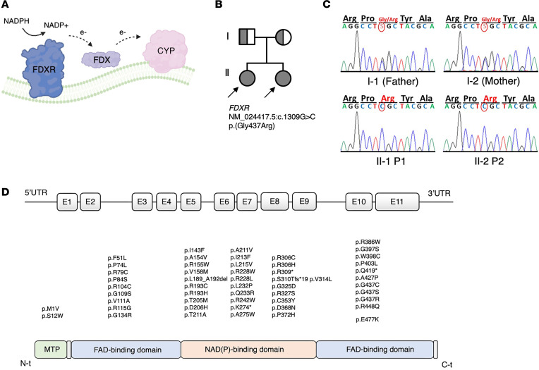
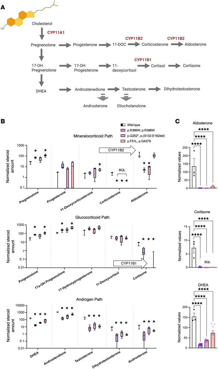
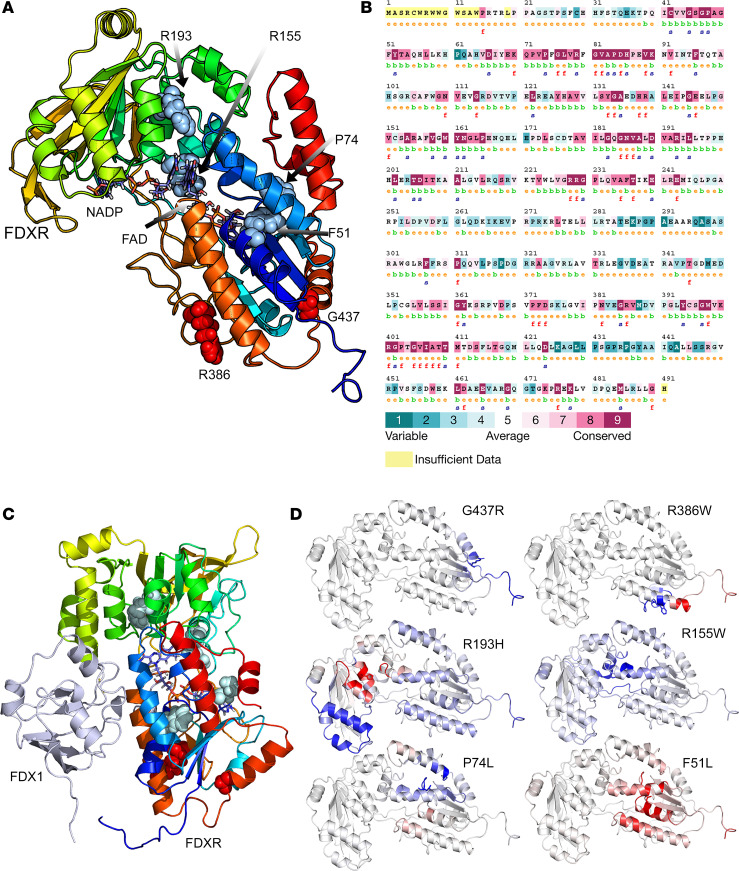
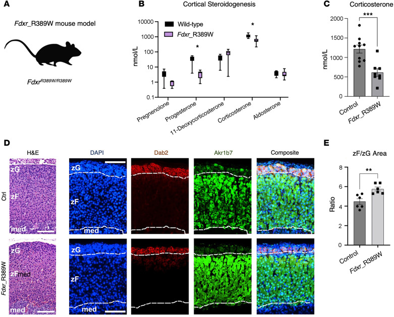
Genetic defects affecting steroid biosynthesis cause cortisol deficiency and differences of sex development; among these defects are recessive mutations in the steroidogenic enzymes CYP11A1 and CYP11B, whose function is supported by reducing equivalents donated by ferredoxin reductase (FDXR) and ferredoxin. So far, mutations in the mitochondrial flavoprotein FDXR have been associated with a progressive neuropathic mitochondriopathy named FDXR-related mitochondriopathy (FRM), but cortisol insufficiency has not been documented. However, patients with FRM often experience worsening or demise following stress associated with infections. We investigated 2 female patients with FRM carrying the potentially novel homozygous FDXR mutation p.G437R with ambiguous genitalia at birth and sudden death in the first year of life; they presented with cortisol deficiency and androgen excess compatible with 11-hydroxylase deficiency. In addition, steroidogenic FDXR-variant cell lines reprogrammed from 3 patients with FRM fibroblasts displayed deficient mineralocorticoid and glucocorticoid production. Finally, Fdxr-mutant mice allelic to the severe p.R386W human variant showed reduced progesterone and corticosterone production. Therefore, our comprehensive studies show that human FDXR variants may cause compensated but possibly life-threatening adrenocortical insufficiency in stress by affecting adrenal glucocorticoid and mineralocorticoid synthesis through direct enzyme inhibition, most likely in combination with disturbed mitochondrial redox balance.

Keywords: Endocrinology; Genetic diseases; Genetics; Mitochondria; Molecular genetics.


Figures

Figure 1
Figure 1. Role of FDXR and genetic characteristics of the FDXR variants identified in the index patients and of the reported patients manifesting with FDXR-related mitochondriopathy (FRM).
(A) Schematic representation of the role of the flavoprotein ferredoxin–NADP(+) reductase (FDXR) as electron acceptor from nicotinamide adenine dinucleotide phosphate (NADPH), and electron donor for ferredoxin proteins (FDX), from where electrons are finally donated to effector Cytochrome P450 (CYP) enzymes associated to the inner mitochondrial membrane. (B) Pedigree of a family in which the 2 daughters are affected by neuropathy and adrenal insufficiency caused by the homozygous c.1309G > C (p.G437R) variant in FDXR. (C) displays the result of Sanger sequencing around the c.139 region for the members of the family in B. (D) reviews the FDXR variants that have been described in patients with FRM as of June 2023, including the novel p.G437R described in the index patients in this manuscript, aligned to the relevant protein domain. Domain annotation is based on a crystallography analysis of the Bos taurus FDXR ortholog (36).
Figure 2
Figure 2. Cells from FDXR patient display low CYP11B1 and CYP11B2 enzymatic activity.
(A) Classical steroids and steroidogenic pathways, all initiated from cholesterol (top left), occurring in the human adrenal cortex. In red, the official names of the 3 FDXR-dependent mitochondrial steroidogenic enzymes, namely CYP11A1 (also known as Cholesterol side-chain cleavage enzyme), CYP11B2 (Aldosterone Synthase), and CYP11B1 (Steroid 11β-hydroxylase). (B) Steroid amounts in culture media conditioned by reprogrammed fibroblasts from FDXR patients compared with control values (representing steroid amounts in culture media conditioned by reprogrammed fibroblasts from a single nonaffected individual; i.e., Control). Steroids are split among 3 graphs according to their belonging to a specific steroid class. Arrows containing the names of enzymes indicate the enzymatic reaction carried out by the enzyme. Asterisks reflect discoveries found using a multiple unpaired t test assuming individual variance for each steroid. *P < 0.05. (C) The endpoint or most representative steroids for each pathway on the left, on a linear scale. Statistical analysis was conducted using a 1-way ANOVA followed by Dunnett’s multiple comparisons test. All values in B and C are normalized by GAPDH transcripts contained within the cell monolayer, used as a proxy for cell number, as reported in Supplemental Figure 1D. Below quantification level (BQL) indicates the samples in which steroid levels were not measurable above the lowest quantification limit using LC-MS. DHEA, Dehydroepiandrosterone; 11-DOC, 11-deoxycorticosterone. ****Padj < 0.0001.
Figure 3
Figure 3. Sequence and structure analysis of mutations in FDXR.
(A) A 3D model of human FDXR displayed as a ribbon diagram. The positions of the phenylalanine 51, proline 74, arginine 155, arginine 193, arginine 386, and glycine 437 residues are indicated. The structural model of human FDXR is based on a known 3D structure of the bovine protein as described in the Methods section. The diagram is colored using a rainbow palette with blue at N-terminus and red at C-terminus. Cofactors (NADP, FAD) are shown as stick models, while amino acids phenylalanine 51, proline 74, arginine 155, arginine 193, arginine 386, and glycine 437 are shown as spheres. (B) The evolutionary sequence conservation of FDXR. Most of the mutations reported in this study are highly conserved across species and are predicted to have structural roles. Sequences are colored based on amino acid conservation, with dark blue being the least conserved and dark red being the most conserved, while yellow indicates that no prediction could be made. (C) A complex of FDXR and FDX1 proteins showing the locations of mutated residues, which are not at the FDXR-FDX interface and are predicted not to have a direct effect on FDX-FDXR interaction. (D) Stability and flexibility analysis of mutated FDXR structures compared with WT FDXR. An increased flexibility was observed for amino acid changes F51L and R193H (shown in red), indicating decreased stability that was supported by differential free energy calculations. Decreased flexibility due to P74L, R155W, R386W, and G537R mutations is shown in blue.
Figure 4
Figure 4. The FdxrR389W mouse model shows no impairment of adrenal structure and zonation.
(A) Schematic of the novel mouse model (FdxrR389W) carrying homozygous R389W mutations, allelic to the hotspot R386W variant in FDXR patients. (B) Serum steroid profile of the FdxrR389W mice compared with control animals. Asterisks reflect discoveries found using a multiple unpaired t test assuming individual variance for each steroid, with FDR, and a 2-stage step-up method (Benjamini, Krieger, and Yekutieli). *P < 0.01. (C) Serum levels of corticosterone, the main glucocorticoids in mice, in control and FdxrR389W mice. Significance was tested using an unpaired t test. (D) Micrographs of representative adrenal sections, either stained with H&E (left) or immunoassayed with Dab2 (zona glomerulosa, zG), Akr1b7 (zona fasciculata, zF), and DAPI (for nuclei; right panels). Scale bar: 200 μm. Dotted white lines outline the zG region as identified using Dab2 staining, and the corticomedullary (med) region (below) as marked by the lower boundary of the Akr1b7 staining. (E) Ratio values calculated as zF area normalized by zG area, measured on 6 independent entire adrenal coronal sections for either controls or FdxrR389W samples. An unpaired t test was used to calculate significance. **P < 0.01; ***P < 0.001.

References

    1. Miller WL, et al. The Adrenal Cortex and Its Disorders. In: Sperling MA, ed. Sperling Pediatric Endocrinology, 5th Edition. Elsevier; 2020:425–490.
    1. Pignatti E, Flück CE. Adrenal cortex development and related disorders leading to adrenal insufficiency. Mol Cell Endocrinol. 2021;527:111206. doi: 10.1016/j.mce.2021.111206. - DOI - PubMed
    1. Miller WL. Steroidogenic electron-transfer factors and their diseases. Ann Pediatr Endocrinol Metab. 2021;26(3):138–148. doi: 10.6065/apem.2142154.077. - DOI - PMC - PubMed
    1. Griffin A, et al. Ferredoxin 1b (Fdx1b) Is the Essential Mitochondrial Redox Partner for Cortisol Biosynthesis in Zebrafish. Endocrinology. 2016;157(3):1122–1134. - PMC - PubMed
    1. Oakes JA, et al. Ferredoxin 1b deficiency leads to testis disorganization, impaired spermatogenesis, and feminization in zebrafish. Endocrinology. 2019;160(10):2401–2416. doi: 10.1210/en.2019-00068. - DOI - PubMed

Publication types