Review
. 2025 Mar 3;17(3):a041520.
doi: 10.1101/cshperspect.a041520.

The Mechanics of Building Functional Organs

Affiliations
Review

The Mechanics of Building Functional Organs

Toby G R Andrews et al. Cold Spring Harb Perspect Biol. .

Abstract

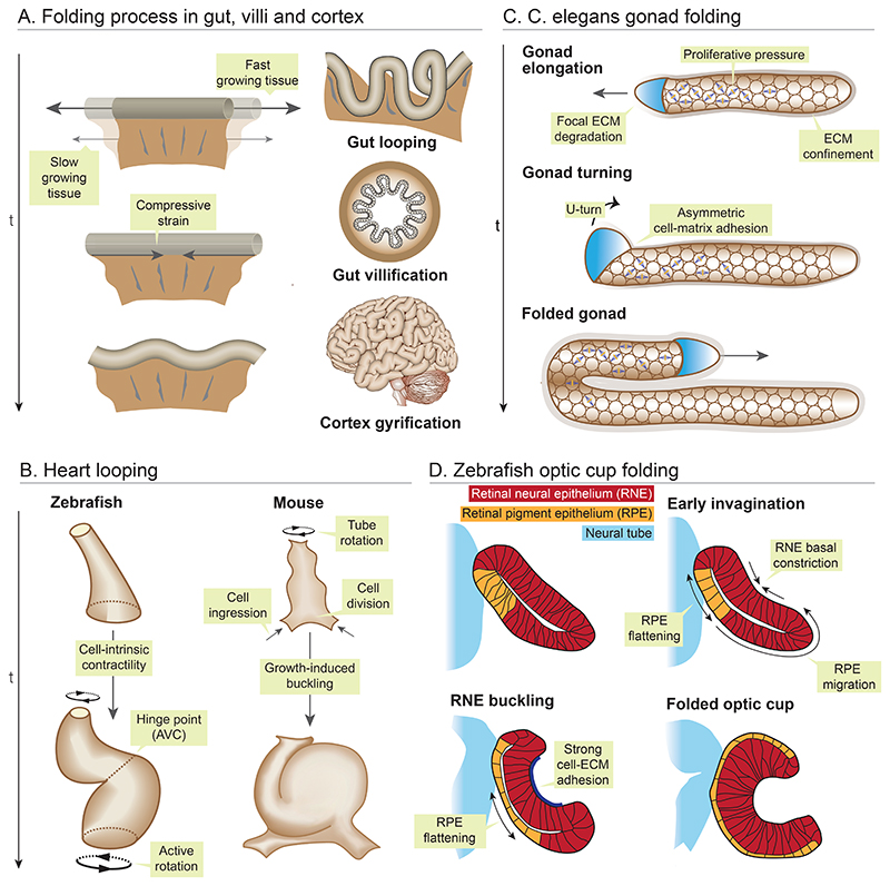
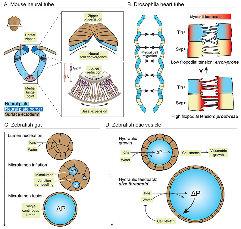
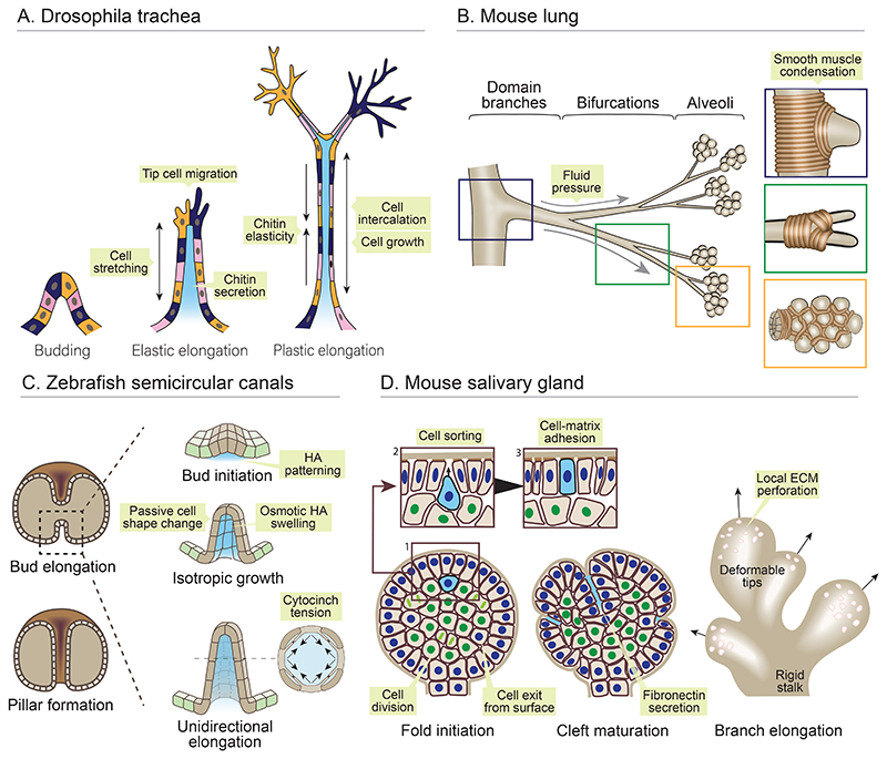
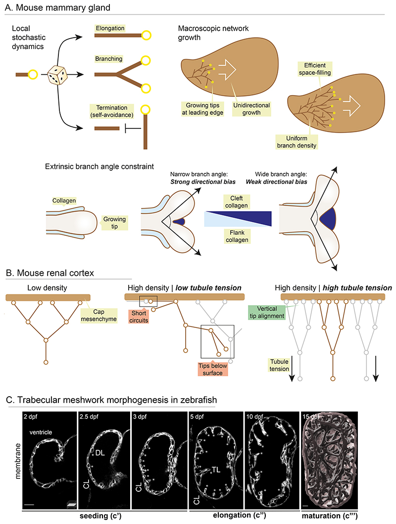
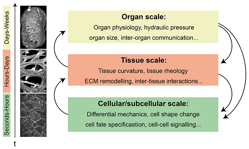
Organ morphogenesis is multifaceted, multiscale, and fundamentally a robust process. Despite the complex and dynamic nature of embryonic development, organs are built with reproducible size, shape, and function, allowing them to support organismal growth and life. This striking reproducibility of tissue form exists because morphogenesis is not entirely hardwired. Instead, it is an emergent product of mechanochemical information flow, operating across spatial and temporal scales-from local cellular deformations to organ-scale form and function, and back. In this review, we address the mechanical basis of organ morphogenesis, as understood by observations and experiments in living embryos. To this end, we discuss how mechanical information controls the emergence of a highly conserved set of structural motifs that shape organ architectures across the animal kingdom: folds and loops, tubes and lumens, buds, branches, and networks. Moving forward, we advocate for a holistic conceptual framework for the study of organ morphogenesis, which rests on an interdisciplinary toolkit and brings the embryo center stage.


Figures

Figure 1
Figure 1. Physical principles of folding and looping morphogenesis.
(A) A faster growing tissue confined by slower growing adjacent tissue experiences compressive forces, which generates mechanical instabilities, driving brain cortex folding, mid gut lopping and gut villification. (B) In zebrafish, actomyosin forces twists the two chambers of the heart around the atrioventricular (AV) canal, resulting in torsion of the linear heart tube into S-shape. In mice, asymmetric rotation, preferential cell ingression and proliferation at the ventral pole buckles the linear heart tube rightwards to form a loop. (C) In C. elegans proliferative pressure from germ cells and ECM confinement propels gonad elongation, while direction of folding is driven by polarised cell-ECM interaction. (D) Optic cup morphogenesis in zebrafish is a multifaceted process. RPE cells migrate and get incorporated into the inner RNE layer, which compresses and buckle the RNE. The basal surface of the RNE layer constricts and is constrained by ECM adhesion which further accentuates the buckling process. The outer RPE layer stretches and flattens to drive the optic cup folding.
Figure 2
Figure 2. Physical principles of tube morphogenesis.
(A) The vertebrate neural tube forms through folding of the epithelial neural plate. Schematic shows a transverse section of an amniote embryo, with the neural folds elevating and converging at the dorsal midline. (Top inlay) Cell junction contraction and remodelling zippers the neural folds together and separates them from the surface ectoderm. (Bottom inlay) Protraction of S-phase at the ventral midline prolongs basal nuclear localisation, driving medial hinge point formation. (B) The drosophila heart tube is formed by bilateral rows of cardioblasts (CBs) that converge at the dorsal midline and form new cell-cell adhesions. CBs extend filopodia that form homotypic contacts with contralateral CBs owing to differential adhesion profiles. Myosin II oscillates between the front and rear of each CB, operating a proof-reading system. At the rear, filopodia can extend and form nascent adhesions. At the front, filopodial tension increases, severing heterotypic adhesions and reinforcing homotypic adhesions. (C) The zebrafish gut forms a lumen de novo through cord hollowing. Active ion transport builds an osmotic gradient, followed by water. Positive hydraulic pressure (ΔP) inflates small microlumens, which fuse through junctional remodelling. (D) The zebrafish otic vesicle similarly inflates through generation of hydraulic pressure. As the lumen grows, surrounding cells stretch and undergo viscoelastic shape change. High hydraulic pressure inhibits ion transport, allowing self-organisation of lumen size.
Figure 3
Figure 3. Morphogenesis of epithelial buds and branches.
(A) The drosophila trachea branches through epithelial remodelling in post-mitotic cells. Buds elongate through active migration of tip cells, which increase tension in the stalk, leading to elastic cell stretch and branch outgrowth. Area growth and intercalation of stalk cells enables plastic elongation, restricted by elasticity of a lumenal chitin matrix. (B) Domain branches, bifurcations and alveoli in the mouse lung are sculpted by smooth muscle fibres which locally condense at branch tips and form a physical growth barrier. The airway epithelium buds between smooth muscle fibres under positive hydraulic force. (C) Zebrafish semi-circular canals appear as buds extending into the otic vesicle lumen. Hyaluronic acid (HA) is secreted locally beneath the prospective bud, and osmotically swells, leading to budding through passive cellular deformations. Cytocinches between cells increase circumferential tension, translating isotropic HA swelling into anisotropic bud elongation. (D) The mouse salivary gland buds through folding of the epithelial surface and basement membrane. Cells exit the surface layer (blue nuclei) and divide in the core of the bud (green nuclei). Daughter cells (blue cell) then sort from inner cells owing to differential adhesion, return to the outer, and restore adhesion with the basement membrane. Growth of the surface layer folds the basement membrane, forming clefts stabilised by fibronectin secretion. Outgrowth of new branches is aided by focal ECM degradation, converting it from a stiff shell to a deformable lattice.
Figure 4
Figure 4. Mechanical control of network growth dynamics.
(A) The mouse mammary gland epithelium branches with stochastic dynamics. (Left) Branches make stochastic decisions to elongate or branch and terminate through local self-avoidance. (Right) These dynamics allow robust network growth with uniform density, effectively filling space. (Bottom) Collagen accumulates on the flanks of branches and limits branch angle. Branch angle is defined by relative collagen enrichment in the flank and cleft, and dictates global network directionality. (B) In the mouse renal cortex, branch tips interface with the cap mesenchyme for nephron induction. At high density, this topology can be disrupted, with tips short-circuiting or falling beneath the surface. Tubule tension ensures a vertical alignment of tip families at high density, allowing a high packing density with robust mesenchymal interfaces. (C) Trabecular meshwork morphogenesis in zebrafish. Mid-sagittal section (c’, c”) and 3D surface rendered (c”’) images of zebrafish hearts expressing membrane marker. Compact layer (CL), delaminating, (DL, asterisks) and trabecular (TL, asterisks) cells. Stochastic single cell delamination from the outer compact layer seeds trabecular layer cells, and this seeding is triggered by mechanical heterogeneity (c’). These single trabecular cells transform into multicellular ridges (c”), and these ridges remodel and coalesce to form a mature 3D topological meshwork filling the ventricle lumen (c”’). Scale bar = 50 μm.
Fig. 5
Fig. 5. Bridging scales in Organ Morphogenesis.
The robust shape and function of organs emerge through reciprocal mechanical interactions operating across spatiotemporal scales: from seconds to weeks, and between cells to tissues to organs and back.

References

    1. What’s next for bioimage analysis? Nat Methods. 2023;20:945–946. - PubMed
    1. Agarwal P, Shemesh T, Zaidel-Bar R. Directed cell invasion and asymmetric adhesion drive tissue elongation and turning in C. elegans gonad morphogenesis. Dev Cell. 2022;57:2111–2126.:e6. - PubMed
    1. Akhtar N, Streuli CH. An integrin-ILK-microtubule network orients cell polarity and lumen formation in glandular epithelium. Nat Cell Biol. 2013;15:17–27. doi: 10.1038/ncb2646. - DOI - PMC - PubMed
    1. Alt S, Ganguly P, Salbreux G. Vertex models: From cell mechanics to tissue morphogenesis. Philos Trans R Soc Lond B Biol Sci. 2017;372 doi: 10.1098/rstb.2015.0520. - DOI - PMC - PubMed
    1. Alvers AL, Ryan S, Scherz PJ, Huisken J, Bagnat M. Single continuous lumen formation in the zebrafish gut is mediated by smoothened-dependent tissue remodeling. Development. 2014;141:1110–1119. doi: 10.1242/dev.100313. - DOI - PMC - PubMed

LinkOut - more resources