. 2024 Jun 17;14(1):13920.
doi: 10.1038/s41598-024-63565-z.

The matricellular protein CCN5 prevents anti-VEGF drug-induced epithelial-mesenchymal transition of retinal pigment epithelium

Affiliations

The matricellular protein CCN5 prevents anti-VEGF drug-induced epithelial-mesenchymal transition of retinal pigment epithelium

Sora Im et al. Sci Rep. .

Abstract

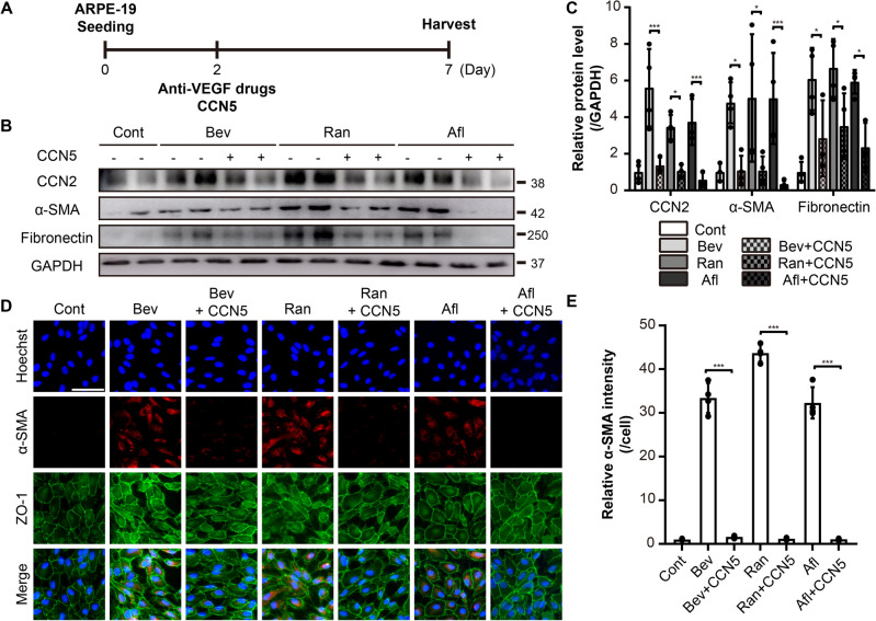
Age-related macular degeneration (AMD) is one of the major causes of blindness in the elderly worldwide. Anti-vascular endothelial growth factor (VEGF) drugs have been widely used to treat the neovascular type of AMD (nAMD). However, VEGF acts not only as a pro-angiogenic factor but also as an anti-apoptotic factor in the eyes. In this study, we found that anti-VEGF drugs, including bevacizumab (Bev), ranibizumab (Ran), and aflibercept (Afl), induced epithelial-mesenchymal transition (EMT) in ARPE-19 cells in vitro, accompanied by the induction of CCN2, a potent pro-fibrotic factor. Similarly, intravitreal injection of Afl into mouse eyes resulted in EMT in the retinal pigmented epithelium (RPE). Co-treatment with CCN5, an anti-fibrotic factor that down-regulates CCN2 expression, significantly attenuated the adverse effects of the anti-VEGF drugs both in vitro and in vivo. Inhibition of the VEGF signaling pathway with antagonists of VEGF receptors, SU5416 and ZM323881, induced EMT and up-regulated CCN2 in ARPE-19 cells. Additionally, knock-down of CCN2 with siRNA abolished the adverse effects of the anti-VEGF drugs in ARPE-19 cells. Collectively, these results suggest that anti-VEGF drugs induce EMT in RPE through the induction of CCN2 and that co-treatment with CCN5 attenuates the adverse effects of anti-VEGF drugs in mouse eyes.


Conflict of interest statement

K.M.W. and W.J.P. share co-ownership of Olives Biotherapeutics. No potential conflicts of interest exist for other authors.

Figures

Figure 1
Figure 1
Anti- VEGF drugs induce EMT in ARPE-19 cells. (A) An experimental scheme. The ARPE-19 cells were treated with anti-VEGF drugs, bevacizumab (Bev), ranibizumab (Ran), and aflibercept (Afl), at 2× clinical doses. (B) Cell lysates from ARPE-19 cells were immunoblotted with antibodies against CCN2, α-smooth muscle actin (α-SMA), fibronectin, and GAPDH. Data are representatives of more than three independent experiments. (C) Protein levels were quantified using Image-J software and plotted. Bars show the mean ± SD. (n = 4–6 per group; one-way ANOVA; *p < 0.05, **p < 0.01, ***p < 0.001) (D) Immunofluorescence images of ARPE-19 cells stained with anti-α-SMA and anti-zonula occludens-1 (ZO-1) antibodies, and Hoechst 33342. Representative images are shown. Scale bar, 50 μm. (E) The intensity of α-SMA per cell was quantified and plotted using Image-J software. Bars show the mean ± SD. (n = 3 per group; one-way ANOVA; ***p < 0.001).
Figure 2
Figure 2
Recombinant CCN5 protein inhibits EMT induced by anti-VEGF drugs in ARPE-19 cells. (A) An experimental scheme. The ARPE-19 cells treated with anti-VEGF drugs, Bev, Ran, and Afl, at 2× clinical doses in the absence or presence of recombinant CCN5 protein (500 ng/mL). (B) Cell lysates from ARPE-19 cells were immunoblotted with antibodies against CCN2, α-SMA, fibronectin, and GAPDH. Data are representatives of more than three independent experiments. (C) Protein levels were quantified using Image-J software and plotted. Bars show the mean ± SD. (n = 4 per group; one-way ANOVA; *p < 0.05, ***p < 0.001) (D) Immunofluorescence images of ARPE-19 cells stained with anti-α-SMA and anti-ZO-1 antibodies, and Hoechst 33342. Representative images are shown. Scale bar, 50 μm. (E) The intensity of α-SMA per cell was quantified and plotted using Image-J software. Bar show the mean ± SD. (n = 3–4 per group; one-way ANOVA; ***p < 0.001).
Figure 3
Figure 3
Recombinant CCN5 protein inhibits EMT induced by aflibercept (Afl) in mouse eyes. (A) An experimental scheme. Normal human IgG (40 mg/eye), Afl (40 μg/eye), and Afl and CCN5 (40 ng/eye) were intravitreally injected. Mice were sacrificed, and eyes were enucleated at 14 days post-injection. (B) Flat mounts of RPE/choroid complex were stained with anti-α-SMA antibody. Representative images are shown. Scale bar, 1000, 500, and 250 μm. (C) The intensity of α-SMA positive areas was quantified by Image-J software and plotted. (D) Flat mounts of RPE/choroid complex were stained with anti-α-SMA and anti-ZO-1 antibodies, and Hoechst 33342. Representative images are shown. Scale bar, 50 μm. (E) The mean number of RPE cells in 100 × 100 μm area was manually counted and plotted. (F) The mean size of RPE cells in 100 × 100 μm area was manually calculated and plotted. Error bars = SEM. (n = 8, Cont; n = 8, IgG; n = 10, Afl; n = 13, Afl+CCN5; one-way ANOVA; ***p < 0.001).
Figure 4
Figure 4
Antagonists of VEGF receptors induce EMT in ARPE-19 cells. (A) An experimental scheme. The ARPE-19 cells were treated with 5 μM of SU5416, an antagonist of VEGFR-1/2, or 10 nM of ZM323881, an antagonist of VEGFR-2. (B) Cell lysates from ARPE-19 cells were immunoblotted with antibodies against CCN2, α-SMA, fibronectin, and GAPDH. Data are representatives of more than three independent experiments. (C) Protein levels were quantified using Image-J software and plotted. Bars show the mean ± SD. (n = 4 per group; one-way ANOVA; **p < 0.01, ***p < 0.001) (D) Immunofluorescence images of ARPE-19 cells stained with anti-α-SMA and anti-CCN2 antibodies, and Hoechst 33342. Representative images are shown. Scale bar, 50 μm. (E) The intensity of CCN2 or α-SMA per cell was quantified and plotted using Image-J software. Bars show the mean ± SD. (n = 4 per group; one-way ANOVA; ***p < 0.001).
Figure 5
Figure 5
Afl-induced EMT is ameliorated by CCN2 siRNA in ARPE-19 cells. (A) An experimental scheme. The ARPE-19 cells were treated with CCN2 siRNA (1 μM) or CCN5 (500 ng/mL) in the presence of 2× Afl for 5 days. (B) Cell lysates from ARPE-19 cells were immunoblotted with antibodies against CCN2, α-SMA, fibronectin, and GAPDH. Data are representatives of more than three independent experiments. (C) Protein levels were quantified using Image-J software and plotted. Bars show the mean ± SD. (n = 5 per group; one-way ANOVA; ***p < 0.001) (D) Immunofluorescence images of ARPE-19 cells were stained with anti- α-SMA and anti-CCN2 antibodies, and Hoechst 33342. Representative images are shown. Scale bar, 50 μm. (E) The intensity of CCN2 or α-SMA per cell was quantified and plotted using Image-J software. Bars show the mean ± SD. (n = 4 per group; one-way ANOVA; ***p < 0.001).

References

    1. Pedrosa AC, et al. Treatment of neovascular age-related macular degeneration with anti-VEGF agents: Predictive factors of long-term visual outcomes. J. Ophthalmol. 2017;2017:4263017. doi: 10.1155/2017/4263017. - DOI - PMC - PubMed
    1. Little K, Ma JH, Yang N, Chen M, Xu H. Myofibroblasts in macular fibrosis secondary to neovascular age-related macular degeneration—The potential sources and molecular cues for their recruitment and activation. EBioMedicine. 2018;38:283–291. doi: 10.1016/j.ebiom.2018.11.029. - DOI - PMC - PubMed
    1. Campa C, et al. Inflammatory mediators and angiogenic factors in choroidal neovascularization: Pathogenetic interactions and therapeutic implications. Mediat. Inflamm. 2010 doi: 10.1155/2010/546826. - DOI - PMC - PubMed
    1. Zhang H, Liu Z-L. Transforming growth factor-β neutralizing antibodies inhibit subretinal fibrosis in a mouse model. Int. J. Ophthalmol. 2012;5:307–311. doi: 10.3980/j.issn.2222-3959.2012.03.11. - DOI - PMC - PubMed
    1. Ishikawa K, Kannan R, Hinton DR. Molecular mechanisms of subretinal fibrosis in age-related macular degeneration. Exp. Eye Res. 2016;142:19–25. doi: 10.1016/j.exer.2015.03.009. - DOI - PMC - PubMed

MeSH terms