

3D-printed ultra-small Brownian viscometers
- PMID: 38886461
- PMCID: PMC11183119
- DOI: 10.1038/s41598-024-64792-0
3D-printed ultra-small Brownian viscometers
Abstract
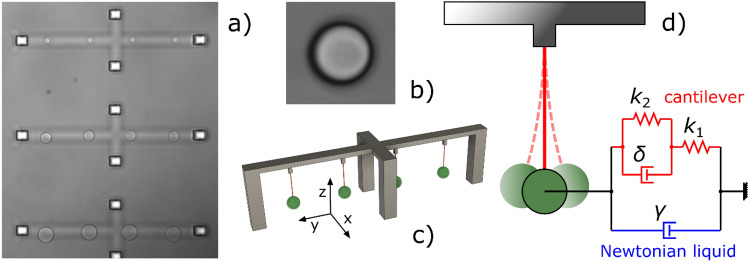
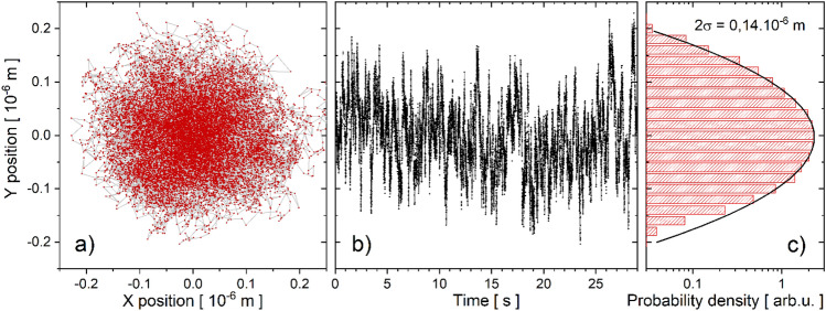
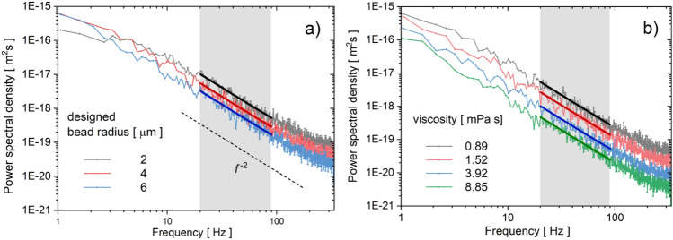
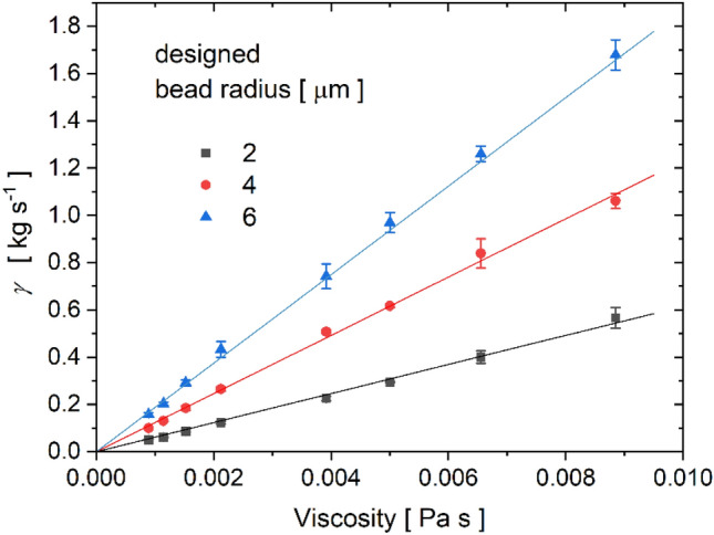
Measuring viscosity in volumes smaller than a microliter is a challenging endeavor. A new type of microscopic viscometers is presented to assess the viscosity of Newtonian liquids. Micron-sized flexible polymer cantilevers are created by two-photon polymerization direct laser writing. Because of the low stiffness and high elasticity of the polymer material the microcantilevers exhibit pronounced Brownian motion when submerged in a liquid medium. By imaging the cantilever's spherically shaped end, these fluctuations can be tracked with high accuracy. The hydrodynamic resistance of the microviscometer is determined by fitting the power spectral density of the measured fluctuations with a theoretical frequency dependence. Validation measurements in water-glycerol mixtures with known viscosities reveal excellent linearity of the hydrodynamic resistance to viscosity, allowing for a simple linear calibration. The stand-alone viscometer structures have a characteristic size of a few tens of microns and only require a very basic external instrumentation in the form of microscopic imaging at moderate framerates (~ 100 fps). Thus, our results point to a practical and simple to use ultra-low volume viscometer that can be integrated into lab-on-a-chip devices.
Keywords: Brownian fluctuations; Power spectral density; Two-photon polymerization; Viscoelastic polymer nanowire; Viscometer.
© 2024. The Author(s).
Conflict of interest statement
The authors declare no competing interests.
Figures
References
-
- Zhang ZH, Liu Y. Recent progresses of understanding the viscosity of concentrated protein solutions. Curr. Opin. Chem. Eng. 2017;16:48–55. doi: 10.1016/j.coche.2017.04.001. - DOI
Grants and funding
- BO/00290/21/11/Magyar Tudományos Akadémia
- ÚNKP-23-5-SZTE-717/Innovációs és Technológiai Minisztérium
- FK 138520/Nemzeti Kutatási Fejlesztési és Innovációs Hivatal
- APVV-21-0333/Agentúra na Podporu Výskumu a Vývoja
- APVV-21-0333/Agentúra na Podporu Výskumu a Vývoja
- APVV-21-0333/Agentúra na Podporu Výskumu a Vývoja
- APVV-21-0333/Agentúra na Podporu Výskumu a Vývoja
- APVV-21-0333/Agentúra na Podporu Výskumu a Vývoja
- APVV-21-0333/Agentúra na Podporu Výskumu a Vývoja
- VEGA 2/0101/22/Ministerstvo školstva, vedy, výskumu a športu Slovenskej republiky
- VEGA 2/0101/22/Ministerstvo školstva, vedy, výskumu a športu Slovenskej republiky
- ITMS2014+: 313011V455/European Regional Development Fund
- ITMS2014+: 313011V455/European Regional Development Fund
- ITMS2014+: 313011V455/European Regional Development Fund
- ITMS2014+: 313011V455/European Regional Development Fund
LinkOut - more resources
Full Text Sources
Miscellaneous
