. 2024 Aug;43(15):3240-3255.
doi: 10.1038/s44318-024-00137-x. Epub 2024 Jun 17.

The SMC5/6 complex prevents genotoxicity upon APOBEC3A-mediated replication stress

Affiliations

The SMC5/6 complex prevents genotoxicity upon APOBEC3A-mediated replication stress

Dylan F Fingerman et al. EMBO J. 2024 Aug.

Abstract

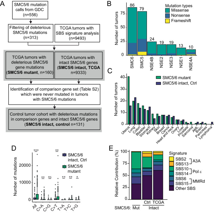
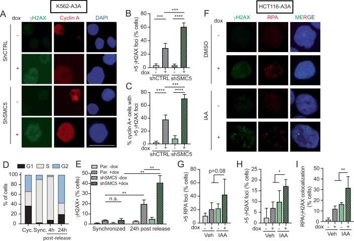
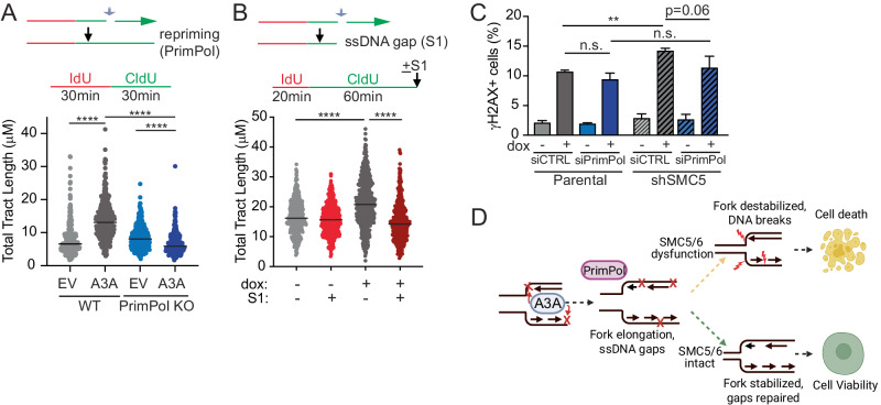
Mutational patterns caused by APOBEC3 cytidine deaminase activity are evident throughout human cancer genomes. In particular, the APOBEC3A family member is a potent genotoxin that causes substantial DNA damage in experimental systems and human tumors. However, the mechanisms that ensure genome stability in cells with active APOBEC3A are unknown. Through an unbiased genome-wide screen, we define the Structural Maintenance of Chromosomes 5/6 (SMC5/6) complex as essential for cell viability when APOBEC3A is active. We observe an absence of APOBEC3A mutagenesis in human tumors with SMC5/6 dysfunction, consistent with synthetic lethality. Cancer cells depleted of SMC5/6 incur substantial genome damage from APOBEC3A activity during DNA replication. Further, APOBEC3A activity results in replication tract lengthening which is dependent on PrimPol, consistent with re-initiation of DNA synthesis downstream of APOBEC3A-induced lesions. Loss of SMC5/6 abrogates elongated replication tracts and increases DNA breaks upon APOBEC3A activity. Our findings indicate that replication fork lengthening reflects a DNA damage response to APOBEC3A activity that promotes genome stability in an SMC5/6-dependent manner. Therefore, SMC5/6 presents a potential therapeutic vulnerability in tumors with active APOBEC3A.

Keywords: Cancer Mutagenesis; Cytidine Deaminase; Genome Integrity; Mutational Signatures; Replication Stress.


Conflict of interest statement

The authors declare no competing interests.

Figures

Figure 1
Figure 1. Functional genomics screen identifies synthetic lethality between loss of the SMC5/6 complex and expression of APOBEC3A.
(A) Schematic for functional genomics screen to identify synthetic lethality with APOBEC3A. The Brunello CRISPR-Cas9 guide RNA (sgRNA) library was used in THP1 cells expressing a doxycycline (dox)-inducible, HA-tagged APOBEC3A transgene (THP1-A3A). sgRNAs were identified and quantified by sequencing at day 0 (baseline library integration) and day 15 after dox treatment. Depletion of sgRNAs at day 15 in dox-treated cells relative to untreated controls represents potential synthetic lethal genes. (B) The top 250 genes identified as potentially synthetic lethal with APOBEC3A are grouped by Gene Ontology (GO) terms. (C) Negatively selected sgRNAs in dox-treated relative to untreated cells at day 15. SMC5/6 complex genes in red. Previously defined synthetic lethal interactions are denoted in blue. (D) THP1-A3A cells were depleted of SMC5 by stable integration of shRNA. Cell lysates were probed with antibodies to HA and SMC5. Tubulin was used as a loading control. The viability of cells treated with dox for 72 h or untreated was determined by FACS after staining for fluorescent-labeled calcein AM (live) and DNA (dead). The mean and SD of triplicate experiments are shown. p values by two-tailed t-test. ****p < 0.0001 **p < 0.01. Source data are available online for this figure.
Figure 2
Figure 2. SMC5/6 loss potentiates APOBEC3A-mediated genotoxicity.
K562 cells engineered to express doxycycline-inducible HA-tagged APOBEC3A (K562-A3A) were depleted of SMC5 by RNAi (shSMC5) and compared to parental K562-A3A cells. All results are representative of three independent biological replicates. (A) Immunoblot shows APOBEC3A expression (HA antibody) and SMC5 depletion. Tubulin was used as a loading control. (B) Cell proliferation was measured by counting cells over the course of 7 days. Bars are SEM, p value by sum-of-squares F-test. (C) DNA damage response signaling was assessed after 72 h of dox treatment by intracellular staining and flow cytometry analysis of the phosphorylated form of the histone variant H2AX (γH2AX). The mean and SD of triplicate experiments are shown. (D) Comet assay results are shown as a dot plot of individual values, the bar is the median of olive moments. (E) Cell viability was assessed by WST8 live cell quantitation of K562 cells after 7 days of dox treatment. Mean and SD are shown. (F) Intracellular staining and flow cytometry analysis of γH2AX in K562 cells induced with dox for 72 h to express the catalytically inactive A3A*C106S mutant. Mean and SD are shown. (G) The viability of K562-A3A cells was assessed by WST8 quantitation after treatment with ATR inhibitor (AZD6738) for 5 days at indicated doses. Legend as in (B). Data shown as mean and SEM. Data information: for panels (BF), data were representative of n = 3 biological replicates, for panel (G), data were representative of n = 2 biological replicates. For panels (CF), p values by two-tailed t-test. ****p < 0.0001, ***p < 0.001, **p < 0.01, *p < 0.05 Source data are available online for this figure.
Figure 3
Figure 3. The DNA binding function of SMC5/6 is essential in yeast cells that express APOBEC3A.
Wild-type (WT) and mutant yeast cells containing mutations in the DNA binding sites of Smc5 or Nse4 (DNAm) were examined. Cells were transformed with APOBEC3A expression plasmid (pA3A) or control vector. (A, B) APOBEC3A protein levels from extracts of the indicated cells were assessed by immunoblot using an antibody specific to APOBEC3A. PGK1 was used as a loading control. (C, D) Cells with indicated genotypes containing either vector or pA3A were analyzed for growth by spotting tenfold serial dilutions of cells. Plates were grown for 24 h at indicated temperatures. Source data are available online for this figure.
Figure 4
Figure 4. APOBEC3 mutational signatures are absent in tumors with dysfunctional SMC5/6.
(A) Pipeline for mutational signature analysis in tumors with SMC5/6 dysfunction. Tumor genomes in the GDC data portal were evaluated for missense, nonsense, or frameshift mutations in the coding regions of SMC5/6 complex genes, which would predict dysfunction. After matching with tumors in TCGA in which SBS signatures were defined, 160 tumors were identified as having mutant SMC5/6 genes. Control genes (n = 40, listed in Table EV1) were defined by those which were never mutated in tumors with deleterious mutations in SMC5/6 genes. Tumors in which deleterious mutations in control genes were identified constituted a control (Ctrl) cohort. Bold text in gray squares indicates tumor groups included in panels (BE). (B) The number of tumors with mutations in each SMC5/6 subunit gene. (C) The type of tumors represented in both SMC5/6 mutant and SMC5/6 intact control groups. (D) Total mutation burden in each tumor genome from SMC5/6 mutant (n = 160) and SMC5/6 intact control (n = 131) cohorts. The bar is the median. p value by two-tailed t-test, *p < 0.05, **p < 0.01, ***p < 0.001. (E) The relative contribution of each single base substitution (SBS) mutational signature (COSMIC v3.2) identified within the SMC5/6 mutant (Mut) and SMC5/6 intact cohorts. The latter is divided by the control cohort and all other tumor genomes within TCGA. SBS signatures that comprise >4% relative contribution to mutations within each cohort are included, along with their proposed etiology. Pol ε polymerase epsilon, MMRd mismatch repair deficiency. Source data are available online for this figure.
Figure 5
Figure 5. DNA damage caused by the combination of APOBEC3A activity and SMC5/6 loss occurs during DNA replication.
(AC) K562-A3A cells were treated with dox for 72 h and then analyzed by immunofluorescent staining of cyclin A and γH2AX. (A) Representative images are shown. The scale bar is 25 μm. DAPI stains nuclei. (B) Quantification of nuclei with ≥5 γH2AX foci. (C) Quantification of cyclin A-positive cells that had ≥5 nuclear γH2AX foci. (D) K562-A3A cells were synchronized by double thymidine block (sync.) and then released. Dox was added after the first thymidine block. Cells were collected 4 and 24 h after release and compared to asynchronously cycling (cyc.) cells. The cell cycle was analyzed by propidium iodide (PI) staining. Bars are the mean of three biological replicates. (E) Cells were analyzed for intracellular γH2AX staining by flow cytometry after synchronization and release. (FI) HCT116 cells with integrated E3 OsTIR1 ligase and mAID-tagged NSE4A and SMC6 subunits were treated with IAA to degrade SMC5/6 components and with dox to induce APOBEC3A expression for 72 h. (F) Representative images showing RPA foci, γH2AX foci, and DAPI staining (blue). (GI) Quantification of cells with >5 RPA foci (G), >5 γH2AX foci (H), and co-localized foci (I). At least 200 nuclei were analyzed per condition. Data information: for panels (B, C, E, H, I) n = 3 biological replicates, For panels (B, C, E), p value by two-tailed t-test. For panels (GI) p value by nested Anova ****p < 0.001, ***p < 0.001, **p < 0.01, *p < 0.05. Error bars are SEM in panels (B, C) and Error bars are SD in panels (E, GI). Source data are available online for this figure.
Figure 6
Figure 6. SMC5/6 is required for APOBEC3A-mediated replication fork elongation.
(A) Schematic of DNA fiber assay and representative fiber tract. (B, C) Total tract length (μM) of DNA fibers (CldU + IdU) representing complete replication tracts from U2OS-A3A cells treated with 1 ug/ml dox for indicated time points (B) and indicated dox doses for 72 h (C). (DG) Total tract length (μM) of complete fibers (CldU + IdU) in K562-A3A (D), USOS-A3A (E), HCT116-A3A (F), and Jurkat-A3A cells (G) treated with dox for 72 h. (H) Total tract length (μM) of DNA fibers from K562 and U2OS cells induced with dox for 72 h to express catalytically inactive APOBEC3A (A3A*C106S). (I) Total tract length of DNA fibers from NCI-H2347 cells transfected with siRNA targeting SMC5 or control and treated with type I IFN for 72 h. Data information: for panels (B–I) DNA fiber assays were performed in biological triplicate and analyzed by Kruskal–Wallis test. Bars are median. ****p < 0.0001, ***p < 0.001, **p < 0.01, *p < 0.05. Source data are available online for this figure.
Figure 7
Figure 7. APOBEC3A-mediated replication fork elongation is dependent on PrimPol.
(A) Depiction of replication fork lesion bypassed by PrimPol-mediated repriming downstream of the lesion (gray arrow), resulting in longer DNA fiber. Schematic of fiber assay. U2OS cells depleted of PrimPol by CRISPR-Cas9 editing were compared to isogenic controls. Total tract length (CldU+IdU) of DNA fibers was measured 24 h after transfection of empty vector (EV) or APOBEC3A (A3A). (B) Depiction of replication fork lesion with ssDNA gap (gray arrow) which would be expected to result in shorter DNA fiber upon cleavage of the ssDNA gap by S1 nuclease. Schematic of S1 fiber assay. Total tract length (CldU+IdU) of U2OS-A3A cells treated with dox for 72 h. DNA fiber assays in panels (A, B) were performed in biological triplicate and analyzed by Kruskal–Wallis test. Bars are median, ****p < 0.0001. (C) K562-A3A cells were depleted of SMC5 by integrated shRNA and/or PrimPol by siRNA transfection. Cells were treated with dox for 72 h then analyzed for γH2AX by flow cytometry. Error bars are SD of n = 3 biological replicates. p value by two-tailed t-test, **p < 0.01. (D) Proposed model of APOBEC3A-induced genotoxicity enabled by SMC5/6 dysfunction. Source data are available online for this figure.

Update of

References

    1. Agashe S, Joseph CR, Reyes TAC, Menolfi D, Giannattasio M, Waizenegger A, Szakal B, Branzei D (2021) Smc5/6 functions with Sgs1-Top3-Rmi1 to complete chromosome replication at natural pause sites. Nat Commun 12:2111 - PMC - PubMed
    1. Alabert C, Bukowski-Wills JC, Lee SB, Kustatscher G, Nakamura K, de Lima Alves F, Menard P, Mejlvang J, Rappsilber J, Groth A (2014) Nascent chromatin capture proteomics determines chromatin dynamics during DNA replication and identifies unknown fork components. Nat Cell Biol 16:281–293 - PMC - PubMed
    1. Alexandrov LB, Kim J, Haradhvala NJ, Huang MN, Tian Ng AW, Wu Y, Boot A, Covington KR, Gordenin DA, Bergstrom EN et al (2020) The repertoire of mutational signatures in human cancer. Nature 578:94–101 - PMC - PubMed
    1. Alt A, Dang HQ, Wells OS, Polo LM, Smith MA, McGregor GA, Welte T, Lehmann AR, Pearl LH, Murray JM et al (2017) Specialized interfaces of Smc5/6 control hinge stability and DNA association. Nat Commun 8:14011 - PMC - PubMed
    1. Ampatzidou E, Irmisch A, O’Connell MJ, Murray JM (2006) Smc5/6 is required for repair at collapsed replication forks. Mol Cell Biol 26:9387–9401 - PMC - PubMed

LinkOut - more resources