. 2024 Jun 17;12(1):99.
doi: 10.1186/s40478-024-01813-z.

Cleaved TMEM106B forms amyloid aggregates in central and peripheral nervous systems

Affiliations

Cleaved TMEM106B forms amyloid aggregates in central and peripheral nervous systems

Mehtap Bacioglu et al. Acta Neuropathol Commun. .

Erratum in

Abstract

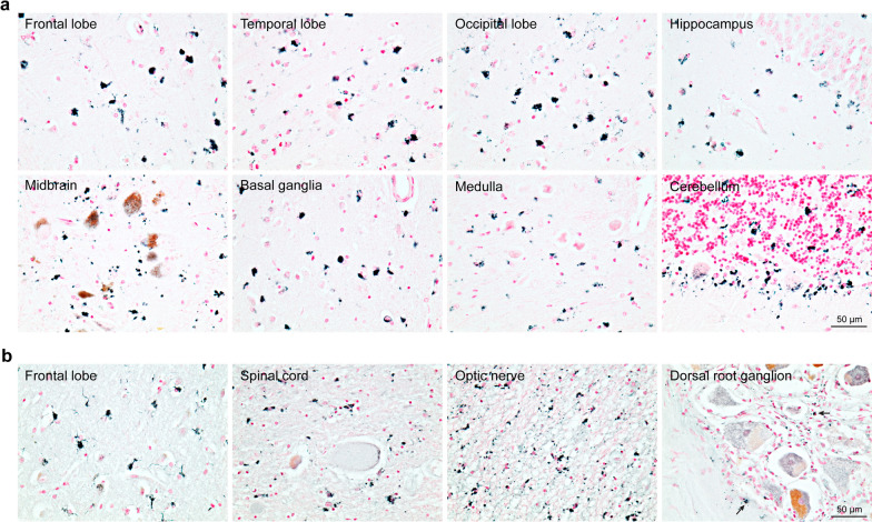
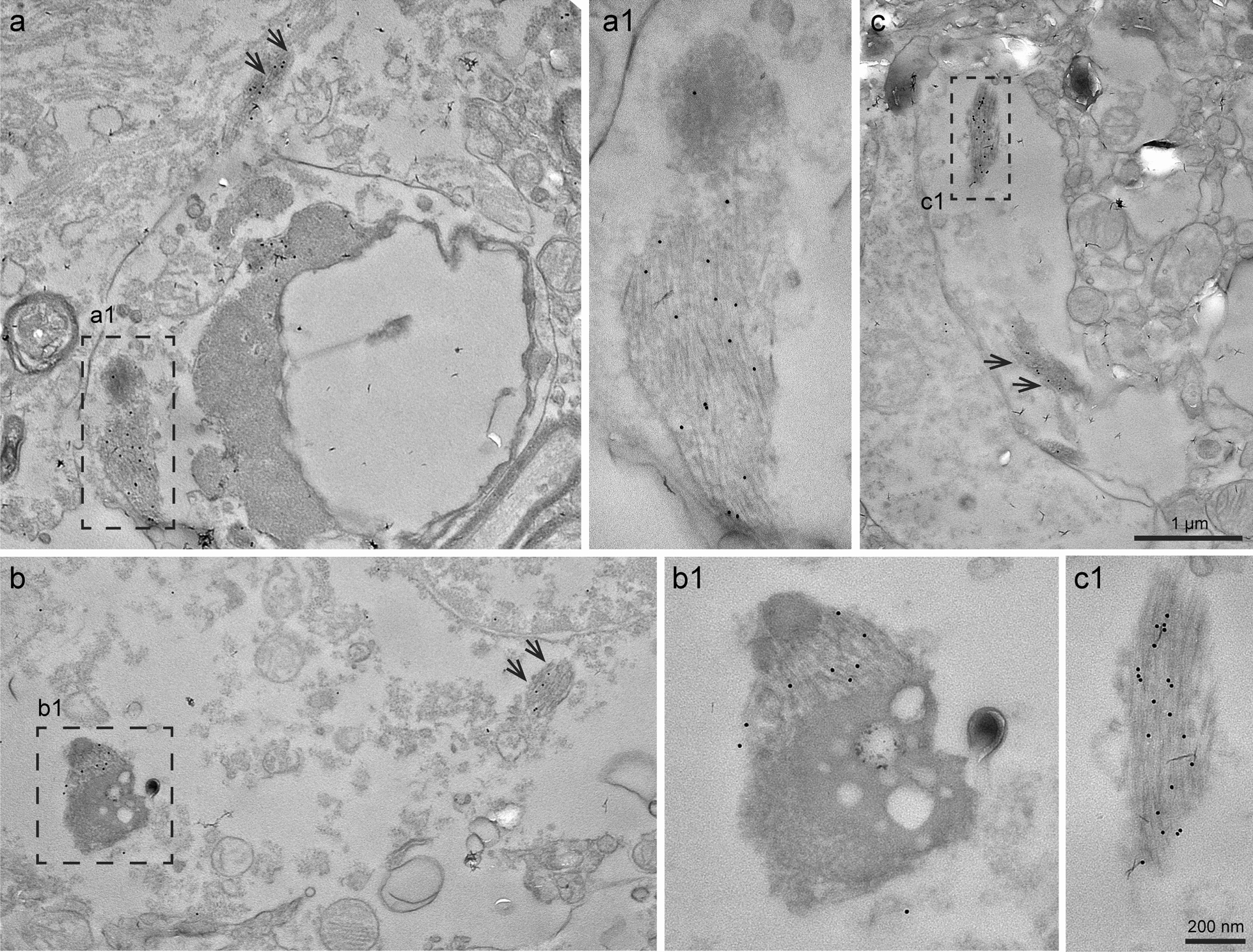
Filaments made of residues 120-254 of transmembrane protein 106B (TMEM106B) form in an age-dependent manner and can be extracted from the brains of neurologically normal individuals and those of subjects with a variety of neurodegenerative diseases. TMEM106B filament formation requires cleavage at residue 120 of the 274 amino acid protein; at present, it is not known if residues 255-274 form the fuzzy coat of TMEM106B filaments. Here we show that a second cleavage appears likely, based on staining with an antibody raised against residues 263-274 of TMEM106B. We also show that besides the brain TMEM106B inclusions form in dorsal root ganglia and spinal cord, where they were mostly found in non-neuronal cells. We confirm that in the brain, inclusions were most abundant in astrocytes. No inclusions were detected in heart, liver, spleen or hilar lymph nodes. Based on their staining with luminescent conjugated oligothiophenes, we confirm that TMEM106B inclusions are amyloids. By in situ immunoelectron microscopy, TMEM106B assemblies were often found in structures resembling endosomes and lysosomes.

Keywords: Amyloid; Astrocytes; Peripheral nervous system; TMEM106B filaments; Vacuoles.


Conflict of interest statement

The authors declare that they have no competing interests.

Figures

Fig. 1
Fig. 1
Immunohistochemical staining with anti-TMEM106B antibodies A303-439A, TMEM239 and TMEM263. a, Schematic of human TMEM106B, with the epitopes of anti-TMEM106B antibodies indicated. b, Staining of frontal cortex sections from neurologically normal individuals aged 75 (case 1) and 25 (case 2) years (yo = years-old). Nuclei are counterstained in red. Scale bars: 50 µm and 20 µm in the inset. Note similar cytoplasmic staining by A303-439A and TMEM263 in 25 yo and 75 yo individuals; TMEM239 stained inclusions in the 75 yo individual, with no staining in the 25 yo individual
Fig. 2
Fig. 2
Presence of TMEM106B inclusions in central and peripheral nervous systems. a, Staining with antibody TMEM239 of different brain regions from an individual aged 78 years with progressive supranuclear palsy (case 3). Scale bar, 50 µm. b, Staining with antibody TMEM239 of brain, spinal cord and dorsal root ganglion sections from an individual aged 90 years with sporadic Alzheimer’s disease (case 4). Scale bar, 50 µm. Arrows in the dorsal root ganglion picture indicate TMEM106B aggregates
Fig. 3
Fig. 3
Absence of TMEM106B inclusions in peripheral organs. a Staining with TMEM239 of heart, liver, hilar lymph node, spleen and brain sections from neurologically normal individuals aged 71 (case 4), 75 (case 1) or 76 (case 6) years (yo = years-old). Nuclei are counterstained in red. Note the presence of TMEM106B inclusions in brain, but not in peripheral organs. Lipofuscin was observed as brown-coloured grainy material in some peripheral organs. Immunostaining of brain for the 75 yo subject is shown in Fig. 1. b Immunoblot analysis with TMEM239 of sarkosyl-insoluble extracts from heart, liver, hilar lymph node, spleen and brain from the same neurologically normal individuals aged 71, 75 or 76 years (yo = years-old) as in a. Note the presence of a 29-kDa band only in brain (red arrow). No brain tissue from the 71 yo individual was available for immunoblotting, but the presence of TMEM106B inclusions in brain is shown by immunostaining in a
Fig. 4
Fig. 4
Age-dependent formation of TMEM106B inclusions, irrespective of the presence of disease. Staining with TMEM239 of frontal cortex sections from individuals with Sanfilippo syndrome (case 7), juvenile-onset synucleinopathy (JOS) (case 8), Alper’s disease (case 9), multiple sclerosis (case 10), cerebellar degeneration (case 11), Huntington’s disease (case 12), Friedreich’s ataxia (case 13), Down’s syndrome (case 14), frontotemporal dementia and parkinsonism linked to chromosome 17 with P301L mutation in MAPT (FTDP-17T) (case 15), Parkinson’s disease dementia (PDD) with a mutation in the glucocerebrosidase (GBA) gene (case 16), argyrophilic grain disease (AGD) (case 17), Pick’s disease (case 18), frontotemporal lobar degeneration with TDP-43 inclusions (FTLD-TDP) (case 19), progressive supranuclear palsy (PSP) (case 20), and Parkinson’s disease (PD) (case 21). Frontal cortex sections from a neurologically normal individual aged 98 years (case 22) were also used; yo = years-old. Scale bar, 50 µm and 20 µm (inset). TMEM106B inclusions were only detected in individuals older than 50 years
Fig. 5
Fig. 5
TMEM106B inclusions in brain cells. a Double-immunostaining of TMEM239 (black) and neuronal marker NeuN (red) of cortical sections from a neurologically normal individual aged 84 years (case 23); a1, a2, a3, close-up images of insets in a. TMEM106B inclusions were sometimes present in NeuN-positive nerve cells. b Double-immunostaining of TMEM239 (black) and astrocytic marker glial fibrillary acidic protein (GFAP) (red) of cortical sections from a neurologically normal individual aged 76 years (case 24); b1, b2, b3, close-up images of insets in b. Many TMEM106B inclusions were present in GFAP-positive astrocytes. c Double-immunostaining of TMEM239 (black) and oligodendrocytic marker APC/CC1 (red) of cortical sections from a neurologically normal individual aged 76 years (case 24); c1, c2, c3, close-up images of insets in c. TMEM106B inclusions were occasionally present in APC/CC1-positive oligodendrocytes. d Double-immunostaining of TMEM239 (black) and microglial marker Iba-1 (red) of cortical sections from a neurologically normal individual aged 84 years (case 23); d1, d2, d3, close-up images of insets in d. TMEM106B inclusions were occasionally present in Iba-1-positive microglia
Fig. 6
Fig. 6
Double-labelling immunofluorescence of TMEM106B antibodies and luminescent conjugated oligothiophenes (LCOs), which are amyloid dyes. a Double-labelling immunofluorescence of sections from the frontal cortex of neurologically normal individuals aged 83 (case 25) and 84 (case 23) years with antibody TMEM239 (red) and LCOs pFTAA, hFTAA, HS68 and Amytracker 540 (green). Inclusions were labelled by both TMEM239 and LCOs (double labelling in yellow). b, c Double-labelling immunofluorescence of sections from the frontal cortex of a neurologically normal individual aged 84 years (case 26) using pFTAA and anti-TMEM106B antibodies TMEM263 (b) and A303-439A (c) (recognising TMEM106B C-terminus and N-terminus respectively). Inclusions were only labelled by pFTAA. d Double-labelling immunofluorescence of sections from the dorsal root ganglion of an individual aged 90 years with sporadic Alzheimer’s disease (case 4) with antibody TMEM239 (red) and pFTAA, (green). Inclusions were labelled by both TMEM239 and pFTAA (double labelling in yellow)
Fig. 7
Fig. 7
Immunogold electron microscopy on brain tissue sections. Immunogold EM on frontal cortex of an individual aged 90 years with Alzheimer’s disease (case 4) revealed the presence of bundles of filaments within the cell body and processes of cells, most likely astrocytes. The bundles of filaments decorated by gold particles were seen in association with dense osmiophilic structures with the morphological characteristics of secondary lysosomes. Bundles of filaments decorated by the gold particles, as well as the dense osmiophilic structures, may be present in vacuoles and were surrounded by a membrane. a Low power transversal view of a cell process containing immunogold labelled filaments enlarged in Box (a1). Adjacent to the transversal view of the cell process, another bundle of filaments decorated by gold particles is indicated by two arrows; a1, higher magnification of the smaller osmiophilic structure associated with the bundle of gold-decorated filament. b Low power view of a portion of a cell body. Part of the nucleus is seen in the upper right. Box (b1) shows an electron-dense osmiophilic structure with vacuoles that is associated with a bundle of filaments decorated by gold particles. Another bundle of filaments is indicated by two arrows; b1 higher magnification of the electron-dense osmiophilic structure associated with filaments decorated by gold particles. c Low power view of a cell process containing two bundles of filaments, one in box (c1) and the other indicated by two arrows; c1, higher magnification of filaments seen in box (c). Scale bar in c, 1 µm (also for a and b) and in c1, 200 nm (also for a1 and b1)

References

    1. Allen B, Ingram E, Takao M, Smith MJ, Jakes R, Virdee K et al (2002) Abundant tau filaments and nonapoptotic neurodegeneration in transgenic mice expressing human P301S tau protein. J Neurosci 22:9340–9351 10.1523/JNEUROSCI.22-21-09340.2002 - DOI - PMC - PubMed
    1. Arseni D, Hasegawa M, Murzin AG, Kametani F, Arai M, Yoshida M et al (2022) Structure of pathological TDP-43 filaments from ALS with FTLD. Nature 601:139–143 10.1038/s41586-021-04199-3 - DOI - PMC - PubMed
    1. Arseni D, Chen R, Murzin AG, Peak-Chew SY, Garringer HJ, Newell KL et al (2023) TDP-43 forms amyloid filaments with a distinct fold in type A FTLD-TDP. Nature 620:898–903 10.1038/s41586-023-06405-w - DOI - PMC - PubMed
    1. Åslund A, Sigurdson CJ, Klingstedt T, Grathwohl S, Bolmont T, Dickstein DL et al (2009) Novel pentameric thiophene derivatives for in vitro and in vivo imaging of a plethora of protein aggregates in cerebral amyloidoses. ACS Chem Biol 4:673–683 10.1021/cb900112v - DOI - PMC - PubMed
    1. Baggen J, Jacquemyn M, Persoons L, Vanstreels E, Pye VE, Wrobel AG et al (2023) TMEM106B is a receptor mediating ACE2-independent SARS-CoV-2 cell entry. Cell 186:3427–3442 10.1016/j.cell.2023.06.005 - DOI - PMC - PubMed

Publication types