. 2025 Mar 1;20(3):845-857.
doi: 10.4103/1673-5374.392889. Epub 2024 Jan 8.

Maintaining moderate levels of hypochlorous acid promotes neural stem cell proliferation and differentiation in the recovery phase of stroke

Affiliations

Maintaining moderate levels of hypochlorous acid promotes neural stem cell proliferation and differentiation in the recovery phase of stroke

Lin-Yan Huang et al. Neural Regen Res. .

Abstract

JOURNAL/nrgr/04.03/01300535-202503000-00029/figure1/v/2024-06-17T092413Z/r/image-tiff It has been shown clinically that continuous removal of ischemia/reperfusion-induced reactive oxygen species is not conducive to the recovery of late stroke. Indeed, previous studies have shown that excessive increases in hypochlorous acid after stroke can cause severe damage to brain tissue. Our previous studies have found that a small amount of hypochlorous acid still exists in the later stage of stroke, but its specific role and mechanism are currently unclear. To simulate stroke in vivo, a middle cerebral artery occlusion rat model was established, with an oxygen-glucose deprivation/reoxygenation model established in vitro to mimic stroke. We found that in the early stage (within 24 hours) of ischemic stroke, neutrophils produced a large amount of hypochlorous acid, while in the recovery phase (10 days after stroke), microglia were activated and produced a small amount of hypochlorous acid. Further, in acute stroke in rats, hypochlorous acid production was prevented using a hypochlorous acid scavenger, taurine, or myeloperoxidase inhibitor, 4-aminobenzoic acid hydrazide. Our results showed that high levels of hypochlorous acid (200 μM) induced neuronal apoptosis after oxygen/glucose deprivation/reoxygenation. However, in the recovery phase of the middle cerebral artery occlusion model, a moderate level of hypochlorous acid promoted the proliferation and differentiation of neural stem cells into neurons and astrocytes. This suggests that hypochlorous acid plays different roles at different phases of cerebral ischemia/reperfusion injury. Lower levels of hypochlorous acid (5 and 100 μM) promoted nuclear translocation of β-catenin. By transfection of single-site mutation plasmids, we found that hypochlorous acid induced chlorination of the β-catenin tyrosine 30 residue, which promoted nuclear translocation. Altogether, our study indicates that maintaining low levels of hypochlorous acid plays a key role in the recovery of neurological function.


Conflict of interest statement

Conflicts of interest: The authors declare that they have no competing interests.

Figures

Figure 1
Figure 1
Clearance of accumulated hypochlorous acid protects from acute ischemic stroke. (A, B) Representative images and quantitative analysis of cerebral infarct size detected by TTC staining in a rat MCAO model following intraperitoneal administration of 50 mg/kg Tau and 30 mg/kg 4-ABAH (n = 5). Tau and 4-ABAH significantly reduced the infarct area in MCAO rats. White region indicates the infarct area. (C, D) Representative images and statistical analysis of BBB permeability determined by EB staining (n = 3). Tau and 4-ABAH significantly reduced EB extravasation in MCAO rats. (E) Effect of different concentrations of hypochlorous acid on cleaved caspase-3 expression in HT22 cells under O/R conditions. Hypochlorous acid was administrated upon reoxygenation for 24 hours (n = 3). (F) Effect of Tau on hypochlorous acid (200 μM)-induced caspase-3 in HT22 cells under O/R conditions (n = 3). (G, H) Effect of Tau on hypochlorous acid-induced apoptosis in HT22 cells under O/R conditions. Apoptosis was observed by Hoechst 33258 staining (n = 3). Hypochlorous acid (200 μM) increased apoptosis rate, while Tau (200 μM) reversed the pro-apoptotic effect of hypochlorous acid. Tau markedly inhibited apoptosis in O/R-challenged HT22 cells. Scale bar: 100 μm. Data are expressed as mean ± SD. *P < 0.05, **P < 0.01, ***P < 0.001, ****P < 0.0001 (B, D, E: one-way analysis of variance followed by Dunnett’s post hoc test; F, H: one-way analysis of variance followed by Tukey’s post hoc test). 4-ABAH: 4-Aminobenzohydrazide; EB: Evans blue; MCAO: middle cerebral artery occlusion; O/R: oxygen-glucose deprivation/reoxygenation; Tau: taurine; TTC: 2,3,5-triphenyl-2H-tetrazolium chloride.
Figure 2
Figure 2
Microglia are a crucial source of hypochlorous acid in the stroke recovery phase. (A–C) Co-localization and statistical analysis of Iba-1+ microglia (Cy3, red) and hypochlorous acid (HKOCl-3, green) in ischemic SVZ of MCAO rats. Hypochlorous acid was visualized using a HKOCl-3 probe. Hypochlorous acid levels (normalized to day 1) increased on the 3rd and 5th days after MCAO. Fluorescence intensity of Iba-1 increased on the 5th day, but decreased on the 7th and 14th days after MCAO. Scale bar: 100 μm. (D) Hypochlorous acid production in BV2 cells and neutrophils with or without O/R challenge. (E) BV2/C17.2 co-culture system to examine the effect of hypochlorous acid produced by BV2 cells, with or without O/R challenge. (F) Neutrophil/C17.2 co-culture system to examine the effect of hypochlorous acid produced by neutrophils on C17.2 cell viability under O/R conditions. Taurine (100 μM) and 4-ABAH (100 μM) were used to scavenge or prevent hypochlorous acid production, respectively, in O/R-challenged neutrophils. Data are expressed as mean ± SD (n = 3). *P < 0.05, **P < 0.01, ***P < 0.001, ****P < 0.0001 (B, C: one-way analysis of variance followed by Dunnett’s post hoc test; D–F: one-way analysis of variance followed by Tukey’s post hoc test). DAPI: 4,6-Diamidino-2-phenylindole; Iba1: ionized calcium binding adapter molecule 1; O/R: oxygen-glucose deprivation/reoxygenation; SVZ: subventricular zone.
Figure 3
Figure 3
Moderate hypochlorous acid is beneficial for maintaining NSC proliferation in the recovery phase of stroke. (A) Timeline of the experimental design. Rats were subjected to 2 hours focal ischemia, followed by reperfusion for 10 days. On the 5th day, 5 μM/10 μL hypochlorous acid and 5 μM/10 μL HKOCl-3 were injected into the lateral ventricle. (B–G) Immunostaining and statistical analysis of Nestin+ (Alexa Fluor 488, green) and Sox2+ (Cy3, red) NSCs in ischemic SVZ. Hypochlorous acid decreased Nestin+ and Sox2+ intensity on the 7th day, but increased their intensity on the 10th day. Data were normalized to the MCAO group. (H) Effect of different concentrations of hypochlorous acid on C17.2 cell viability measured by CCK8 assay. (I, J) BrdU staining (Cy3, red) examined the effect of hypochlorous acid on C17.2 cell proliferation under O/R conditions. The ratio of BrdU+ to DAPI+ cells was increased by 5 μM hypochlorous acid in O/R-challenged HT22 cells, but inhibited by 200 μM hypochlorous acid. Data are expressed as mean ± SD (n = 3). *P < 0.05, **P < 0.01, ***P < 0.001, ****P < 0.0001 (D, G: one-way analysis of variance followed by Tukey’s post hoc test, H, J: one-way analysis of variance followed by Dunnett’s post hoc test). BrdU: 5-Bromo-2′-deoxyuridine; CCK8: cell counting kit 8; DAPI: 4,6-diamidino-2-phenylindole; NSCs: neural stem cells; O/R: oxygen-glucose deprivation.
Figure 4
Figure 4
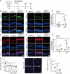
Moderate hypochlorous acid promotes neural stem cell differentiation into astrocytes and neurons under O/R conditions. (A, B) Effect and statistical analysis of low concentration hypochlorous acid on the expression of GFAP, Tuj-1, and CNP detected by western blotting in C17.2 cells. Data were normalized to the Control (Con) group. (C, D) Effect and statistical analysis of low concentration hypochlorous acid on expression of GFAP, Tuj-1, and CNP detected by western blotting in O/R-challenged C17.2 cells. Data were normalized to the Con group. (E–H) Representative images and statistical analysis of low concentration hypochlorous acid on immunopositivity of GFAP (PE, red), Tuj-1 (FITC, green), and CNP (FITC, green) in O/R-challenged primary NSCs. 5 μM hypochlorous acid increased the ratio of GFAP+ to DAPI+ cells, but reduced the ratio of CNP+ to DAPI+ cells. However, 100 μM hypochlorous acid increased the ratio of Tuj-1+ to DAPI+ cells and GFAP+ to DAPI+ cells, but reduced the ratio of CNP+ to DAPI+ cells. (I, J) Representative images and statistical analysis of low concentration hypochlorous acid on immunopositivity of Tuj-1 (FITC, green) in the SVZ of MCAO rats. Low-concentration hypochlorous acid increased Tuj-1 immunopositivity. Data were normalized to the MCAO group. Scale bar: 50 μm. Data are expressed as mean ± SD (n = 5). *P < 0.05, **P < 0.01, ***P < 0.001 (B, F–H: one-way analysis of variance followed by Dunnett’s post hoc test, D, J: one-way analysis of variance followed by Tukey’s post hoc test). CNP: 2′,3′-Cyclic nucleotide 3′-phosphodiesterase; DAPI: 4′,6-diamidino-2-phenylindole; GFAP: glial fibrillary acidic protein; MCAO: middle cerebral artery occlusion; NSC: neural stem cell; O/R: oxygen-glucose deprivation/reoxygenation; SVZ: subventricular zone; Tuj-1: beta-III tubulin.
Figure 5
Figure 5
Low concentration of hypochlorous acid promotes nuclear translocation of β-catenin in C17.2 cells under O/R conditions. (A–C) Expression and statistical analysis of β-catenin detected by western blotting in the cytoplasm and nucleus of C17.2 cells. Data were normalized to the control (Con) group. (D, E) Translocation of β-catenin (Cy3, red) in C17.2 cells treated with 5 and 100 μM hypochlorous acid under O/R conditions visualized by immunofluorescence staining. Nuclear translocation of β-catenin was promoted by 5 μM hypochlorous acid and inhibited by 100 μM hypochlorous acid. Scale bar: 100 μm, 25 μm for enlarged images. Data are expressed as mean ± SD (n = 3). *P < 0.05, **P < 0.01, ***P < 0.001, ****P < 0.0001 (B, C, E: one-way analysis of variance followed by Tukey’s post hoc test). O/R: Oxygen-glucose deprivation/reoxygenation; Tau: taurine.
Figure 6
Figure 6
Tyrosine residue chlorination of β-catenin is a critical modification induced by low concentration hypochlorous acid. (A) Illustration of tyrosine chlorination and di-chlorination structures. (B) MS/MS spectra of tyrosine residue 30 of β-catenin peptides derived from O/R-challenged C17.2 cells with (labeled as HOCl-Tyr30) or without hypochlorous acid (labeled as Ctrl-Tyr30) treatment. The b ion refers to the N-terminal part of the peptide, and the y ion refers to the C-terminal part of the peptide. O/R: Oxygen-glucose deprivation/reoxygenation; HOCI: hypochlorous acid; MS/MS: tandem mass spectrometry.
Figure 7
Figure 7
Chlorination of Tyr30 may contribute to nuclear translocation of β-catenin induced by low-concentration hypochlorous acid. (A) Construction strategy for wildtype and Y30A, Y331A, Y333A, and Y30/331/333A mutant (triple Mut) plasmids. Gray boxes indicate GFP sequence. (B, C) The effect of Y30A, Y331A, Y333A, and Y30/331/333A mutant plasmids on nuclear translocation of β-catenin in C17.2 cells stimulated by 100 μM hypochlorous acid. Data normalized by WT group are expressed as mean ± SD (n = 3). *P < 0.05, **P < 0.01 (one-way analysis of variance followed by Dunnett’s post hoc test). A: Alanine; Mut: mutation; WT: wildtype; Y: tyrosine.

Similar articles

Cited by

References

    1. Augsburger F, Filippova A, Jaquet V. Methods for detection of NOX-derived superoxide radical anion and hydrogen peroxide in cells. Methods Mol Biol. 20191982:233–241. - PubMed
    1. Baliou S, Adamaki M, Ioannou P, Pappa A, Panayiotidis MI, Spandidos DA, Christodoulou I, Kyriakopoulos AM, Zoumpourlis V. Protective role of taurine against oxidative stress (Review) Mol Med Rep. 2021;24:605. - PMC - PubMed
    1. Borhani S, Corciulo C, Larranaga-Vera A, Cronstein BN. Adenosine A(2A) receptor (A2AR) activation triggers Akt signaling and enhances nuclear localization of β-catenin in osteoblasts. FASEB J. 2019;33:7555–7562. - PMC - PubMed
    1. Breckwoldt MO, Chen JW, Stangenberg L, Aikawa E, Rodriguez E, Qiu S, Moskowitz MA, Weissleder R. Tracking the inflammatory response in stroke in vivo by sensing the enzyme myeloperoxidase. Proc Natl Acad Sci U S A. 2008;105:18584–18589. - PMC - PubMed
    1. Chaker Z, Codega P, Doetsch F. A mosaic world: puzzles revealed by adult neural stem cell heterogeneity. Wiley Interdiscip Rev Dev Biol. 2016;5:640–658. - PMC - PubMed