. 2025 Mar 1;20(3):858-872.
doi: 10.4103/NRR.NRR-D-23-01925. Epub 2024 May 17.

Inhibiting SHP2 reduces glycolysis, promotes microglial M1 polarization, and alleviates secondary inflammation following spinal cord injury in a mouse model

Affiliations

Inhibiting SHP2 reduces glycolysis, promotes microglial M1 polarization, and alleviates secondary inflammation following spinal cord injury in a mouse model

Xintian Ding et al. Neural Regen Res. .

Abstract

JOURNAL/nrgr/04.03/01300535-202503000-00030/figure1/v/2024-06-17T092413Z/r/image-tiff Reducing the secondary inflammatory response, which is partly mediated by microglia, is a key focus in the treatment of spinal cord injury. Src homology 2-containing protein tyrosine phosphatase 2 (SHP2), encoded by PTPN11, is widely expressed in the human body and plays a role in inflammation through various mechanisms. Therefore, SHP2 is considered a potential target for the treatment of inflammation-related diseases. However, its role in secondary inflammation after spinal cord injury remains unclear. In this study, SHP2 was found to be abundantly expressed in microglia at the site of spinal cord injury. Inhibition of SHP2 expression using siRNA and SHP2 inhibitors attenuated the microglial inflammatory response in an in vitro lipopolysaccharide-induced model of inflammation. Notably, after treatment with SHP2 inhibitors, mice with spinal cord injury exhibited significantly improved hind limb locomotor function and reduced residual urine volume in the bladder. Subsequent in vitro experiments showed that, in microglia stimulated with lipopolysaccharide, inhibiting SHP2 expression promoted M2 polarization and inhibited M1 polarization. Finally, a co-culture experiment was conducted to assess the effect of microglia treated with SHP2 inhibitors on neuronal cells. The results demonstrated that inflammatory factors produced by microglia promoted neuronal apoptosis, while inhibiting SHP2 expression mitigated these effects. Collectively, our findings suggest that SHP2 enhances secondary inflammation and neuronal damage subsequent to spinal cord injury by modulating microglial phenotype. Therefore, inhibiting SHP2 alleviates the inflammatory response in mice with spinal cord injury and promotes functional recovery postinjury.


Conflict of interest statement

Conflicts of interest: All the authors declare that there is no conflict of interest in this paper.

Figures

Figure 1
Figure 1
SHP2 expression in mouse tissue from the site of SCI and in LPS-stimulated microglia. (A) Establishment of SCI mouse model. Created with Adobe Illustrator 2023. (B, C) Immunofluorescence staining for SHP2 (green-CoraLite488) and Iba-1 (red-CoraLite594) in the mouse spine 14 days after SCI. Quantitative analysis showing that SHP2 and Iba-1 expression levels increased significantly relative to the Sham group (n = 5 slices from three mice per group). Scale bar: 50 μm. (D, E) Western blotting was used to detect SHP2 expression 1, 7, and 14 days after SCI in mice. (F–M) The protein expression levels of inflammation markers and SHP2 in microglia were detected by western blotting. The experiment was repeated three times. Data are expressed as the mean ± SD. A P-value below 0.05 was considered to be statistically significant, *P < 0.05, **P < 0.01, ***P < 0.001 (unpaired t-test [C]; one-way analysis of variance with Bonferroni’s post hoc test [E, G–I, K–M]). BBB: Basso–Beattie–Bresnahan; CD86: cluster of differentiation 86; DAPI: 4′,6-diamidino-2′-phenylindole; Iba-1: ionized calcium-binding adapter molecule 1; IF: immunofluorescence staining; iNOS: inducible nitric oxide synthase; LPS: lipopolysaccharide; NS: not significant; SCI: spinal cord injury; SHP2: Src homology 2 domain-containing protein tyrosine phosphatase 2.
Figure 2
Figure 2
SHP2 downregulation reduces microglia-mediated inflammation. (A, B) mRNA expression levels of SHP2, iNOS, CD86, TNF-α, IL-1β, CD206, and Arg-1 in microglia treated with NSC87877 and siSHP2, as assessed by quantitative polymerase chain reaction. (C–E) Immunoreactivities of CD163 (green-CoraLite488) and CD86 (red-CoraLite594) in microglia treated with NSC87877, as detected by immunofluorescence staining. Microglia treated with LPS showed a significant increase in expression of the inflammatory marker CD86, while BV2 cells treated with LPS + NSC87877 exhibited a significant decrease in CD86 expression. Additionally, there was a significant increase in expression of the anti-inflammatory marker CD163 in the LPS + NSC87877 group relative to the LPS group. Scale bars: 50 μm. (F–I) Western blot for the protein expressions of SHP2, iNOS, CD86, CD206, Arg-1, and other proteins in microglia treated with NSC87877 and siSHP2. Data are expressed as the mean ± SD. The experiment was repeated three times. *P < 0.05, **P < 0.01, ***P < 0.001 (one-way analysis of variance with Bonferroni’s post hoc test). Arg-1: Arginase-1; CD163: cluster of differentiation 163; CD206: cluster of differentiation 206; CD86: cluster of differentiation 86; DAPI: 4′,6-diamidino-2′-phenylindole; IF: immunofluorescence staining; IL-1β: interleukin-1 beta; LPS: lipopolysaccharide; NS: not significant; NSC87877: SHP2 inhibitor; TNF-α: tumor necrosis factor-alpha; SHP2: Src homology 2 domain-containing protein tyrosine phosphatase 2; siNC: small interfering RNA negative control; siSHP2: small interfering RNA of SHP2.
Figure 3
Figure 3
SHP2 inhibition reduces tissue damage after SCI, facilitating functional recovery. (A, B) Iba-1 (red-CoraLite594), CD163 (green-CoraLite488), and CD86 (green-CoraLite488) immunoreactivities were detected by immunofluorescence staining. Quantitative analysis showed that CD86 fluorescence intensity near the SCI site was significantly decreased in the SCI + NSC87877 group compared with the SCI group. Conversely, there was a significant increase in CD163 expression (n = 5 slices from three mice per group). Scale bars: 50 μm. (C) HE staining of mouse spinal cord sections 14 days after SCI. HE staining of transverse and longitudinal spinal cord sections showed that the injured area in the SCI + NSC87877 group was significantly smaller than that in the SCI group (n = 5 slices from three mice per group). Scale bars: 100 μm (left), 600 μm (right). (D, E) BBB scores and residual urine volumes at 1, 3, 5, 7, and 14 days post-SCI (n = 6 mice per group). (F) Footprint analysis 14 days after SCI. (G, H) Stride length and width in mice subjected to SCI and treated with NSC87877. Relative to the SCI group, the SCI + NSC87877 group showed increased stride length and decreased stride width (n = 6 mice per group). (I, J) The fall latency and rotational speed in the rotarod experiment were evaluated on the 14th day after SCI. Data are expressed as mean ± SD. *P < 0.05, **P < 0.01, ***P < 0.001 (one-way analysis of variance with Bonferroni’s post hoc test [B, G–J]; two-way analysis of variance with Bonferroni’s post hoc test [D and E]). BBB score: Basso, Beattie, and Bresnahan locomotor rating scale; CD163: cluster of differentiation 163; CD86: cluster of differentiation 86; DAPI: 4′,6-diamidino-2′-phenylindole; HE: hematoxylin and eosin; Iba-1: ionized calcium-binding adapter molecule 1; IF: immunofluorescence staining; ns: not significant; NSC87877: SHP2 inhibitor; SCI: spinal cord injury; SHP2: Src homology 2 domain-containing protein tyrosine phosphatase 2.
Figure 4
Figure 4
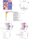
Transcriptome sequencing indicates that SHP2 is related to inflammation and energy metabolism. (A) Heatmap of the transcriptome sequencing results from SCI and SCI + NSC87877 mice 14 days after SCI (n = 3 mice per group). (B) Volcano map of the transcriptome sequencing results from SCI and SCI + NSC87877 mice 14 days after SCI. (C–G) GO term and KEGG pathway enrichment analyses of the transcriptome sequencing results from the SCI and SCI + NSC87877 mice 14 days after SCI. GO: Gene Ontology; KEGG: Kyoto encyclopedia of genes and genomes; NSC87877: SHP2 inhibitor; SCI: spinal cord injury; HP2: Src homology 2 domain-containing protein tyrosine phosphatase 2.
Figure 5
Figure 5
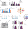
SHP2 regulates glycolysis/OXPHOS in LPS-treated microglia. (A, B) GLUT3, PKM2, HK-1, and LDHA protein expression levels in microglia treated with siSHP2 were detected by western blot. (C) The mRNA expression levels of GLUT3, PKM2, HK-1, and LDHA in microglia treated with NSC87877 were detected by qRT-PCR. (D) Schematic diagram illustrating blockade of the glucose metabolism pathway by 2-DG and oligomycin. (E–G) Intracellular ATP production and lactic acid production in microglia treated with LPS, oligomycin, 2-DG, and NSC87877, as detected using a glycolysis/OXPHOS kit. (H–L) SHP2, CD163, CD86, and iNOS expression levels in microglia treated with LPS, oligomycin, 2-DG, and NSC87877 were detected by western blot. Data are expressed as the mean ± SD. The experiment was repeated three times. *P < 0.05, **P < 0.01, ***P < 0.001 (one-way analysis of variance with Bonferroni’s post hoc test). 2-DG: 2-Deoxy-D-glucose; CD163: cluster of differentiation 163; CD86: cluster of differentiation 86; GLUT3: glucose transporter 3; HK-1: hexokinase-1; iNOS: inducible nitric oxide synthase; LDHA: lactate dehydrogenase A; LPS: lipopolysaccharide; ns: not significant; NSC87877: SHP2 inhibitor; OXPHOS: oxidative phosphorylation; PKM2: pyruvate kinase M2; RLU: relative light unit; SHP2: Src homology 2 domain-containing protein tyrosine phosphatase 2; siNC: small interfering RNA negative control; siSHP2: small interfering RNA of SHP2.
Figure 6
Figure 6
SHP2 inhibition reduces neuronal apoptosis in the context of SCI. (A, B) NeuN (red-CoraLite594) and SHP2 (green-CoraLite488) immunoreactivities in mice treated with NSC87877 14 days after SCI, as detected by immunofluorescence. Quantitative analysis showing that a decrease in SHP2 fluorescence intensity and an increase in the fluorescence intensity of NeuN, a neuronal marker, in SCI + NSC87877 group mice near the injury site (n = 5 slices from three mice per group). Scale bars: 50 μm. (C) Nissl staining of mouse spinal cord transverse and longitudinal sections 14 days after SCI showing that, compared with the SCI group, the SCI + NSC87877 group exhibited more Nissl bodies with a more uniform distribution (n = 5 slices from three mice per group). Scale bars: 100 μm (left), 600 μm (right). (D, E) Western blot analysis of Caspase3, C-caspase3, Bax, and Bcl-2 protein expression in PC12 cells co-cultured with BV2 cells treated with LPS or LPS + NSC87877 for 24 hours. The experiment was repeated three times. (F, G) C-caspase3 (red-CoraLite594) expression in PC12 cells was detected by immunofluorescence staining. Quantitative analysis showed decreased immunofluorescence intensity of the apoptosis marker C-caspase3 in neurons co-cultured with BV2 cells from the LPS + NSC87877 group. The experiment was repeated three times. Scale bars: 50 μm. Data are expressed as the mean ± SD. *P < 0.05, **P < 0.01, ***P < 0.001 (one-way analysis of variance with Bonferroni’s post hoc test). Bax: Bcl-2-associated X protein; Bcl-2: B-cell lymphoma 2; Caspase3: cysteine-aspartic acid protease 3; C-caspase3: cleaved caspase-3; DAPI: 4′,6-diamidino-2′-phenylindole; IF: immunofluorescence staining; LPS: lipopolysaccharide; NeuN: neuronal nuclei; NSC87877: SHP2 inhibitor; SCI: spinal cord injury; SHP2: Src homology 2 domain-containing protein tyrosine phosphatase 2.
Figure 7
Figure 7
SHP2 overexpression exacerbates tissue damage following SCI, further impairing neurological function. (A–H) SHP2 (red-CoraLite594), Iba-1 (green-CoraLite488), PKM2 (red-CoraLite594), NeuN (red-CoraLite594), and C-caspase3 (green-CoraLite488) immunoreactivities in vivo were detected by immunofluorescence staining assays. Compared with the SCI group, the SCI + AVV-PTPN11 group showed a significant increase in SHP2 fluorescence intensity at the site of SCI. This increase was accompanied by increased Iba-1 and PKM2 fluorescence intensity and co-localization at the injury site. NeuN fluorescence intensity of at the site of SCI decreased, but C-caspase3 fluorescence intensity increased, and their co-localization increased (n = 5 slices from three mice per group). Scale bars: 50 μm. (I) HE and Nissl staining of transverse and longitudinal mouse spinal cord sections showed that, compared with the SCI group, the SCI + AAV-PTPN11 group displayed greater loss of spinal cord tissue. Additionally, fewer Nissl bodies were observed in spinal cord sections from the SCI + AAV-PTPN11 group (n = 5 slices from three mice per group). Scale bars: 200 μm (upper) and 600 μm (lower). (J, K) BBB scores and residual urine volumes at 1, 3, 5, 7, and 14 days post-SCI (n = 6 mice per group). Data are expressed as mean ± SD. *P < 0.05, **P < 0.01, ***P < 0.001 (unpaired t-test [B, D, E, G and H]; two-way analysis of variance with Bonferroni’s post hoc test [J and K]). AAV: Adeno-associated virus; BBB: Basso–Beattie–Bresnahan; C-Caspase3: cleaved caspase-3; DAPI: 4′,6-diamidino-2′-phenylindole; HE: hematoxylin and eosin; Iba-1: ionized calcium-binding adapter molecule 1; IF: immunofluorescence staining; NeuN: neuronal nuclei; NS: not significant; KM2: pyruvate kinase M2; SCI: spinal cord injury; SHP2: Src homology 2 domain-containing protein tyrosine phosphatase 2.
Figure 8
Figure 8
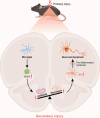
The mechanism by which microglial polarization leads to neuronal apoptosis after SCI. OXPHOS: Oxidative phosphorylation; SCI: spinal cord injury; SHP2: Src homology 2 domain-containing protein tyrosine phosphatase 2.

References

    1. Ahuja CS, Nori S, Tetreault L, Wilson J, Kwon B, Harrop J, Choi D, Fehlings MG. Traumatic spinal cord injury-repair and regeneration. Neurosurgery. 2017;80:S9–S22. - PubMed
    1. Al Mamun A, Chauhan A, Qi S, Ngwa C, Xu Y, Sharmeen R, Hazen AL, Li J, Aronowski JA, McCullough LD, Liu F. Microglial IRF5-IRF4 regulatory axis regulates neuroinflammation after cerebral ischemia and impacts stroke outcomes. Proc Natl Acad Sci U S A. 2020;117:1742–1752. - PMC - PubMed
    1. Ashburner M, Ball CA, Blake JA, Botstein D, Butler H, Cherry JM, Davis AP, Dolinski K, Dwight SS, Eppig JT, Harris MA, Hill DP, Issel-Tarver L, Kasarskis A, Lewis S, Matese JC, Richardson JE, Ringwald M, Rubin GM, Sherlock G. Gene ontology: tool for the unification of biology. The Gene Ontology Consortium. Nat Genet. 2000;25:25–29. - PMC - PubMed
    1. Beattie MS. Inflammation and apoptosis: linked therapeutic targets in spinal cord injury. Trends Mol Med. 2004;10:580–583. - PubMed
    1. Beck KD, Nguyen HX, Galvan MD, Salazar DL, Woodruff TM, Anderson AJ. Quantitative analysis of cellular inflammation after traumatic spinal cord injury: evidence for a multiphasic inflammatory response in the acute to chronic environment. Brain. 2010;133:433–447. - PMC - PubMed

LinkOut - more resources