. 2024 Jun 3:15:1387945.
doi: 10.3389/fimmu.2024.1387945. eCollection 2024.

A local drug delivery system prolongs graft survival by dampening T cell infiltration and neutrophil extracellular trap formation in vascularized composite allografts

Affiliations

A local drug delivery system prolongs graft survival by dampening T cell infiltration and neutrophil extracellular trap formation in vascularized composite allografts

Isabel Arenas Hoyos et al. Front Immunol. .

Abstract

Introduction: The standard treatment for preventing rejection in vascularized composite allotransplantation (VCA) currently relies on systemic immunosuppression, which exposes the host to well-known side effects. Locally administered immunosuppression strategies have shown promising results to bypass this hurdle. Nevertheless, their progress has been slow, partially attributed to a limited understanding of the essential mechanisms underlying graft rejection. Recent discoveries highlight the crucial involvement of innate immune components, such as neutrophil extracellular traps (NETs), in organ transplantation. Here we aimed to prolong graft survival through a tacrolimus-based drug delivery system and to understand the role of NETs in VCA graft rejection.

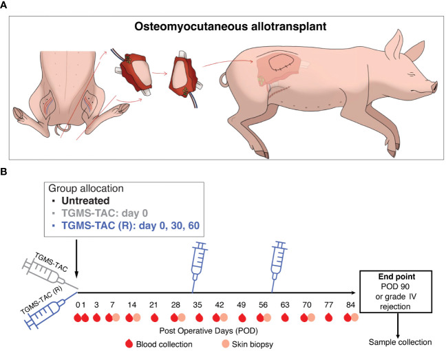
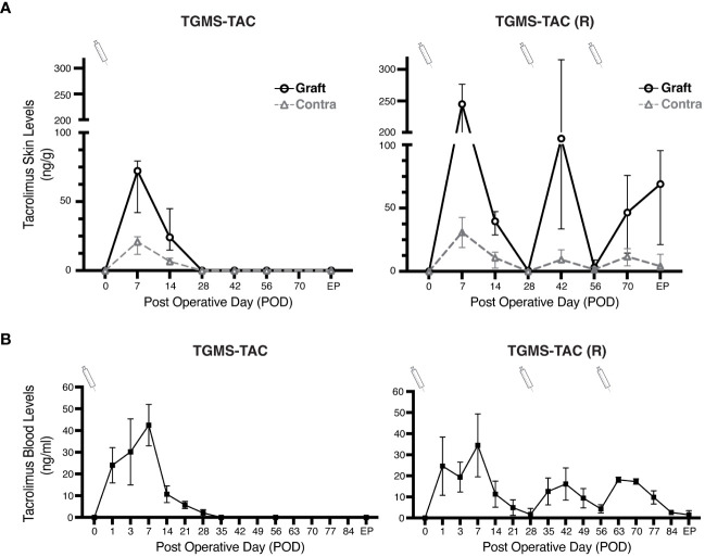
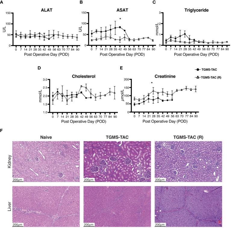
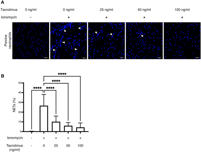
Methods: To prevent off-target toxicity and promote graft survival, we tested a locally administered tacrolimus-loaded on-demand drug delivery system (TGMS-TAC) in a multiple MHC-mismatched porcine VCA model. Off-target toxicity was assessed in tissue and blood. Graft rejection was evaluated macroscopically while the complement system, T cells, neutrophils and NETs were analyzed in graft tissues by immunofluorescence and/or western blot. Plasmatic levels of inflammatory cytokines were measured using a Luminex magnetic-bead porcine panel, and NETs were measured in plasma and tissue using DNA-MPO ELISA. Lastly, to evaluate the effect of tacrolimus on NET formation, NETs were induced in-vitro in porcine and human peripheral neutrophils following incubation with tacrolimus.

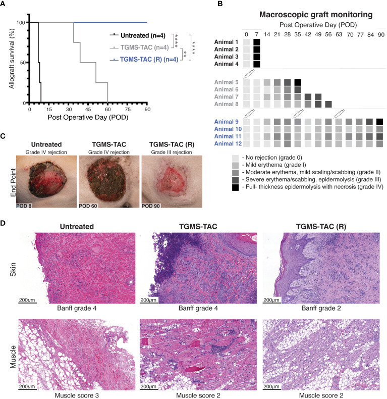
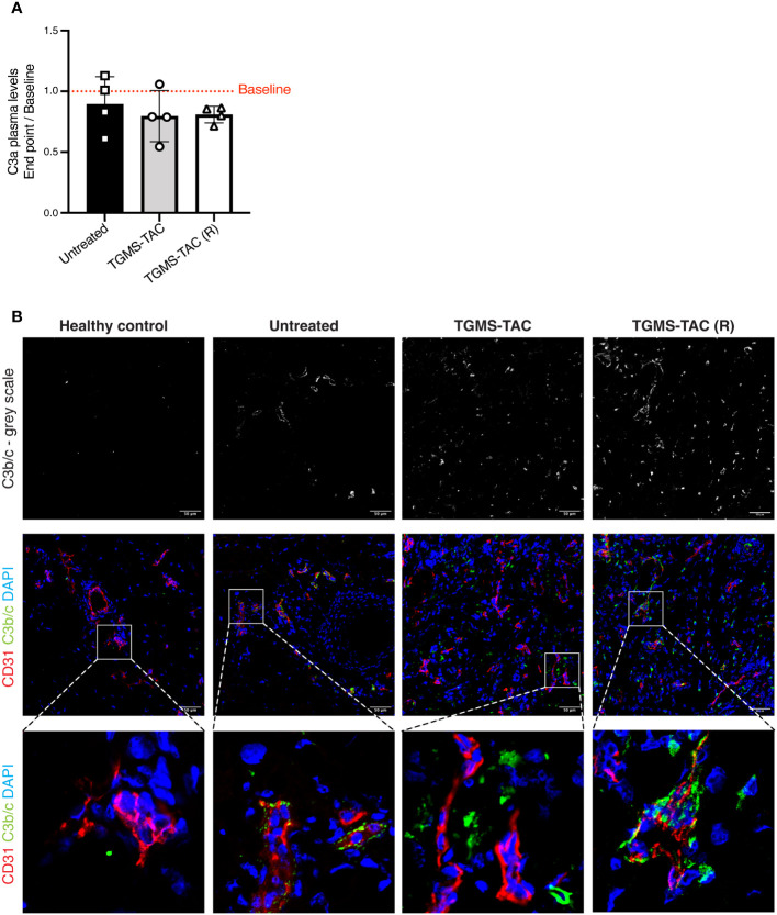
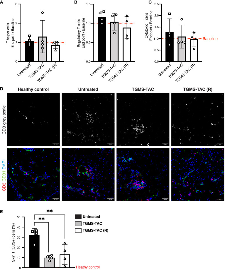
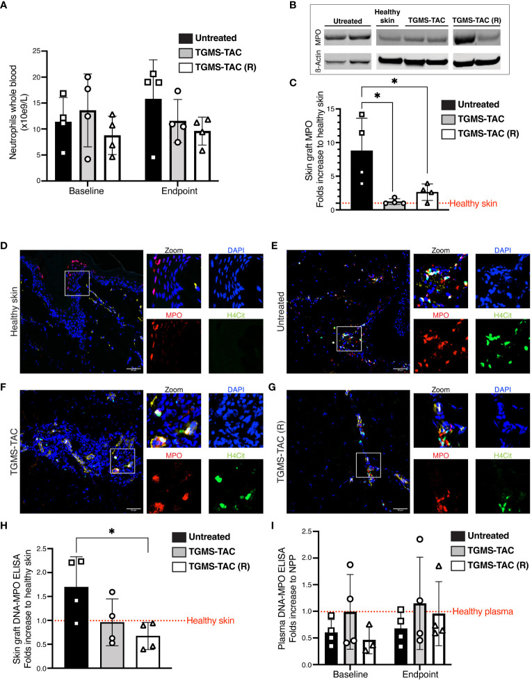
Results: Repeated intra-graft administrations of TGMS-TAC minimized systemic toxicity and prolonged graft survival. Nevertheless, signs of rejection were observed at endpoint. Systemically, there were no increases in cytokine levels, complement anaphylatoxins, T-cell subpopulations, or neutrophils during rejection. Yet, tissue analysis showed local infiltration of T cells and neutrophils, together with neutrophil extracellular traps (NETs) in rejected grafts. Interestingly, intra-graft administration of tacrolimus contributed to a reduction in both T-cellular infiltration and NETs. In fact, in-vitro NETosis assessment showed a 62-84% reduction in NETs after stimulated neutrophils were treated with tacrolimus.

Conclusion: Our data indicate that the proposed local delivery of immunosuppression avoids off-target toxicity while prolonging graft survival in a multiple MHC-mismatch VCA model. Furthermore, NETs are found to play a role in graft rejection and could therefore be a potential innovative therapeutic target.

Keywords: calcineurin inhibitors (CNIs); drug delivery systems (DDSs); local immunosuppression; neutrophil extracellular traps (NETs); porcine model; tacrolimus; transplantation immunology; vascularized composite allotransplantation (VCA).


Conflict of interest statement

The authors declare that the research was conducted in the absence of any commercial or financial relationships that could be construed as a potential conflict of interest.

Figures

Figure 1
Figure 1
Experimental design. (A) Schematic representation of the modified heterotopic hind limb allotransplantation model, where an osteomyocutaneous flap containing graft-draining lymph nodes was transplanted into the flank of a recipient pig. (B) Recipient timeline. After transplantation, pigs were allocated to 3 groups: Group 1, untreated, with no immunosuppression. Group 2, subcutaneous intra-graft injection of TGMS-TAC, 20 mL/kg graft weight, only at POD 0 immediately after transplantation. Group 3, TGMS-TAC intra-graft injection, same dosing as in group 2, repeated injections at POD 0, 30 and 60. During follow-up, blood and tissue samples were collected at defined time points.
Figure 2
Figure 2
TGMS-TAC administration prolongs graft survival. (A) Kaplan-Meier survival curve of the three different groups (n = 4/group): untreated, TGMS-TAC single administration, and TGMS-TAC multiple administration (TGMS-TAC (R)). **p<0.01, ****p<0.0001 calculated by Log-rank (Mantel-Cox) test. (B) Macroscopic evaluation of graft survival during follow-up was graded as follows: 0 = no difference between graft skin and native skin, I = mild erythema, II = moderate erythema with the beginning of scaling and scabbing, III = severe erythema and scabbing with areas of epidermolysis, IV = full-thickness graft epidermolysis with areas of necrosis. Animals 5–8 were euthanized when reaching grade IV rejection at POD 35, 39, 51, and 60, respectively. A syringe indicates the time points of TGMS-TAC injection (C) Representative images of graft changes at endpoint (macroscopic grade IV rejection or POD90) with their corresponding grading. (D) Representative histological H&E staining of graft skin and muscle samples collected at endpoint. Skin was scored according to the Banff 2007 working classification. Muscle changes were graded as 0 = none, 1 = minimal, 2 = moderate, and 3 = extensive damage.
Figure 3
Figure 3
Local TGMS-TAC injections result in higher concentrations of the drug in grafts. (A) Comparison of TAC tissue levels in grafts and contralateral side skin for both single administration of TGMS-TAC (left), and multiple administration TGMS-TAC (R) (right). TAC levels were measured in skin biopsies collected on different postoperative days (POD). Tissue concentrations are represented as ng of TAC per g of skin. (B) Whole-blood levels of tacrolimus in pigs from the TGMS-TAC and (TGMS-TAC (R) groups were measured at POD 0, 1, 3, and then on a weekly basis. Blood levels are expressed as ng/ml. TGMS-TAC injection time points are indicated with a syringe. TAC was measured in both skin and whole blood using LC-MS/MS. Data are represented as means ± SD of n=4 samples.
Figure 4
Figure 4
TGMS-TAC does not lead to off-target toxicity. Plasma levels of (A) alanine aminotransferase (ALAT), (B) aspartate aminotransferase (ASAT), (C) triglyceride, (D) cholesterol, and (E) creatinine as markers for liver and kidney off-target toxicity, respectively, during follow-up. Data are represented as means ± SD. *p<0.05 calculated by two-way ANOVA with Bonferroni correction for multiple comparisons. (F) Representative histological sections (H&E) of kidney and liver samples collected at endpoint from single-injection TGMS-TAC and multiple-injection TGMS-TAC (R) groups, respectively, compared with samples from a naive healthy pig.
Figure 5
Figure 5
Complement activation is observed in rejected skin tissues but is not detected systemically. (A) Plasma levels of C3a quantified by ELISA. Data are represented as fold-changes at endpoint compared to baseline (endpoint/baseline). Data are individual values and means ± SD. Average baseline levels are denoted as a red dotted line. Statistical significance was assessed by one-way ANOVA with Tukey correction for multiple comparisons, p>0.05. (B) Representative immunofluorescence images of skin cryosections from grafts at endpoint compared to skin sections from a naive healthy pig. Skin was stained using DAPI for nuclei (blue), an anti-CD31 antibody as a marker for endothelial cells (red), and anti-C3b/c for complement deposition (green).
Figure 6
Figure 6
TGMS-TAC administration decreases local T-cell infiltration, with minimal systemic effects. (A-C) Flow cytometry identification of T-cell subsets from peripheral blood. Data are represented as fold changes at endpoint compared to baseline (composite baselines denoted as a red dotted line). T cells were identified as T helper (CD3+, CD4+, CD8-), Regulatory T cells (CD3+, CD4+, FoxP3+) and Cytotoxic T cells (CD3+, CD8+, CD4-). Data are shown as individual values with indication of mean ± SD. Statistical significance was assessed by one-way ANOVA with Tukey correction for multiple comparisons, p>0.05. (D) Representative immunofluorescence images of skin cryosections from grafts at endpoint compared to skin samples from a naive healthy pig. Skin was stained using DAPI for nuclei (blue), anti-CD31 for endothelial cells (green), and anti-CD3 for infiltrating T cells (red). (E) Quantification of T cells. The percentage of T cells in healthy naive skin is denoted as a red dotted line. Data are shown as individual values and mean ± SD. Statistical significance was assessed by one-way ANOVA with Tukey’s multiple-comparison test, **p<0.01.
Figure 7
Figure 7
Graft-infiltrating neutrophils and NETs are found in skin tissue of rejected VCA grafts and are more prominent in untreated pigs. (A) Absolute frequency of circulating neutrophils measured from whole blood at baseline and at endpoint. Statistical significance was assessed by two-way ANOVA with Tukey correction for multiple comparisons, p>0.05. Data are shown as individual values and mean ± SD. (B) Representative western blot for myeloperoxidase (MPO) protein as a marker for neutrophil infiltration in graft skin. (C) Quantification of western blot data for MPO levels (ß actin control). *p<0.05 by two-way ANOVA with Tukey’s multiple-comparison test. (D-G) Representative immunofluorescence images of skin cryosections from grafts at endpoint compared to skin samples from a naive healthy pig. Skin was stained using DAPI for nuclei (blue), anti-MPO antibody as a marker for neutrophils (red), and anti-citrullinated Histone 4 (H4Cit) antibody (green) for NETs. (H-I) NET plasma levels in (H) graft skin and, (I) plasma, quantified by DNA-MPO ELISA. Statistical significance was assessed by two-way and one-way ANOVA, respectively. Data are shown as individual values and mean ± SD. **p<0.05. The dotted red lines represent composite baseline levels for naive healthy skin and plasma.
Figure 8
Figure 8
In-vitro inhibition of NET formation by tacrolimus: Isolated neutrophils from healthy pigs (n = 3) were stimulated using ionomycin to induce NET formation in the presence of different concentrations of TAC (0, 25, 50 and 100 ng/ml). (A) NETs were identified using DAPI (blue) and imaged through confocal microscopy. White arrowheads indicate extruded DNA content from neutrophils, confirming the presence of NETs. (B) NET percentages were calculated as the number of NETs/total number of neutrophils per field, using 5 representative images per condition. ****p<0.0001 by two-way ANOVA with Tukey’s multiple-comparison test.

References

    1. Gilbert R. Transplant is successful with a cadaver forearm. Med Trib Med News. (1964) 5:20–2.
    1. Dubernard JM, Owen E, Herzberg G, Lanzetta M, Martin X, Kapila H, et al. . Human hand allograft: report on first 6 months. Lancet. (1999) 353:1315–20. doi: 10.1016/S0140-6736(99)02062-0 - DOI - PubMed
    1. Diep GK, Berman ZP, Alfonso AR, Ramly EP, Boczar D, Trilles J, et al. . The 2020 facial transplantation update: A 15-year compendium. Plast Reconstr Surg - Glob Open. (2021) 9:e3586. doi: 10.1097/GOX.0000000000003586 - DOI - PMC - PubMed
    1. Petruzzo P, Lanzetta M, Dubernard JM, Landin L, Cavadas P, Margreiter R, et al. . The international registry on hand and composite tissue transplantation. Transplantation. (2010) 90:1590–4. doi: 10.1097/TP.0b013e3181ff1472 - DOI - PubMed
    1. Hernandez JA, Miller J, Oleck NC, Porras-Fimbres D, Wainright J, Laurie K, et al. . OPTN/SRTR 2020 annual data report: VCA. Am J Transpl. (2022) 22:623–47. doi: 10.1111/ajt.16980 - DOI - PubMed

MeSH terms