. 2024 Jun 14;13(1):2367777.
doi: 10.1080/2162402X.2024.2367777. eCollection 2024.

CD28-CD57+ T cells from head and neck cancer patients produce high levels of cytotoxic granules and type II interferon but are not senescent

Affiliations

CD28-CD57+ T cells from head and neck cancer patients produce high levels of cytotoxic granules and type II interferon but are not senescent

Brendan L C Kinney et al. Oncoimmunology. .

Abstract

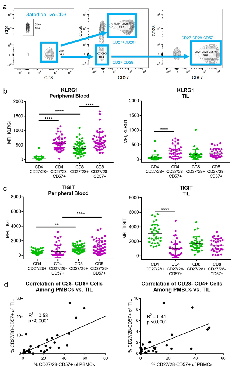
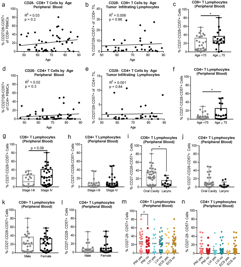
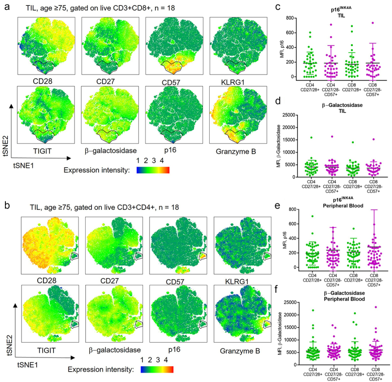
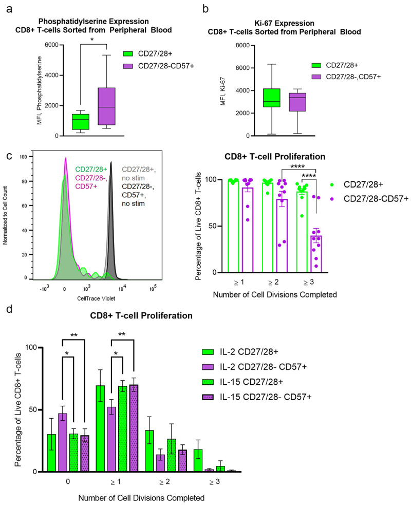
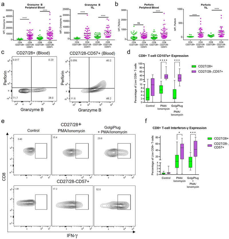
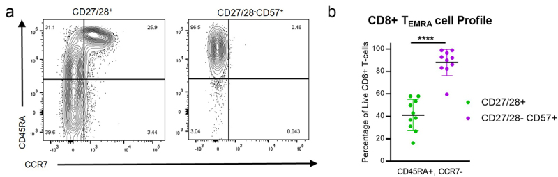
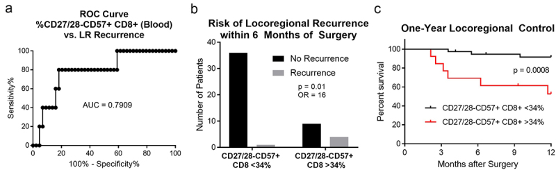
T lymphocytes expressing CD57 and lacking costimulatory receptors CD27/CD28 have been reported to accumulate with aging, chronic infection, and cancer. These cells are described as senescent, with inability to proliferate but enhanced cytolytic and cytokine-producing capacity. However, robust functional studies on these cells taken directly from cancer patients are lacking. We isolated these T cells and their CD27/28+ counterparts from blood and tumor samples of 50 patients with previously untreated head and neck cancer. Functional studies confirmed that these cells have enhanced ability to degranulate and produce IFN-γ. They also retain the ability to proliferate, thus are not senescent. These data suggest that CD27/28-CD57+ CD8+ T cells are a subset of highly differentiated, CD45RA+ effector memory (TEMRA) cells with retained proliferative capacity. Patients with > 34% of these cells among CD8+ T cells in the blood had a higher rate of locoregional disease relapse, suggesting these cells may have prognostic significance.

Keywords: CD28; CD57; KLRG1; TEMRA cells; cellular senescence; costimulation; head and neck cancer.


Conflict of interest statement

NCS discloses consulting fees from Sensorion, Regeneron, and GeoVax in addition to research funding from Astex Pharmaceuticals. The authors have no conflicting financial interests related to this work.

Figures

Figure 1.
Figure 1.
CD27/28-CD57+ T cells and their CD27/28+ counterparts were isolated from the peripheral blood and tumor tissue of HNSCC patients. (a) Flow cytometry gating strategy used for CD8+ and CD4+ subsets. (b) KLRG1 expression among T cell subsets in the peripheral blood (left) and tumor infiltrating lymphocytes (TIL, right). MFI, mean fluorescence intensity. (c) TIGIT expression among T cell subsets in the peripheral blood (left) and TIL (right). (d) Correlation (Pearson correlation coefficient, squared) between percent of CD27/28-CD57+ T cells in the peripheral blood versus TIL for CD8+ subsets (left) and CD4+ subsets (right). Data in B and C represent mean ± one SD. **p < .01, ***p < .001, ****p < .0001 by two-way ANOVA with post hoc Tukey analyses.
Figure 2.
Figure 2.
CD27/28-CD57+ T cells are more abundant in the peripheral blood of HNSCC patients over age 75. (a) Correlation between percent of CD27/28-CD57+ CD8+ T cells in the peripheral blood versus age. (b) Correlation between percent of CD27/28-CD57+ CD8+ T cells in the tumor versus age. (c) Percent of CD27/28-CD57+ CD8+ T cells in the peripheral blood of patients
Figure 3.
Figure 3.
CD27/28-CD57+ T cells are not enriched for markers of senescence. (a, b) Flow cytometry data gated on live CD3+CD8+ (a) and CD3+CD4+ (b) cells from tumor samples of 18 patients ≥ age 75 were concatenated and tSNE plots created. A cluster of CD27/28-CD57+ T cells was identified that was enriched for KLRG1 and granzyme B but not for senescence markers β-galactosidase or p16. (c,d) p16INK4A (c) and β-galactosidase (d) expression among T cell subsets in the TIL. (e, f) p16INK4A (e) and β-galactosidase (f) expression among T cell subsets in the peripheral blood. MFI, mean fluorescence intensity.
Figure 4.
Figure 4.
CD27/28-CD57+ T cells have mildly increased expression of phosphatidylserine but are not senescent. (a) Box plots for surface phosphatidylserine expression by flow cytometry on CD8+ T cells sorted from the peripheral blood into CD27/28-CD57+ and CD27/28+ populations. (b) Box plots for Ki-67 expression by flow cytometry on CD8+ T cells sorted from the peripheral blood into CD27/28-CD57+ and CD27/28+ populations. (c) CellTraceTrace Violet dilution to depict cell division among CD27/28-CD57+ and CD27/28+ populations of CD8+ T cells from the peripheral blood stimulated with CD2/3/28 beads and cultured with IL-2, as shown by representative histograms (left) and quantification (right). (d) Comparison of proliferation achieved with IL-2 versus IL-15 when all lymphocytes were stimulated with CD2/3/28 beads and cultured together for 4 days. Data are combined from four independent experiments using blood from nine individual patients, except for D (two experiments using blood from three patients). Data represent mean ± one SD, box plot whiskers in a and B represent min and max. *p < .05, **p < .01, ****p < .0001 by student’s t test (a, b) or two-way ANOVA with post-hoc Tukey analyses (c, d).
Figure 5.
Figure 5.
CD27/28-CD57+ T cells secrete high amounts of cytolytic enzymes and cytokines upon stimulation. (a) Granzyme B expression among T cell subsets in the peripheral blood (left) and TIL (right). MFI, mean fluorescence intensity. (b) Perforin expression among T cell subsets in the peripheral blood (left) and TIL (right). (c) Representative flow plots showing baseline expression of granzyme B and perforin among CD8+ T cell subsets in the peripheral blood. (d) Box plots for surface CD107a expression among CD8+ T cell subsets in the peripheral blood after no treatment (control) versus treatment with PMA/ionomycin with or without GolgiPlug. (e) Representative flow plots of IFN-γ expression in CD8+ T cell subsets from the peripheral blood treated with PMA/ionomycin with or without GolgiPlug vs. control. (f) Box plots for quantification of IFN-γ expression among CD8+ T cell subsets from the peripheral blood. *p < .05, ***p < .001, ****p < .0001 by two-way ANOVA with post hoc Tukey analyses.
Figure 6.
Figure 6.
CD27/28-CD57+ T cells are a subset of TEMRA cells. (a) Representative flow plots for CD45RA and CCR7 expression among CD8+ T cell subsets from the peripheral blood. (b) Quantification of TEMRA cells (CD45RA+CCR7-) among CD8+ T cell subsets from the peripheral blood. Data represent mean ± one SD. Data represent 4 independent experiments with PBMCs from a total of 10 individual patients. ****p < .0001 by student’s t test.
Figure 7.
Figure 7.
CD27/28-CD57+ T cells are associated with early locoregional disease relapse in surgically-treated HNSCC. We performed a receiver operating curve (ROC) analysis (a) comparing the proportion of CD8+ T cells in the blood that are CD27/28-CD57+ versus the incidence of locoregional (LR) recurrence within 6 months after surgery. After selecting 34% as the cutoff with 80% sensitivity and specificity for LR recurrence, we then found that patients with > 34% of these cells within the peripheral blood were more likely to have LR disease recurrence within 6 months (b). On Kaplan-Meier analysis, patients with > 34% of these cells within the peripheral blood (red) had significantly lower locoregional disease control (c). AUC, area under the curve; OR, odds ratio.

References

    1. Kadambi S, Loh KP, Dunne R, Magnuson A, Maggiore R, Zittel J, Flannery M, Inglis J, Gilmore N, Mohamed M. et al. Older adults with cancer and their caregivers — current landscape and future directions for clinical care. Nat Rev Clin Oncol. 2020;17(12):742–12. doi: 10.1038/s41571-020-0421-z. - DOI - PMC - PubMed
    1. Saba NF, Blumenschein G Jr., Guigay J, Licitra L, Fayette J, Harrington KJ, Kiyota N, Gillison ML, Ferris RL, Jayaprakash V. et al. Nivolumab versus investigator’s choice in patients with recurrent or metastatic squamous cell carcinoma of the head and neck: efficacy and safety in CheckMate 141 by age. Oral Oncol. 2019;96:7–14. doi: 10.1016/j.oraloncology.2019.06.017. - DOI - PMC - PubMed
    1. Kared H, Martelli S, Ng TP, Pender SL, Larbi A. CD57 in human natural killer cells and T-lymphocytes. Cancer Immunol Immunother. 2016;65(4):441–452. doi: 10.1007/s00262-016-1803-z. - DOI - PMC - PubMed
    1. Huff WX, Kwon JH, Henriquez M, Fetcko K, Dey M. The evolving role of CD8+CD28−immunosenescent T cells in cancer immunology. Int J Mol Sci. 2019;20(11):2810. doi: 10.3390/ijms20112810. - DOI - PMC - PubMed
    1. Mondal AM, Horikawa I, Pine SR, Fujita K, Morgan KM, Vera E, Mazur SJ, Appella E, Vojtesek B, Blasco MA. et al. p53 isoforms regulate aging- and tumor-associated replicative senescence in T lymphocytes. J Clin Invest. 2013;123(12):5247–5257. doi: 10.1172/JCI70355. - DOI - PMC - PubMed