. 2024 Jun 25;121(26):e2402783121.
doi: 10.1073/pnas.2402783121. Epub 2024 Jun 18.

CaMKII autophosphorylation is the only enzymatic event required for synaptic memory

Affiliations

CaMKII autophosphorylation is the only enzymatic event required for synaptic memory

Xiumin Chen et al. Proc Natl Acad Sci U S A. .

Abstract

Ca2+/calmodulin (CaM)-dependent kinase II (CaMKII) plays a critical role in long-term potentiation (LTP), a well-established model for learning and memory through the enhancement of synaptic transmission. Biochemical studies indicate that CaMKII catalyzes a phosphotransferase (kinase) reaction of both itself (autophosphorylation) and of multiple downstream target proteins. However, whether either type of phosphorylation plays any role in the synaptic enhancing action of CaMKII remains hotly contested. We have designed a series of experiments to define the minimal requirements for the synaptic enhancement by CaMKII. We find that autophosphorylation of T286 and further binding of CaMKII to the GluN2B subunit are required both for initiating LTP and for its maintenance (synaptic memory). Once bound to the NMDA receptor, the synaptic action of CaMKII occurs in the absence of target protein phosphorylation. Thus, autophosphorylation and binding to the GluN2B subunit are the only two requirements for CaMKII in synaptic memory.

Keywords: CaMKII; CaMKII autophosphorylation; LTP; downstream kinase activity; synaptic memory.


Conflict of interest statement

Competing interests statement:The authors declare no competing interest.

Figures

Fig. 1.
Fig. 1.
CaMKII-mediated synaptic enhancement is independent of substrate protein phosphorylation. (A) Schematic diagram showing the transfection, transfected constructs, and electrophysiological recording arrangement. Control represents the WT untransfected neurons. All experiments are from slice culture. (B) Scatterplots showing amplitudes of AMPAR EPSCs for single pairs (open circles) of control cell and cells overexpressing constitutively active CaMKII (T286D/T305A/T306A), referred to throughout as Active CaMKII for 2 to 3 d (short OE) (n = 15 pairs). Filled circle indicate mean ± SEM (Control = 15 ± 2; Active CaMKII short OE = 41 ± 8.8, P < 0.0001). (C) Scatterplots showing amplitudes of NMDAR EPSCs for single pairs (open circles) of control cells and cells transfected with Active CaMKII 2 to 3 d (short OE) (n = 15 pairs). Filled circles indicate mean ± SEM (Control = 15 ± 5; Active CaMKII short OE = 14 ± 5, P = 0.9). (D) Bar graph of ratios normalized to control (%) summarizing the mean ± SEM of AMPAR and NMDAR EPSCs of values represented in B (314 ± 26, P < 0.0001) and C (118 ± 20, P = 0.98). (E) Scatterplots showing amplitudes of AMPAR EPSCs for single pairs (open circles) of control and overexpressing cells of CaMKII D135N, referred to throughout as Dead CaMKII, 2 to 3 d (short OE) (E, n = 30 pairs). Filled circle indicate mean ± SEM (Control = 76.1 ± 13.9; Dead CaMKII short OE = 75.9 ± 15, P =0.98). (F) Dead CaMKII with the additional T286D/T305A/T306A mutation, referred to throughout as Dead (open) CaMKII 2 to 3 d (short OE) (F, n = 25 pairs). Filled circle indicate mean ± SEM [Control = 42.5 ± 8; Dead (open) CaMKII short OE = 120 ± 18.2, P < 0.0001]. (G) Bar graph of ratios normalized to control (%) summarizing the mean ± SEM of AMPAR EPSCs of values represented in E (120 ± 23, P = 0.6) and F (350 ± 50, P < 0.0001). (H) Bar graph of ratios normalized to control (%) summarizing the mean ± SEM of NMDAR EPSCs of Dead CaMKII short OE (101 ± 25, P = 0.6) and Dead (open) CaMKII short OE (95 ± 15, P = 0.7) Raw amplitude data from dual cell recordings were analyzed using Wilcoxon signed rank test (P values indicated above). (I) Representative GST GluN2B pull-down results using various mutants of CaMKIIα and bar graphs summarizing the results. Statistical data are presented as means ± SEM, with results from three independent batches of GST pull-down experiments. ns: not significant using one-way ANOVA with Dunnett’s multiple comparisons test. Normalized data were analyzed using a nonpaired t test followed by the Mann−Whitney test. (Scale bars, 30 ms, 50 pA.)
Fig. 2.
Fig. 2.
CaMKII-mediated synaptic enhancement is independent of substrate protein phosphorylation even in the absence of endogenous CaMKIIα. (A) Schematic diagram showing the transfection, transfected constructs, and electrophysiological recording arrangement. Control represents the untransfected neurons. All experiments are from slice culture prepared from CaMKIIα KO mice. (BD) Scatterplots showing amplitudes of AMPAR EPSCs for single pairs (open circles) of control cells and cells expressing CaMKII WT for 2 to 3 d (short OE) (A, n = 20 pairs), Active CaMKII for 2 to 3 d (short OE) (C, n = 25 pairs), and Dead (open) CaMKII for 2 to 3 d (short OE) (D, n = 22 pairs) all in CaMKIIα KO mice slices. Filled circles indicate mean ± SEM [B, Control = 36.3 ± 4.7; CaMKII WT = 37.1 ± 9, P = 0.9; C, Control = 29.4 ± 6.1; Active CaMKII = 66.1 ± 10.6, P < 0.001; D, Control = 34.1 ± 7.5; Dead (open) CaMKII = 68.2 ± 10.7, P < 0.001]. (EG) Scatterplots showing amplitudes of NMDAR EPSCs for single pairs (open circles) of control cells and cells expressing CaMKII WT for 2 to 3 d (short OE) (E, n = 20 pairs), Active CaMKII for 2 to 3 d (short OE) (F, n = 25 pairs), and Dead (open) CaMKII for 2 to 3 d (short OE) (G, n = 22 pairs) all in CaMKIIα KO mice slices. Filled circles indicate mean ± SEM [E, Control = 35.4 ± 6.4; CaMKII wt = 45.2 ± 12.5, P = 0.6; F, Control = 34.3 ± 5.5; Active CaMKII = 32.1 ± 3.8, P = 0.9; G, Control = 16.3 ± 3.7; Dead (open) CaMKII = 27.6 ± 5.7, P = 0.3]. (H) Bar graph of ratios normalized to control (%) summarizing the mean ± SEM of AMPAR EPSCs of values represented in B (113 ± 25, P = 0.8); C (263 ± 37, P < 0.001), and D (257 ± 33, P < 0.001). (I) Bar graph of ratios normalized to control (%) summarizing the mean ± SEM of NMDAR EPSCs of values represented in E (130 ± 23, P = 0.1); F (117 ± 18, P = 0.9), and G (121 ± 19, P = 0.2). Raw amplitude data from dual cell recordings were analyzed using Wilcoxon signed rank test (P values indicated above). Normalized data were analyzed using a one-way ANOVA followed by the Brown–Forsythe test and Bartlett's test. (Scale bars, 30 ms, 50 pA.)
Fig. 3.
Fig. 3.
CaMKII binding to GluN2B is necessary for its synaptic action. (A) Schematic diagram showing the transfection, transfected constructs, and electrophysiological recording arrangement. Control represents the WT untransfected neurons. All experiments are from slice culture. (B and C) Scatterplots showing amplitudes of AMPAR EPSCs for single pairs (open circles) of control cells and cells expressing GluN2B*(GluN2B L1298A-R1300Q) 2 to 3 d (short OE) (B, n = 20 pairs) and GluN2B*+ Active CaMKII 2 to 3 d (short OE) (C, n =31 pairs). Filled circles indicate mean ± SEM [B, Control = 73.4 ± 16.8; GluN2B*(GluN2B L1298A-R1300Q) short OE = 44.9 ± 11.6, P < 0.01; C, Control = 74.9 ± 9.4; GluN2B*+ CA CaMKII short OE = 41.5 ± 7.4, P < 0.01]. (D) Bar graph of ratios normalized to control (%) summarizing the mean± SEM of AMPAR EPSCs (61.5 ± 7, P < 0.001). OE CA CaMKIIα 2 to 3 d data from Fig. 1D were included in the graph. (E) Plots show mean ± SEM AMPAR EPSC amplitude of control cells (filled circles, n = 12) and cells transfected with GluN2B* (green circles, n = 5, three simultaneous recordings included). Raw amplitude data from dual cell recordings were analyzed using Wilcoxon signed rank test (P values indicated above). Normalized data were analyzed using a one-way ANOVA followed by the Brown–Forsythe test and Bartlett’s test. (Scale bars, 30 ms, 50 pA.)
Fig. 4.
Fig. 4.
CaMKII T286A and Dead CaMKII fail to support LTP. (A) Schematic diagram showing the transfection approach and gRNA construct which were used only for the CRISPR DKO experiment. All experiments are from acute slices. 40 Hz, 15 s pairing protocol generates robust LTP in WT CA1 hippocampal cells (n = 19 control cells, filled circles). LTP is largely blocked by 100 μM D-APV (red circles, n = 8) and CRISPR DKO CaMKII (CaMKIIα and CaMKIIβ) (gray circles, n = 10, six simultaneous recordings included). (B) Schematic diagram showing the transfection approach and constructs. All experiments involved expressing either T286A or Dead CaMKII on a CaMKII null background. NMDAR-dependent LTP is absent in cells expressing Dead CaMKII (Yellow circles, n = 11, seven simultaneous recordings included) or CaMKII T286A (green circles, n = 10, six simultaneous recordings included). For all LTP graphs, control cells are shown as filled circles ±SEM and transfected cells are shown as colored circles ±SEM as defined. Traces show representative currents before and after LTP induction. (Scale bar, 50 pA/30 ms.)
Fig. 5.
Fig. 5.
Only CaMKII T286 phosphorylation is required to maintain CaMKII synaptic memory. (A) Plot (filled circles) shows that transient application of 1 μM myr-CN27 (filled circles, n = 20) causes a long-lasting decrease in AMPAR EPSCs (±SEM). When this experiment is repeated in the presence of phosphatase inhibitors (open circles, n = 23), the AMPAR responses rapidly return to baseline. (B) shows the transfection approach and the electrophysiological recording arrangement, which applies to (C and D). (C) Plot shows mean AMPAR EPSC (±SEM) amplitude of control cells (filled circles, n = 8) and cells transfected with Active CaMKII (green circles, n = 12, five simultaneous recordings included). Note the recovery of the synaptic responses. (D) Plot shows mean AMPAR EPSC (±SEM) amplitude of control cells (filled circles, n = 13) and cells transfected with Dead (open) CaMKII (green circles, n = 7, six simultaneous recordings included). Note the rapid recovery of the synaptic responses, confirming that phosphorylation is not required for maintaining the synaptic action of CaMKII.
Fig. 6.
Fig. 6.
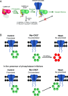
Autophosphorylation of CaMKII T286 is required for synaptic memory, but not phosphorylation of downstream synaptic proteins. (A) Autophosphorylation of CaMKII T286 is required for synaptic memory by stabilizing the binding of CaMKII to GluN2B. The numerous proposed downstream synaptic protein targets of CaMKII are not required for synaptic memory. Rather, the CaMKII/GluN2B complex serves as a structural signaling hub to enhance and maintain synaptic strength. (B) The stability of the CaMKII/GluN2B complex requires the continued phosphorylation of T286 to maintain the synaptic memory. Transient application of myr-CN27 (Fig. 5) dissociates active CaMKII from GluN2B. The released active CaMKII is rapidly dephosphorylated by phosphatases, erasing the synaptic memory. In the presence of phosphatase inhibitors, the active CaMKII rapidly rebinds to GluN2B restoring the synaptic memory.

Update of

Similar articles

Cited by

References

    1. Shonesy B. C., Jalan-Sakrikar N., Cavener V. S., Colbran R. J., CaMKII: A molecular substrate for synaptic plasticity and memory. Prog. Mol. Biol. Transl. Sci. 122, 61–87 (2014). - PubMed
    1. Hell J. W., CaMKII: Claiming center stage in postsynaptic function and organization. Neuron 81, 249–265 (2014). - PMC - PubMed
    1. Lisman J., Raghavachari S., Biochemical principles underlying the stable maintenance of LTP by the CaMKII/NMDAR complex. Brain Res. 1621, 51–61 (2015). - PMC - PubMed
    1. Bhattacharyya M., Karandur D., Kuriyan J., Structural Insights into the regulation of Ca2+/calmodulin-dependent protein kinase II (CaMKII) Cold Spring Harb. Perspect. Biol. 12, a035147 (2020). - PMC - PubMed
    1. Bayer K. U., Schulman H., CaM Kinase: Still Inspiring at 40. Neuron 103, 380–394 (2019). - PMC - PubMed

Substances