

Changes in subchondral bone morphology with osteoarthritis in the ankle
- PMID: 38889162
- PMCID: PMC11185451
- DOI: 10.1371/journal.pone.0290914
Changes in subchondral bone morphology with osteoarthritis in the ankle
Abstract
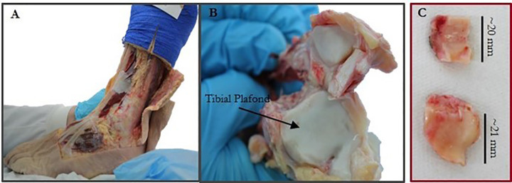
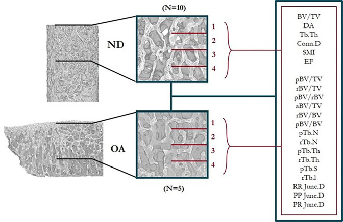
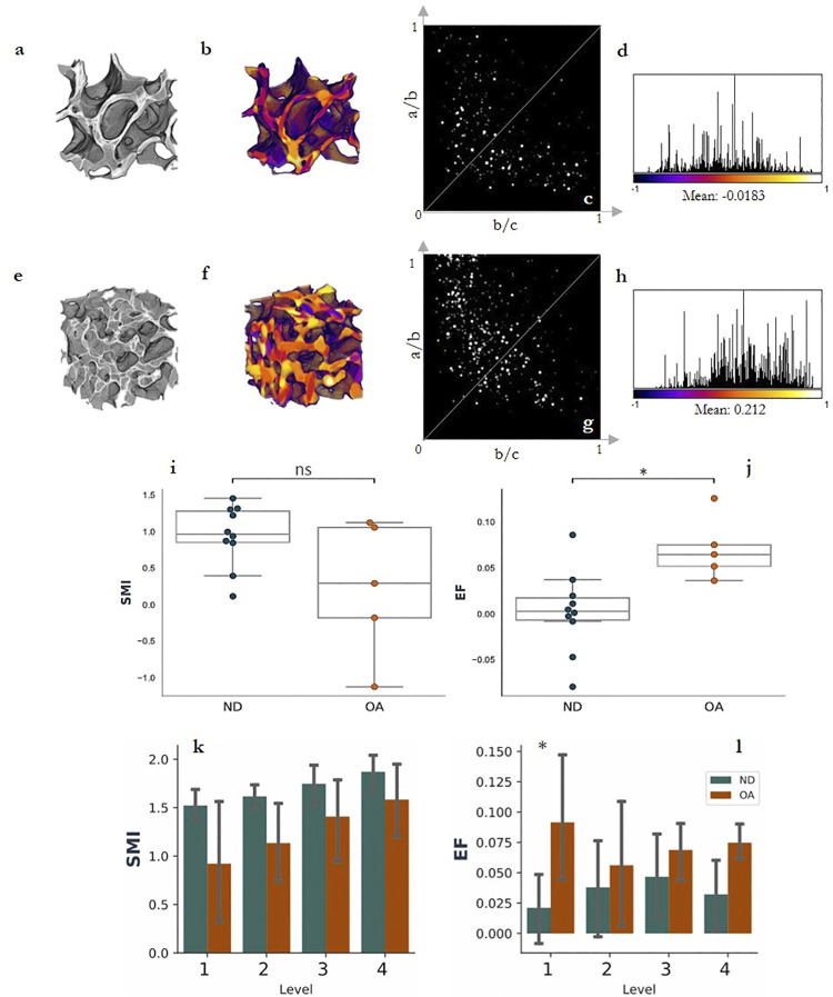
Significant alterations to subchondral trabecular bone microarchitecture are observed in late-stage osteoarthritis (OA). However, detailed investigation of these changes to bone in the ankle are under-reported. This study aimed to fully characterise the trabecular morphology in OA ankle bone specimens compared to non-diseased (ND) controls using both standard and individual-trabecular segmentation-based (ITS) analyses. Ten ND tibial bone specimens were extracted from three cadaveric ankles, as well as five OA bone specimens from patients undergoing total ankle arthroplasty surgery. Each specimen was scanned using microcomputed tomography from which a 4 mm cuboidal volume was extracted for analysis. Morphological parameters for the subchondral trabecular bone were measured using BoneJ (NIH ImageJ) and 3D ITS for whole volumes and at each depth level in 1 mm increments. The results show an overall increase in bone volume fraction (p<0.01) and trabecular thickness (p<0.001) with OA, with a decrease in anisotropy (p<0.05). ITS analysis showed OA bone was composed of more rod-like trabeculae and plate-like trabeculae compared to ND bone. Numerous properties were depth dependent, but the results demonstrated that towards the subchondral bone plate, both rod- and plate-like trabeculae were thicker, rods were longer and plates had increased surface area. Overall, this study has verified key microstructural alterations to ankle subchondral bone that are found in other OA lower-limb joints. Depth-based analysis has highlighted differences of interest for further evaluation into the remodelling mechanisms that occur with OA, which is critical to understanding the role of subchondral bone microarchitecture in the progression of the disease.
Copyright: © 2024 Koria et al. This is an open access article distributed under the terms of the Creative Commons Attribution License, which permits unrestricted use, distribution, and reproduction in any medium, provided the original author and source are credited.
Conflict of interest statement
The authors have declared that no competing interests exist.
Figures


Similar articles
-
Subchondral Trabecular Rod Loss and Plate Thickening in the Development of Osteoarthritis.J Bone Miner Res. 2018 Feb;33(2):316-327. doi: 10.1002/jbmr.3313. Epub 2017 Nov 16. J Bone Miner Res. 2018. PMID: 29044705
-
Association between knee alignment, osteoarthritis disease severity, and subchondral trabecular bone microarchitecture in patients with knee osteoarthritis: a cross-sectional study.Arthritis Res Ther. 2020 Sep 4;22(1):203. doi: 10.1186/s13075-020-02274-0. Arthritis Res Ther. 2020. PMID: 32887657 Free PMC article.
-
Correlation between subchondral bone plate thickness and cartilage degeneration in osteoarthritis of the ankle.Foot Ankle Int. 2014 Dec;35(12):1341-9. doi: 10.1177/1071100714548061. Epub 2014 Aug 18. Foot Ankle Int. 2014. PMID: 25136030
-
Hemodynamic stress shapes subchondral bone in osteoarthritis: An emerging hypothesis.J Orthop Translat. 2021 Dec 30;32:85-90. doi: 10.1016/j.jot.2021.11.007. eCollection 2022 Jan. J Orthop Translat. 2021. PMID: 35070712 Free PMC article. Review.
-
Subchondral bone changes in hand and knee osteoarthritis detected by radiography.Osteoarthritis Cartilage. 2004;12 Suppl A:S10-9. doi: 10.1016/j.joca.2003.09.007. Osteoarthritis Cartilage. 2004. PMID: 14698636 Review.
References
-
- NJR, S.C., 2019. National joint registry for england, wales, northern ireland and the isle of man: 16th annual report, 2019. National Joint Registry Centre.
MeSH terms
LinkOut - more resources
Full Text Sources
Medical
