. 2024 Jun 18;9(15):e171102.
doi: 10.1172/jci.insight.171102.

Macrophage IL-1β mediates atrial fibrillation risk in diabetic mice

Macrophage IL-1β mediates atrial fibrillation risk in diabetic mice

Xiaoxu Zhou et al. JCI Insight. .

Abstract

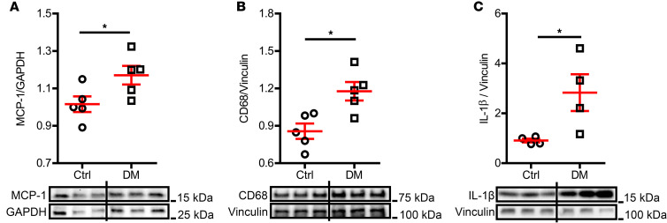
Diabetes mellitus (DM) is an independent risk factor for atrial fibrillation (AF). The mechanisms underlying DM-associated AF are unclear. AF and DM are both related to inflammation. We investigated whether DM-associated inflammation contributed to AF risk. Mice were fed with high-fat diet to induce type II DM and were subjected to IL-1β antibodies, macrophage depletion by clodronate liposomes, a mitochondrial antioxidant (mitoTEMPO), or a cardiac ryanodine receptor 2 (RyR2) stabilizer (S107). All tests were performed at 36-38 weeks of age. DM mice presented with increased AF inducibility, enhanced mitochondrial reactive oxygen species (mitoROS) generation, and activated innate immunity in the atria, as evidenced by enhanced monocyte chemoattractant protein-1 (MCP-1) expression, macrophage infiltration, and IL-1β levels. Signs of aberrant RyR2 Ca2+ leak were observed in the atria of DM mice. IL-1β neutralization, macrophage depletion, and exposure to mitoTEMPO and S107 significantly ameliorated the AF vulnerability in DM mice. Atrial overexpression of MCP-1 increased AF occurrence in normal mice through the same mechanistic signaling cascade as observed in DM mice. In conclusion, macrophage-mediated IL-1β contributed to DM-associated AF risk through mitoROS modulation of RyR2 Ca2+ leak.

Keywords: Arrhythmias; Cardiology; Diabetes; Inflammation; Macrophages.


Figures

Figure 1
Figure 1. AF inducibility in DM mice.
HFD-induced (A) high fasting glucose level and (B) obesity in mice. Echocardiographic evaluation indicated (C) preserved ejection fraction and (D) an increased ratio of transmitral Doppler early filling velocity to tissue Doppler early diastolic mitral annual velocity (E/E′). (E) Left atrial diameter by echocardiography and (F) atrial collagen I level by immunoblotting were comparable between control and DM mice. N = 5–6 mice per group. (G) DM mice had higher AF inducibility compared with the control mice; IL-1β–neutralizing antibody or mitochondrial antioxidant (mitoTEMPO) inhibited AF inducibility in DM mice. N is indicated within the bars. (H) Representative surface and intracardiac ECG traces showing induced AF in DM mice; arrows indicate the end of programmed stimulation. Scale bar: 100 ms. Data are shown as the mean ± SEM. Unpaired t test (AF) or Fisher’s exact test (G) were used. *P < 0.05, **P < 0.01, ***P < 0.001. AF, atrial fibrillation; Ctrl, control; DM, diabetes mellitus; EF, ejection fraction; HFD, high-fat diet; LA, left atrium; MT, mitoTEMPO.
Figure 2
Figure 2. Inflammation was increased in DM atria.
Expression level of (A) MCP-1, (B) macrophage marker CD68, and (C) inflammatory cytokine IL-1β were increased in DM atrial tissue. Representative immunoblotting images are shown. N = 4–5 mice per group. Data are shown as the mean ± SEM. Unpaired t test was used. *P < 0.05. Ctrl, control; DM, diabetes mellitus; MCP-1, monocyte chemoattractant protein-1.
Figure 3
Figure 3. Macrophage-secreted IL-1β mediated DM-associated AF.
(A) Atrial IL-1β level by immunoblotting was significantly lowered in macrophage depleted DM mice treated with clodronate liposomes compared with that in the plain liposome-treated DM mice. Representative immunoblotting images were shown. N = 5–6 mice per group. (B) Macrophage depletion by clodronate liposomes inhibited AF inducibility in DM mice. N is indicated within bars. Data are shown as the mean ± SEM. One-way ANOVA with Bonferroni’s post hoc tests (A) or Fisher’s exact test (B) were used. *P < 0.05, **P < 0.01. AF, atrial fibrillation; Ctrl, control; DM, diabetes mellitus.
Figure 4
Figure 4. A mitochondrial antioxidant reduced AF inducibility.
(A) Representative confocal microscopy images showing mitoROS in atrial cardiomyocytes by MitoSOX Red staining. Scale bar: 20 μm. (B) MitoSOX intensity was increased in DM atrial cardiomyocytes and reversed by mitochondrial antioxidant, mitoTEMPO. The atrial cardiomyocytes were isolated from 3 mice per group. (C) MitoTEMPO reduced the CD68 expression in DM atria. Representative immunoblotting images are shown. N = 3 mice per group. Data are shown as the mean ± SEM. One-way ANOVA with Bonferroni’s post hoc tests (B) or unpaired t test (C) were used. *P < 0.05, ***P < 0.001. Ctrl, control; DM, diabetes mellitus; MT, mitoTEMPO; mitoROS, mitochondrial reactive oxygen species.
Figure 5
Figure 5. RyR2-mediated sarcoplasmic reticulum Ca2+ leak contributed to DM-associated AF.
(A) Oxidized CaMKII and (B) phosphorylated RyR2 (Ser2814) levels determined by immunoblotting were increased in DM mouse atria. N = 5 mice per group. (C) Representative Western blot images of oxidized CaMKII, total CaMKII, p-RyR2-Ser2814, and total RyR2. (D) The RyR-calstabin interaction stabilizer S107 improved AF inducibility. N is indicated within bars. Data are shown as the mean ± SEM. Unpaired t test (A and B) or Fisher’s exact test (D) were used. *P < 0.05. AF, atrial fibrillation; oxi-CaMKII, oxidized Ca2+/calmodulin-dependent protein kinase II; Ctrl, control; DM, diabetes mellitus; RyR2, ryanodine receptor 2.
Figure 6
Figure 6. Atrial MCP-1 overexpression increased AF risk in normal mice.
(A) Representative immunoblotting images of MCP-1 expression in mouse atria and ventricles 1 month after AAV9 vector injection. (B) Atrial but not (C) ventricular MCP-1 levels were elevated in the mice receiving AAV9-ANF-MCP-1 vector injection. (D) Atrial Ccr2 mRNA level was increased in the MCP-1–overexpressing mice. N = 7–8 mice per group. (E) MCP-1 atrial overexpressing mice exhibited high AF inducibility. N is indicated within the bars. Data are shown as the mean ± SEM. Unpaired t test (BD) or Fisher’s exact test (E) were used. *P < 0.05, **P < 0.01. AAV9, adeno-associated virus serotype 9; AF, atrial fibrillation; ANF, atrial natriuretic factor; MCP-1, monocyte chemoattractant protein-1.
Figure 7
Figure 7. Cardiac characterization of atrial MCP-1–overexpressing mice.
(A) MitoSOX intensity was enhanced in the atrial cardiomyocytes overexpressing MCP-1. The atrial cardiomyocytes were isolated from 2 mice per group. (B) Oxi-CaMKII and (C) p-RyR2 (Ser2814) levels were increased in MCP-1–overexpressing atria. (D) Representative immunoblotting images of oxidized CaMKII, total CaMKII, p-RyR2-Ser2814, and total RyR2. (EG) Echocardiographic evaluation of EF, E/E′, and left atrial diameter, respectively. (H) Atrial collagen I level in AAV9-plain and AAV9-MCP-1 vector–treated mice. N = 4–9 mice per group. Data are shown as the mean ± SEM. Unpaired t test (AC and EH) was used. *P < 0.05, **P < 0.01, ***P < 0.001. AAV9, adeno-associated virus serotype 9; AF, atrial fibrillation; E/E′, ratio of transmitral Doppler early filling velocity to tissue Doppler early diastolic mitral annual velocity; EF, ejection fraction; LA, left atrium; MCP-1, monocyte chemoattractant protein-1; oxi-CaMKII, oxidized Ca2+/calmodulin-dependent protein kinase II; RyR2, ryanodine receptor 2.

References

    1. Silva JAD, et al. Diagnosis of diabetes mellitus and living with a chronic condition: participatory study. BMC Public Health. 2018;18(1):699. doi: 10.1186/s12889-018-5637-9. - DOI - PMC - PubMed
    1. Xiong Z, et al. A machine learning aided systematic review and meta-analysis of the relative risk of atrial fibrillation in patients with diabetes mellitus. Front Physiol. 2018;9:835. doi: 10.3389/fphys.2018.00835. - DOI - PMC - PubMed
    1. Bohne LJ, et al. The association between diabetes mellitus and atrial fibrillation: clinical and mechanistic insights. Front Physiol. 2019;10:135. doi: 10.3389/fphys.2019.00135. - DOI - PMC - PubMed
    1. Writing Group M. et al. Heart Disease and Stroke Statistics-2016 Update: a report from the American Heart Association. Circulation. 2016;133(4):e38–360. doi: 10.1161/CIR.0000000000000350. - DOI - PubMed
    1. Andrade J, et al. The clinical profile and pathophysiology of atrial fibrillation: relationships among clinical features, epidemiology, and mechanisms. Circ Res. 2014;114(9):1453–1468. doi: 10.1161/CIRCRESAHA.114.303211. - DOI - PubMed

MeSH terms