Swallowing muscle mass contributes to post-stroke dysphagia in ischemic stroke patients undergoing mechanical thrombectomy
- PMID: 38890004
- PMCID: PMC11294029
- DOI: 10.1002/jcsm.13512
Swallowing muscle mass contributes to post-stroke dysphagia in ischemic stroke patients undergoing mechanical thrombectomy
Abstract
Background: Neurogenic dysphagia is a frequent complication of stroke and is associated with aspiration pneumonia and poor outcomes. Although ischaemic lesion location and size are major determinants of the presence and severity of post-stroke dysphagia, little is known about the contribution of other acute stroke-unrelated factors. We aimed to analyse the impact of swallowing and non-swallowing muscles measurements on swallowing function after large vessel occlusion stroke.
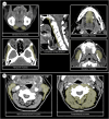
Methods: This retrospective study was based on a prospective registry of consecutive ischaemic stroke patients. Patients who underwent mechanical thrombectomy between July 2021 and June 2022 and received a flexible endoscopic evaluation of swallowing (FEES) within 5 days after admission were included. Demographic, anthropometric, clinical, and imaging data were collected from the registry. The cross-sectional areas (CSA) of selected swallowing muscles (as a surrogate marker for swallowing muscle mass) and of cervical non-swallowing muscles were measured in computed tomography. Skeletal muscle index (SMI) was calculated and used as a surrogate marker for whole body muscle mass. FEES parameters, namely, Functional Oral Intake Scale (FOIS, as a surrogate marker for dysphagia presence and severity), penetration aspiration scale, and the presence of moderate-to-severe pharyngeal residues were collected from the clinical records. Univariate and multivariate ordinal and logistic regression analyses were performed to analyse if total CSA of swallowing muscles and SMI were associated with FEES parameters.
Results: The final study population consisted of 137 patients, 59 were female (43.1%), median age was 74 years (interquartile range 62-83), median baseline National Institutes of Health Stroke Scale score was 12 (interquartile range 7-16), 16 patients had a vertebrobasilar occlusion (11.7%), and successful recanalization was achieved in 127 patients (92.7%). Both total CSA of swallowing muscles and SMI were significantly correlated with age (rho = -0.391, P < 0.001 and rho = -0.525, P < 0.001, respectively). Total CSA of the swallowing muscles was independently associated with FOIS (common adjusted odds ratio = 1.08, 95% confidence interval = 1.01-1.16, P = 0.029), and with the presence of moderate-to-severe pharyngeal residues for puree consistencies (adjusted odds ratio = 0.90, 95% confidence interval = 0.81-0.99, P = 0.036). We found no independent association of SMI with any of the FEES parameters.
Conclusions: Baseline swallowing muscle mass contributes to the pathophysiology of post-stroke dysphagia. Decreasing swallowing muscle mass is independently associated with increasing severity of early post-stroke dysphagia and with increased likelihood of moderate-to-severe pharyngeal residues.
Keywords: Dysphagia; Fiberoptic endoscopic evaluation of swallowing; Ischaemic stroke; Sarcopenia.
© 2024 The Author(s). Journal of Cachexia, Sarcopenia and Muscle published by Wiley Periodicals LLC.
Conflict of interest statement
SC Tauber has served on the scientific advisory boards of Roche and Merck & Co and has received travel and speaker honoraria from Novartis, Teva, Merck & Co, Roche, and Biogen. The remaining authors report no conflict of interests.
Figures


References
-
- Cohen DL, Roffe C, Beavan J, Blackett B, Fairfield CA, Hamdy S, et al. Post‐stroke dysphagia: a review and design considerations for future trials. Int J Stroke 2016;11:399–411. - PubMed
-
- Flowers HL, Skoretz SA, Streiner DL, Silver FL, Martino R. MRI‐based neuroanatomical predictors of dysphagia after acute ischemic stroke: a systematic review and meta‐analysis. Cerebrovasc Dis 2011;32:1–10. - PubMed
-
- Warnecke T, Dziewas R, Wirth R, Bauer JM, Prell T. Dysphagia from a neurogeriatric point of view: pathogenesis, diagnosis and management. Z Gerontol Geriatr 2019;52:330–335. - PubMed
MeSH terms
LinkOut - more resources
Full Text Sources
Medical

