. 2024 Jun 18;15(1):5176.
doi: 10.1038/s41467-024-49135-x.

Immuno-protective vesicle-crosslinked hydrogel for allogenic transplantation

Affiliations

Immuno-protective vesicle-crosslinked hydrogel for allogenic transplantation

Yuqian Wang et al. Nat Commun. .

Abstract

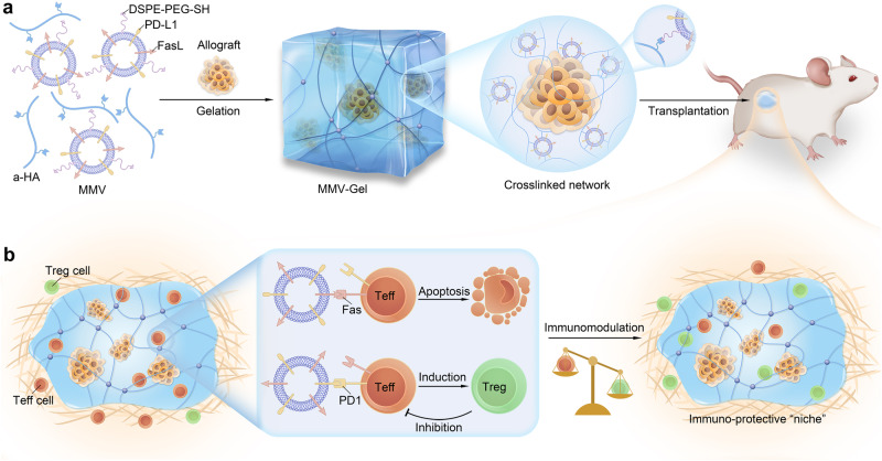
The longevity of grafts remains a major challenge in allogeneic transplantation due to immune rejection. Systemic immunosuppression can impair graft function and can also cause severe adverse effects. Here, we report a local immuno-protective strategy to enhance post-transplant persistence of allografts using a mesenchymal stem cell membrane-derived vesicle (MMV)-crosslinked hydrogel (MMV-Gel). MMVs are engineered to upregulate expression of Fas ligand (FasL) and programmed death ligand 1 (PD-L1). The MMVs are retained within the hydrogel by crosslinking. The immuno-protective microenvironment of the hydrogel protects allografts by presenting FasL and PD-L1. The binding of these ligands to T effector cells, the dominant contributors to graft destruction and rejection, results in apoptosis of T effector cells and generation of regulatory T cells. We demonstrate that implantation with MMV-Gel prolongs the survival and function of grafts in mouse models of allogeneic pancreatic islet cells and skin transplantation.


Conflict of interest statement

R.M., Y.W. and R.H. are applying a patent related to this work. The remaining authors declare that there are no competing interests.

Figures

Fig. 1
Fig. 1. Schematic of local immuno-protective niche implemented by MMV-Gel for prolonged survival and function of allogeneic transplants.
a Schematic of preparation of MMV-Gel for local delivery of allografts. MMV-Gel is formed by mixing thiol-modified FasL/PD-L1-expressing MMVs and a-HA via specific thiol-acrylate Michael addition reaction. As an example, the transplanted grafts can be embedded in the crosslinked network of MMV-Gel. b Schematic of mechanism of MMV-Gel serving as an immuno-protective niche to achieve allograft acceptance. MMV-Gel holds the tethered MMVs to enhance their retention at the implanted site. The local persistence of MMV-Gel improves long-term preservation of the graft. Upon acute immune response to allotransplantation, the Teff cells increasingly infiltrate into the transplanted region with the mission to attack and destruct grafts, ultimately leading to graft rejection, which are characterized to highly express Fas and PD1 following activation. MMV-Gel provides an immuno-protective microenvironment to resist the Teff cell-mediated immune response by the MMV-presenting FasL and PD-L1 binding to their specific receptors, Fas and PD1 on the Teff cells to induce apoptosis of Teff cells and elevation of Treg cells.
Fig. 2
Fig. 2. MMV-Gel triggers apoptosis and suppresses proliferation on the activated T cells.
a Expression of FasL on RBCs, PLTs, aPLTs and MSCs determined by flow cytometry. MFI, mean fluorescent intensity. arb. unit, arbitrary unit. b Expression of PD-L1 on MSCs after treatment with IFN-γ for 12 h determined by flow cytometry. c Particle size and TEM image of MMVs. Scale bar, 50 nm. d Total (early plus late) apoptotic percentages of T cells within the activated splenocytes after treatment with varying amounts of MMVs determined by Annexin V-FITC/PI double-staining assay. e Total apoptotic percentages of T cells within the activated splenocytes after treatment with MMVs (20 μg) in the absence and presence of aFasL. f Proliferation of T cells within the activated splenocytes after treatment with MMVs (10 μg) in the absence and presence of aPD-L1 or aFasL determined by CFSE dilution assay. g Gelation of MMV-Gel by mixing MMVs and a-HA examined by tube inversion assay. h Rheology measurement of MMV-Gel. i SEM images of MMV-Gel. Scale bars, 100 μm (low magnification) and 500 nm (high magnification). j Three-dimensional reconstructed confocal microscopic image of Rho-MMV-Gel. Scale bar, 50 μm. Fluorescent imaging (k) and quantification (l) of viability of the activated T cells infiltrating in L-Gel and MMV-Gel for 48 h examined by calcein AM/PI double-staining assay. Scale bar, 100 μm. Fluorescent imaging (m) and quantification (n) of expression of PD1 on the activated T infiltrating in L-Gel and MMV-Gel for 24 h. Scale bar, 5 μm. Representative is displayed from 3 independent experiments (c, gk, m). Data are shown as mean ± standard deviation (s.d.) (n = 6 independent samples in a, b, df; n = 3 independent samples in l, n). One-way ANOVA with Tukey post-hoc test was used for statistical analysis of a, f. Two-tailed unpaired t-test was used for statistical analysis of b, e, l, n). Source data are provided as a source data file.
Fig. 3
Fig. 3. MMV-Gel protects the encapsulated islet allograft against the activated T cell.
a Cryo-SEM image of islet/MMV-Gel. Pseudocolour processing was used for image display of islet in yellow and hydrogel matrix in blue. Scale bar, 10 μm. b Expression of insulin and glucagon in islet/MMV-Gel examined by immunofluorescent staining. Scale bar, 25 μm. GSIS (c) and stimulation index (d) of free islet and islet/MMV-Gel. GSIS (e) and stimulation index (f) of islet/MMV-Gel within 7 d. Fluorescent imaging (g) and quantification (h) of viability of islet/L-Gel and islet/MMV-Gel after incubation with the activated T cells for 48 h examined by acridine orange/PI double-staining assay. Scale bar, 25 μm. GSIS (i) and stimulation index (j) of islet/L-Gel and islet/MMV-Gel after incubation with the activated T cells for 48 h. k Infiltration of CD8+ and CD4+ T cells in the transplanted site of kidney at 7 d post-transplantation of islet, islet/L-Gel and islet/MMV-Gel examined by immunofluorescent staining. White dotted lines indicate the graft. Scale bar, 50 μm. Proportions of CD8+ Teff (CD8+CD44+CD62L) (l), CD4+ Teff (CD4+CD44+CD62L) (m) and Treg (CD4+FoxP3+) (n) cells in kidney at 7 d post-transplantation of islet, islet/L-Gel and islet/MMV-Gel determined by flow cytometry. Representative is displayed from 3 independent experiments (a, b, g, k). Data are shown as mean ± s.d. (n = 6 independent samples in cf, i, j; n = 3 independent samples in h; n = 6 mice in ln). Two-tailed unpaired t-test was used for statistical analysis of d, hj. One-way ANOVA with Tukey post-hoc test was used for statistical analysis of f, ln. Source data are provided as a source data file.
Fig. 4
Fig. 4. MMV-Gel prolongs survival and function of the transplanted allogeneic islet.
a Changes in the individual BGLs of the diabetic mice after transplantation of islet, islet/L-Gel and islet/MMV-Gel (n = 9 mice). b Expression of insulin in the transplanted site of the kidney at 30 d post-transplantation of islet, islet/L-Gel and islet/MMV-Gel examined by immunofluorescent staining. Representative is displayed from 3 independent experiments. Scale bar, 50 μm. c IPGTT on the normal mice, diabetic mice and diabetic mice at 30 d post-transplantation of islet/MMV-Gel that have survived. d AUC values of glucose obtained from IPGTT. Changes in the individual BGLs of the islet/MMV-Gel-transplanted diabetic mice after graft removal (e) (n = 3 mice) and the islet/MMV-Gel-transplanted FoxP3/DTR diabetic mice after i.p. injection of DT (f) (n = 6 mice). Black arrows indicate graft removal or two i.p. injections of DT at Day 30 and 31. Proportion of CD8+ (g) and CD4+ (h) T cell proliferation in spleen from the mice at 30 d post-transplantation of MMV-Gel to the splenocytes isolated from the BALB/c (recipient-matched), C57BL/6 (donor) and C3H (3rd party) mice determined by flow cytometry. i Changes in the individual BGLs of the diabetic mice after transplantation of islet and islet/MMV-Gel with i.p. injection of RAPA. j Survival of islet and islet/MMV-Gel combined with RAPA (n = 5 mice). Data are shown as mean ± s.d. (n = 3 mice in c, d; n = 6 mice in g, h). One-way ANOVA with Tukey post-hoc test was used for statistical analysis of d. Two-tailed unpaired t-test was used for statistical analysis of g, h. Two-sided log-rank (Mantel–Cox) test was used for statistical analysis of j. Source data are provided as a source data file.
Fig. 5
Fig. 5. MMV-Gel prolongs survival of the transplanted allogeneic skin.
a Infiltration of CD8+ and CD4+ T cells in skin graft at 7 d post-transplantation of skin, skin/L-Gel and skin/MMV-Gel determined by immunofluorescent staining. Representative is displayed from 3 independent experiments. Scale bar, 50 μm. Proportions of CD8+ Teff (CD8+CD44+CD62L), CD4+ Teff (CD4+CD44+CD62L) and Treg (CD4+FoxP3+) cells in the skin graft (bd), SDLN (eg) and spleen (hj) at 7 d post-transplantation of skin, skin/L-Gel and skin/MMV-Gel determined by flow cytometry. k Images of skin graft at 0, 7 and 14 d post-transplantation of skin, skin/L-Gel and skin/MMV-Gel. Histological examination of the transplanted area at 14 d post-transplantation by H&E staining. Representative is displayed from 5 independent experiments. Black dotted lines indicate the graft. Scale bar, 400 μm. l Survival of skin, skin/L-Gel and skin/MMV-Gel (n = 5 mice). Images (m) and weights (n) of SDLN harvested from the mice at 14 d post-transplantation of skin, skin/L-Gel and skin/MMV-Gel. Scale bar, 5 mm. Data are shown as mean ± s.d. (n = 6 mice in bj; n = 5 mice in n). One-way ANOVA with Tukey post-hoc test was used for statistical analysis of bj, n. Two-sided log-rank (Mantel–Cox) test was used for statistical analysis of l. Source data are provided as a source data file.

Similar articles

Cited by

References

    1. Lechler RI, Sykes M, Thomson AW, Turka LA. Organ transplantation–how much of the promise has been realized? Nat. Med. 2005;11:605–613. doi: 10.1038/nm1251. - DOI - PubMed
    1. Chabannon C, et al. Hematopoietic stem cell transplantation in its 60s: a platform for cellular therapies. Sci. Transl. Med. 2018;10:eaap9630. doi: 10.1126/scitranslmed.aap9630. - DOI - PubMed
    1. Yamashita M, Dellorusso PV, Olson OC, Passegue E. Dysregulated haematopoietic stem cell behaviour in myeloid leukaemogenesis. Nat. Rev. Cancer. 2020;20:365–382. doi: 10.1038/s41568-020-0260-3. - DOI - PMC - PubMed
    1. Shapiro AM, Pokrywczynska M, Ricordi C. Clinical pancreatic islet transplantation. Nat. Rev. Endocrinol. 2017;13:268–277. doi: 10.1038/nrendo.2016.178. - DOI - PubMed
    1. Shapiro AMJ, Verhoeff K. A spectacular year for islet and stem cell transplantation. Nat. Rev. Endocrinol. 2023;19:68–69. doi: 10.1038/s41574-022-00790-4. - DOI - PubMed

MeSH terms