Structural basis for IL-33 recognition and its antagonism by the helminth effector protein HpARI2
- PMID: 38890291
- PMCID: PMC11189471
- DOI: 10.1038/s41467-024-49550-0
Structural basis for IL-33 recognition and its antagonism by the helminth effector protein HpARI2
Abstract
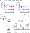
IL-33 plays a significant role in inflammation, allergy, and host defence against parasitic helminths. The model gastrointestinal nematode Heligmosomoides polygyrus bakeri secretes the Alarmin Release Inhibitor HpARI2, an effector protein that suppresses protective immune responses and asthma in its host by inhibiting IL-33 signalling. Here we reveal the structure of HpARI2 bound to mouse IL-33. HpARI2 contains three CCP-like domains, and we show that it contacts IL-33 primarily through the second and third of these. A large loop which emerges from CCP3 directly contacts IL-33 and structural comparison shows that this overlaps with the binding site on IL-33 for its receptor, ST2, preventing formation of a signalling complex. Truncations of HpARI2 which lack the large loop from CCP3 are not able to block IL-33-mediated signalling in a cell-based assay and in an in vivo female mouse model of asthma. This shows that direct competition between HpARI2 and ST2 is responsible for suppression of IL-33-dependent responses.
© 2024. The Author(s).
Conflict of interest statement
The authors declare no competing interests.
Figures


Similar articles
-
HpARI Protein Secreted by a Helminth Parasite Suppresses Interleukin-33.Immunity. 2017 Oct 17;47(4):739-751.e5. doi: 10.1016/j.immuni.2017.09.015. Immunity. 2017. PMID: 29045903 Free PMC article.
-
Vaccination against helminth IL-33 modulators permits immune-mediated parasite ejection.Cell Rep. 2025 May 27;44(5):115721. doi: 10.1016/j.celrep.2025.115721. Epub 2025 May 15. Cell Rep. 2025. PMID: 40378045
-
A helminth-derived suppressor of ST2 blocks allergic responses.Elife. 2020 May 18;9:e54017. doi: 10.7554/eLife.54017. Elife. 2020. PMID: 32420871 Free PMC article.
-
Animal model of Nippostrongylus brasiliensis and Heligmosomoides polygyrus.Curr Protoc Immunol. 2003 Aug;Chapter 19:Unit 19.12. doi: 10.1002/0471142735.im1912s55. Curr Protoc Immunol. 2003. PMID: 18432905 Review.
-
Immune modulation and modulators in Heligmosomoides polygyrus infection.Exp Parasitol. 2012 Sep;132(1):76-89. doi: 10.1016/j.exppara.2011.08.011. Epub 2011 Aug 22. Exp Parasitol. 2012. PMID: 21875581 Free PMC article. Review.
Cited by
-
IL-33-binding HpARI family homologues with divergent effects in suppressing or enhancing type 2 immune responses.Infect Immun. 2024 Mar 12;92(3):e0039523. doi: 10.1128/iai.00395-23. Epub 2024 Jan 31. Infect Immun. 2024. PMID: 38294241 Free PMC article.
-
Heparan sulphate binding controls in vivo half-life of the HpARI protein family.Elife. 2024 Nov 8;13:RP99000. doi: 10.7554/eLife.99000. Elife. 2024. PMID: 39514278 Free PMC article.
-
Cytokines from parasites: manipulating host responses by molecular mimicry.Biochem J. 2025 Apr 29;482(9):433-49. doi: 10.1042/BCJ20253061. Biochem J. 2025. PMID: 40302223 Free PMC article. Review.
-
Modulation of the Immune Response by Nematode Derived Molecules.Int J Mol Sci. 2025 Jun 11;26(12):5600. doi: 10.3390/ijms26125600. Int J Mol Sci. 2025. PMID: 40565065 Free PMC article. Review.
References
-
- McSorley HJ, Smyth DJ. IL-33: A central cytokine in helminth infections. Semin Immunol. 2021;53:101532. - PubMed
-
- Liew FY, Girard JP, Turnquist HR. Interleukin-33 in health and disease. Nat. Rev. Immunol. 2016;16:676–689. - PubMed
-
- Cayrol C, Girard JP. Interleukin-33 (IL-33): a critical review of its biology and the mechanisms involved in its release as a potent extracellular cytokine. Cytokine. 2022;156:155891. - PubMed

