. 2024 Jun 18;15(6):427.
doi: 10.1038/s41419-024-06827-y.

AURKB/CDC37 complex promotes clear cell renal cell carcinoma progression via phosphorylating MYC and constituting an AURKB/E2F1-positive feedforward loop

Affiliations

AURKB/CDC37 complex promotes clear cell renal cell carcinoma progression via phosphorylating MYC and constituting an AURKB/E2F1-positive feedforward loop

Fang Li et al. Cell Death Dis. .

Abstract

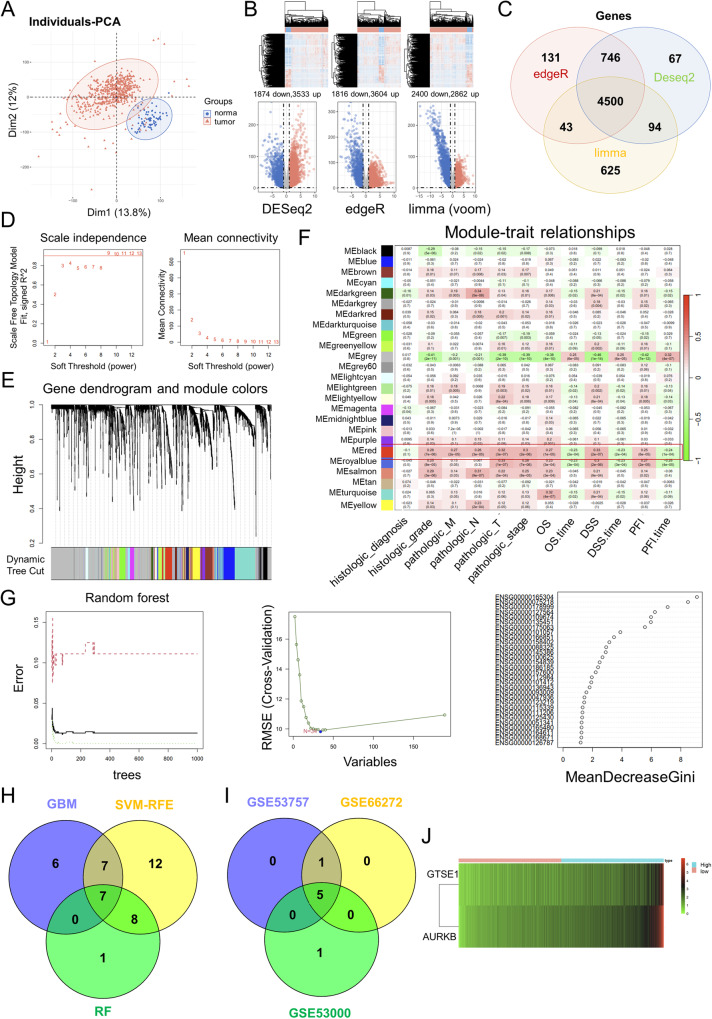
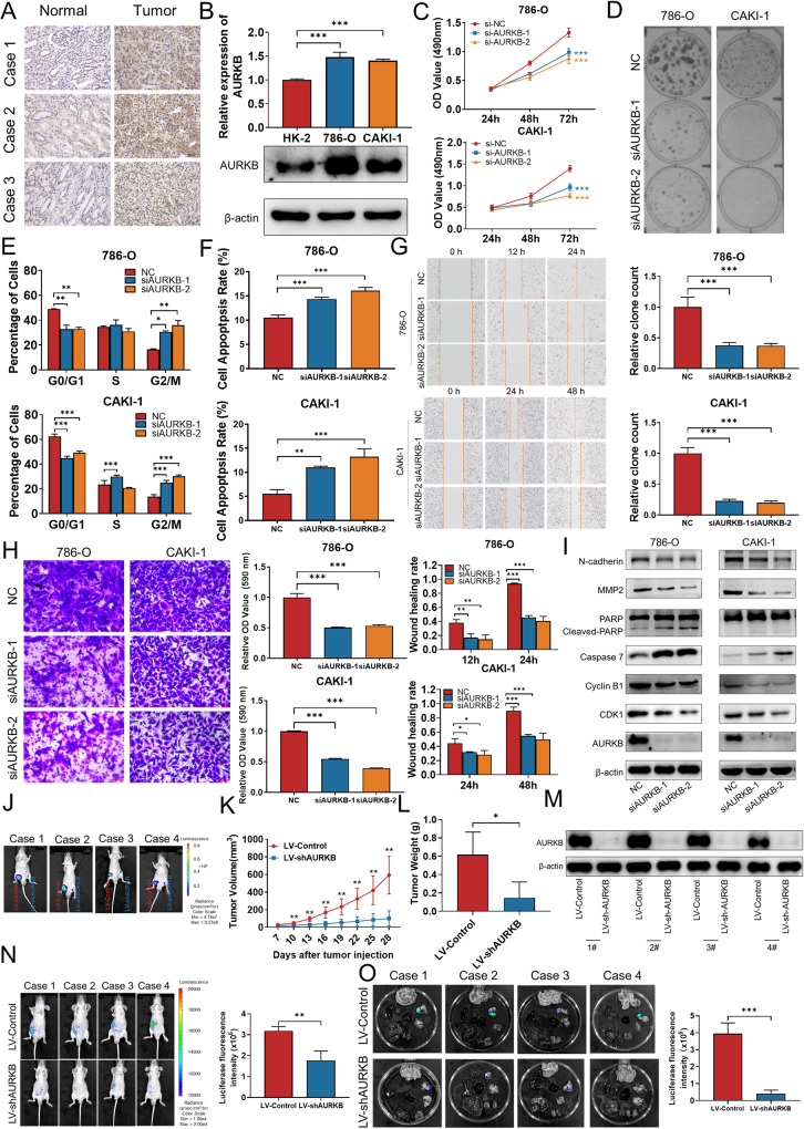
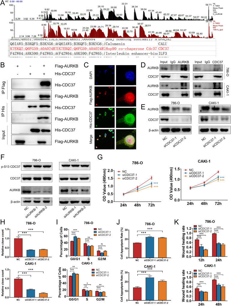
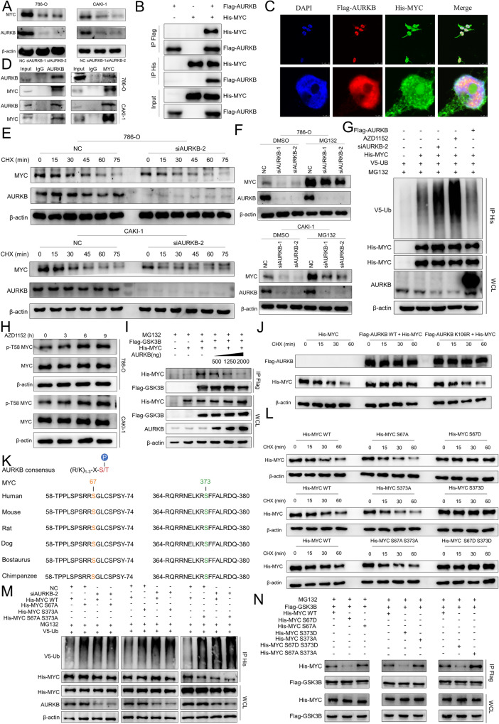
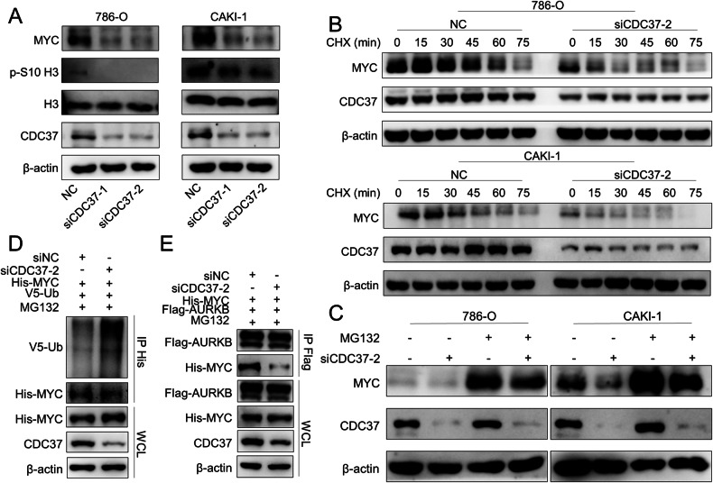
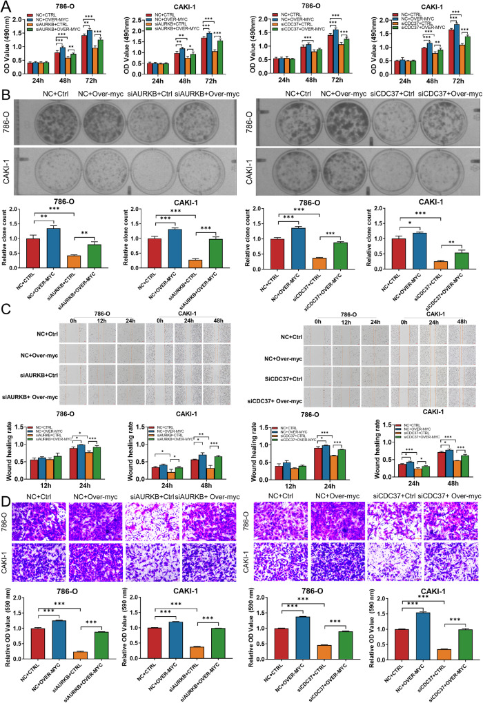
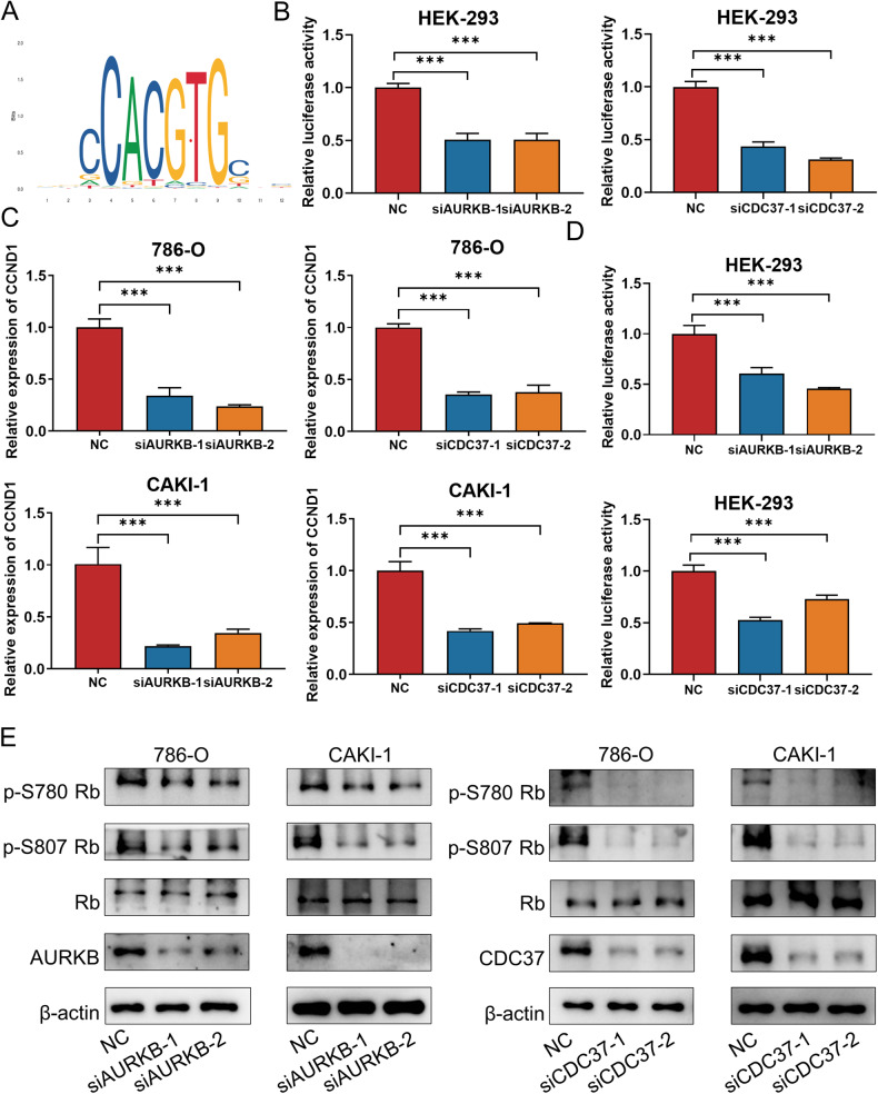
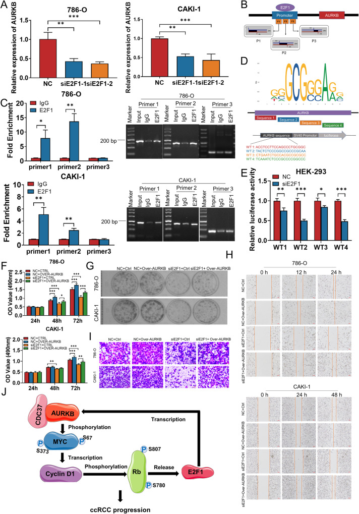
As the second most common malignant tumor in the urinary system, renal cell carcinoma (RCC) is imperative to explore its early diagnostic markers and therapeutic targets. Numerous studies have shown that AURKB promotes tumor development by phosphorylating downstream substrates. However, the functional effects and regulatory mechanisms of AURKB on clear cell renal cell carcinoma (ccRCC) progression remain largely unknown. In the current study, we identified AURKB as a novel key gene in ccRCC progression based on bioinformatics analysis. Meanwhile, we observed that AURKB was highly expressed in ccRCC tissue and cell lines and knockdown AURKB in ccRCC cells inhibit cell proliferation and migration in vitro and in vivo. Identified CDC37 as a kinase molecular chaperone for AURKB, which phenocopy AURKB in ccRCC. AURKB/CDC37 complex mediate the stabilization of MYC protein by directly phosphorylating MYC at S67 and S373 to promote ccRCC development. At the same time, we demonstrated that the AURKB/CDC37 complex activates MYC to transcribe CCND1, enhances Rb phosphorylation, and promotes E2F1 release, which in turn activates AURKB transcription and forms a positive feedforward loop in ccRCC. Collectively, our study identified AURKB as a novel marker of ccRCC, revealed a new mechanism by which the AURKB/CDC37 complex promotes ccRCC by directly phosphorylating MYC to enhance its stability, and first proposed AURKB/E2F1-positive feedforward loop, highlighting AURKB may be a promising therapeutic target for ccRCC.


Conflict of interest statement

The authors declare no competing interests.

Figures

Fig. 1
Fig. 1. Bioinformatics databases identified AURKB as a novel prognostic key gene for ccRCC.
A PCA of ccRCC-related genes in TCGA database. B Three R packages DESeq2, edgeR and limma (voom) were used to screen for differentially expressed genes (DEGs) in ccRCC and genes with p value < 0.05 and log2 fold change (FC)| > 1 were DEGs. C Venn diagram shows the intersection of DEGs screened for ccRCC using three R packages DESeq2, edgeR, and limma (voom), with a total of 4500 DEGs. D Soft-thresholding powers selection. E WGCNA cluster dendrogram and module assignment. F Scale-free gene co-expression network was constructed using the “WGCNA” package, and the red module was identified as the module with the strongest correlation with clinical stage and survival. G Three Machine learning methods, Gradient Boosting Machine, Random Forest and SVM-RFE, were used for binary classification feature screening of the screened red module genes. H Venn diagram shows the intersection of seven genes screened by three machine learning methods, gradient boosting machine, random forest and SVM-RFE. I The Venn diagram shows the intersection of validation results for three GEO databases. J LASSO Cox regression prognosis model was constructed according to the formula riskScore = geneExp*Coef.
Fig. 2
Fig. 2. AURKB is up-regulated in ccRCC and promotes the proliferation and migration of ccRCC cells in vitro in vivo.
A Representative immunohistochemical images of AURKB in human ccRCC tissues and adjacent normal tissues. B qRT-PCR and western blotting were used to detect the mRNA and protein expression of AURKB. C MTT assay was used to detect the effect of AURKB knockdown on the proliferation of ccRCC cells. D Colony formation assay was used to detect the effect of AURKB knockdown on the colony formation ability of ccRCC cells. E The effect of AURKB knockdown on cell cycle of ccRCC cells was detected by flow cytometry. F Flow cytometry was used to detect the effect of AURKB knockdown on cell apoptosis of ccRCC cells. G Wound healing assay was used to detect the effect of AURKB knockdown on the migration of ccRCC cells. H Transwell assay was used to detect the effect of AURKB knockdown on the migration of ccRCC cells, and the OD value of the traversed cells was used for statistical mapping. I Western blotting was used to detect the effect of AURKB knockdown on the expression of cell cycle, apoptosis and migration related molecules at protein level in 786-O and CAKI-1. J On the day 28, tumor was measured by in vivo bioluminescence imaging. K Tumor growth curves of the tumor volumes represent measurements taken every 3 d for 28 d. L The xenograft tumors were weighed and statistically mapped. M The protein levels of AURKB in xenograft tumors were analyzed by western blotting. N Image analysis was performed on nude mice to assess tumor metastasis on the 21th day after injection. O Image analysis was performed on the isolated organs of nude mice on day 21 after injection to assess tumor metastasis. *p < 0.05, **p < 0.01, ***p < 0.001.
Fig. 3
Fig. 3. Identification of CDC37 as a AURKB binding partner and CDC37 phenocopy AURKB in ccRCC.
A Co-IP mass spectrometry identified CDC37 as the binding partner of AURKB. B Lysates of 293T cells overexpressing Flag-AURKB and/or His-CDC37 were subjected to reciprocal Co-IP to detect protein interaction. C Representative images of immunofluorescence staining of DAPI, Flag-AURKB and His-CDC37 in 293T cells. D 786-O and CAKI-1 cell lysates were subjected to Co-IP to detect endogenous CDC37 and AURKB interaction. E Western blotting was used to detect the expression changes of target proteins after CDC37 knockdown in 786-O and CAKI-1 cells, with β-actin as a control. F Western blotting was used to detect the expression changes of target proteins after AURKB knockdown in 786-O and CAKI-1 cells, with β-actin as a control. G MTT assay was used to detect the effect of CDC37 knockdown on the proliferation of 786-0 and CAKI-1 cells. H Colony formation assay was used to detect the effect of CDC37 knockdown on the colony formation ability of 786-0 and CAKI-1 cells. I The effect of CDC37 knockdown on cell cycle of 786-O and CAKI-1 cells was detected by flow cytometry, and the percentage of cells in each period was plotted. J Flow cytometry was used to detect the effect of CDC37 knockdown on cell apoptosis of 786-O and CAKI-1 cells, and the percentage of apoptotic cells was plotted. K Wound healing assay was used to detect the effect of CDC37 knockdown on the migration of 786-O and CAKI-1 cells, and the percentage of wound healing was plotted. *p < 0.05, **p < 0.01, ***p < 0.001.
Fig. 4
Fig. 4. AURKB-Mediated MYC Phosphorylation Contributes to MYC Stability.
A Western blotting was used to detect the expression changes of MYC after AURKB knockdown in ccRCC cells. B Lysates of 293T cells overexpressing Flag-AURKB and/or His-MYC were subjected to reciprocal Co-IP to detect protein interaction. C Representative images of immunofluorescence staining of DAPI, Flag-AURKB and His-CDC37 in 293 T cells. D 786-O and CAKI-1 cell lysates were subjected to Co-IP to detect endogenous MYC and AURKB interaction. E Time course analysis of MYC protein level in AURKB knockdown ccRCC cells. F AURKB knockdown ccRCC cells were treated with MG132 (10 μM) for 6 h before harvest. AURKB and MYC were analyzed by immunoblot. G HEK-293T cells were co-transfected and treated with MG132 (10 μM) for 6 h before harvest. Cell lysates were subjected to Co-IP, ubiquitination, and immunoblot assays. H Time course analysis of MYC and p-T58 MYC by immunoblot upon AZD1152 (786-O: 500 nM; CAKI-1: 300 nM) treatment in ccRCC cells. I HEK-293T cells were co-transfected with Flag-GSK3β, His-MYC, and/or increasing doses of AURKB (0.5,1.25, and 2 μg) and treated with MG132 (10 μM) for 6 h before harvest. Cell lysates were subjected to Co-IP and immunoblot assays. J Time-course analysis of His-MYC levels was performed in 293T cells expressing ectopic His-MYC, Flag-AURKB WT or a mutant (K106R). K Peptide sequence alignment of MYC in different species. L Time-course analysis of His-tagged MYC by immunoblot in 293T cells. M HEK-293T cells were co-transfected and treated with MG132 (10 μM) for 6 h before harvest. Cell lysates were subjected to Co-IP, ubiquitination, and immunoblot assays. N HEK-293T cells were co-transfected and treated with MG132 (10 μM) for 6 h before harvest. Cell lysates were subjected to Co-IP and immunoblot assays.
Fig. 5
Fig. 5. CDC37-mediated binding of AURKB to MYC contributes to MYC Stability.
A Western blotting was used to detect the expression changes of target proteins after CDC37 knockdown in 786-O and CAKI-1 cells, with β-actin as a control. B Time course analysis of MYC protein levels in CDC37 knockdown 786-O and CAKI-1 cells. C CDC37 knockdown 786-O and CAKI-1 cells were treated with MG132 (10 μM) for 6 h before harvest. CDC37 and MYC were analyzed by immunoblot, with β-actin as a control. D HEK-293T cells were co-transfected with NC+His-MYC + V5-UB or siCDC37+His-MYC + V5-UB and treated with MG132 (10 μM) for 6 h before harvest. Cell lysates were subjected to Co-IP, ubiquitination, and immunoblot assays. E HEK-293T cells were co-transfected with NC+His-MYC+Flag-AURKB or siCDC37+His-MYC+Flag-AURKB. Cell lysates were subjected to Co-IP and immunoblot assays.
Fig. 6
Fig. 6. Enhanced MYC expression rescued the effects of AURKB/CDC37 depletion on ccRCC cells.
A, B MTT and colony formation assays were performed to determine the impact of cell viability treated with NC+Ctrl, NC+Over-MYC, siAURKB-2+Ctrl, siAURKB-2+Over-MYC, siCDC37-2+Ctrl, siCDC37-2+ Over-MYC in ccRCC cells. C, D Transwell and wound‐healing analysis represented the migration and metastasis capacity of ccRCC cells co‐transfected with NC+Ctrl, NC+Over-MYC, siAURKB-2+Ctrl, siAURKB-2+ Over-MYC, siCDC37-2+Ctrl, siCDC37-2+ Over-MYC. *p < 0.05, **p < 0.01, ***p < 0.001.
Fig. 7
Fig. 7. AURKB/CDC37 regulates MYC transcriptional program and promotes the phosphorylation of Rb1.
A DNA-binding motif of MYC (JASPAR). B Luciferase assays were performed in HEK-293 cells co-transfected with siNC/siAURKB/siCDC37 and MYC motif. C CCND1 mRNA levels in 786-O and CAKI-1 cells with AURKB or CDC37 knockdown were detected by qRT-PCR. D Luciferase assays were performed in HEK-293 cells co-transfected with siNC/siAURKB/siCDC37 and CCND1 promoter. E Western blotting was used to detect the effect of AURKB or CDC37 knockdown on the expression of phosphorylated Rb in 786-O and CAKI-1 cells. *p < 0.05, **p < 0.01, ***p < 0.001.
Fig. 8
Fig. 8. E2F1 activates AURKB expression directly.
A AURKB mRNA levels in 786-O and CAKI-1 cells with E2F1 knockdown were detected by qRT-PCR. B Schematic diagram of primer design for ChIP-qRT-PCR in the binding site between E2F1 and the promoter region of AURKB. C The binding relationship between E2F1 and the promoter region of AURKB was verified by ChIP-qRT-PCR and agarose gel electrophoresis. D The binding sequence of E2F1 on AURKB predicted by JASPAR was subcloned into the pGL3 promoter luciferase vector. E Luciferase assays were performed in HEK-293 cells co-transfected with siNC/siE2F1-2 and AURKB promoter. F, G MTT and colony formation assays were performed to determine the impact of cell viability treated with NC+ Ctrl, NC + Over-AURKB, siE2F1-2+Ctrl, siE2F1-2+Over-AURKB in ccRCC cells. H, I Transwell and wound‐healing analysis represented the migration and metastasis capacity of ccRCC cells co‐transfected with NC + Ctrl, NC + Over-AURKB, siE2F1-2 + Ctrl, siE2F1-2 + Over-AURKB. J The schematic illustrates that CDC37 and AURKB complexes directly phosphorylate MYC, enhance MYC stability and transcriptional activity, promote Rb phosphorylation and E2F1 release, and in turn activate the transcription of AURKB, forming an AURKB/E2F1 positive feedforward loop that promotes ccRCC progression. *p < 0.05, **p < 0.01, ***p < 0.001.

Similar articles

Cited by

References

    1. Sung H, Ferlay J, Siegel RL, Laversanne M, Soerjomataram I, Jemal A, et al. GlobaL Cancer Statistics 2020: GLOBOCAN estimates of incidence and mortality worldwide for 36 cancers in 185 countries. CA Cancer J Clin. 2021;71:209–49. doi: 10.3322/caac.21660. - DOI - PubMed
    1. Shuch B, Amin A, Armstrong AJ, Eble JN, Ficarra V, Lopez-Beltran A, et al. Understanding pathologic variants of renal cell carcinoma: distilling therapeutic opportunities from biologic complexity. Eur Urol. 2015;67:85–97. doi: 10.1016/j.eururo.2014.04.029. - DOI - PubMed
    1. Rini BI, Campbell SC, Escudier B. Renal cell carcinoma. Lancet. 2009;373:1119–32. doi: 10.1016/S0140-6736(09)60229-4. - DOI - PubMed
    1. Nerich V, Hugues M, Paillard MJ, Borowski L, Nai T, Stein U, et al. Clinical impact of targeted therapies in patients with metastatic clear-cell renal cell carcinoma. Onco Targets Ther. 2014;7:365–74. doi: 10.2147/OTT.S56370. - DOI - PMC - PubMed
    1. Motzer RJ, Bukowski RM, Figlin RA, Hutson TE, Michaelson MD, Kim ST, et al. Prognostic nomogram for sunitinib in patients with metastatic renal cell carcinoma. Cancer. 2008;113:1552–8. doi: 10.1002/cncr.23776. - DOI - PubMed

Publication types

MeSH terms