. 2024 Jun 18;15(6):423.
doi: 10.1038/s41419-024-06811-6.

Expression and functional implications of YME1L in nasopharyngeal carcinoma

Affiliations

Expression and functional implications of YME1L in nasopharyngeal carcinoma

Fuwei Cheng et al. Cell Death Dis. .

Abstract

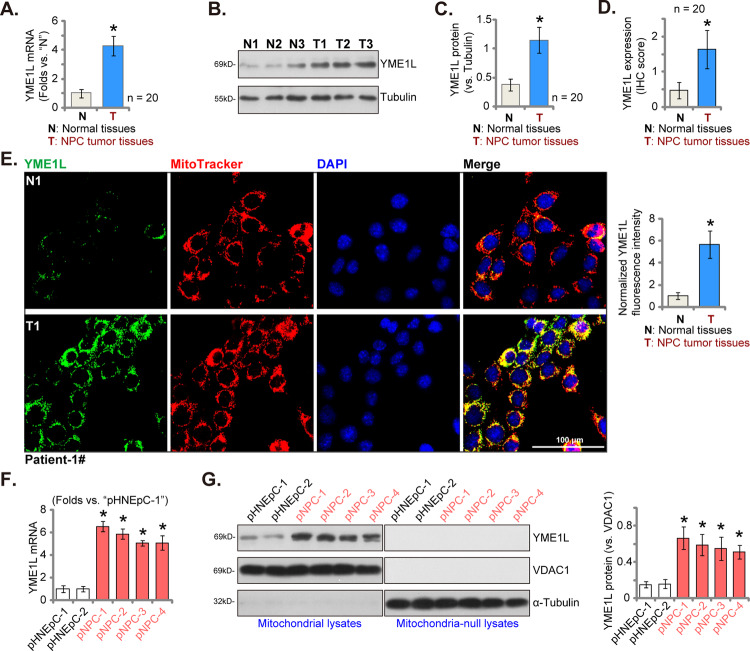
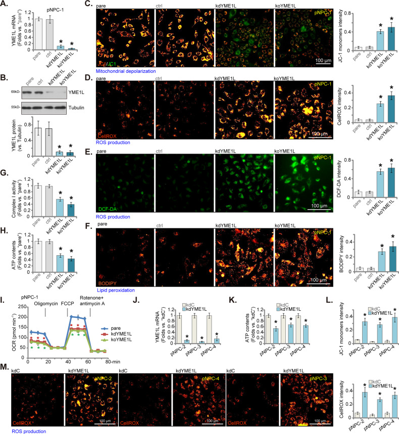
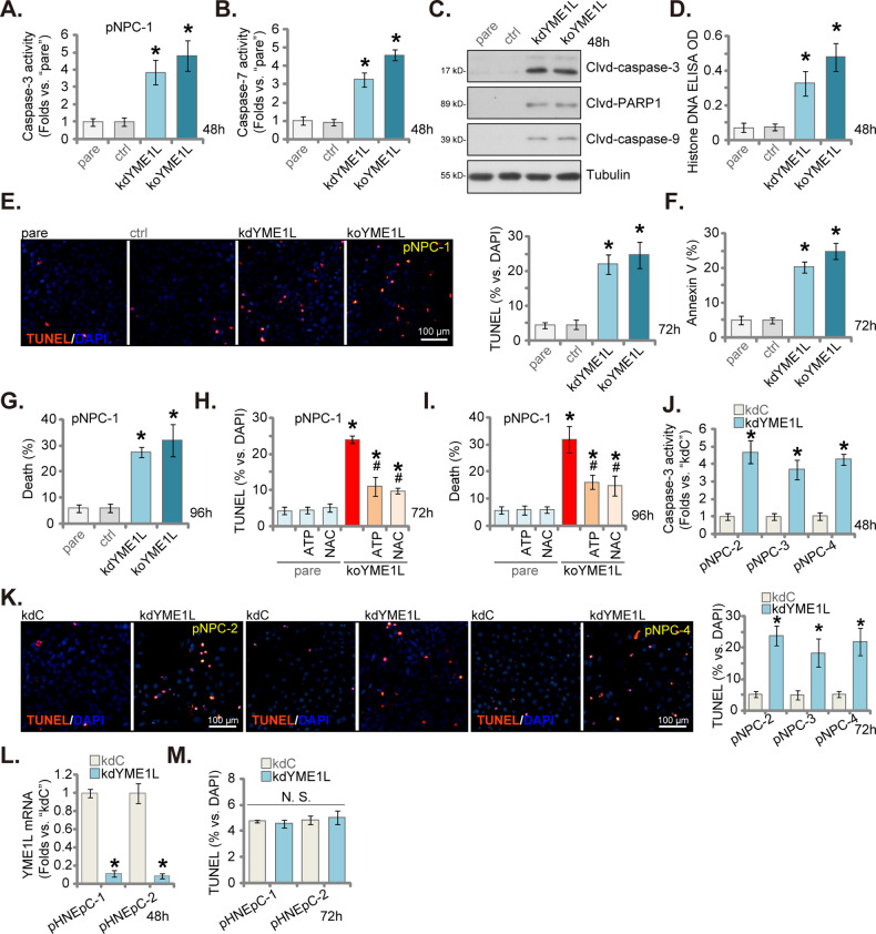
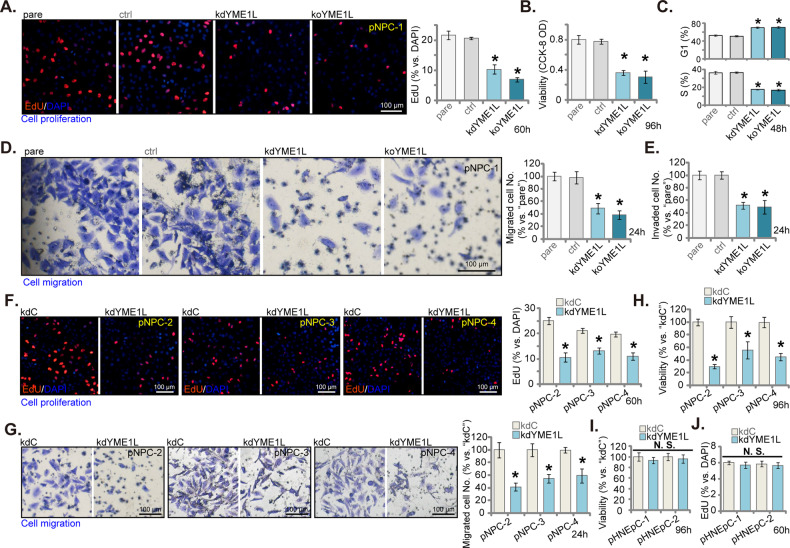
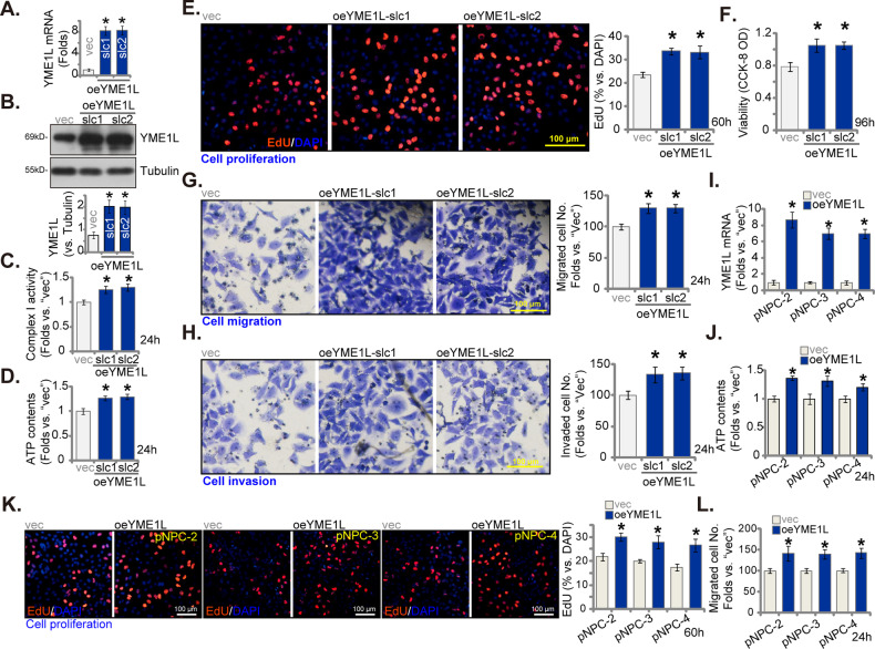
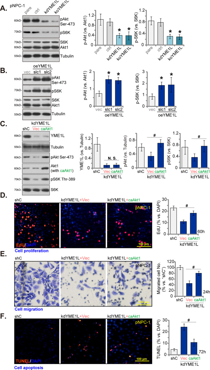
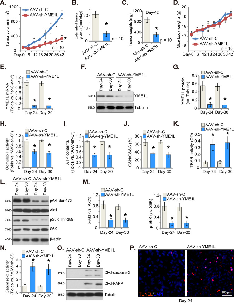
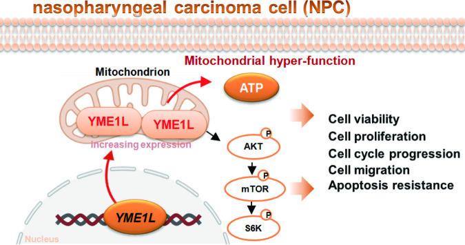
Mitochondria play a crucial role in the progression of nasopharyngeal carcinoma (NPC). YME1L, a member of the AAA ATPase family, is a key regulator of mitochondrial function and has been implicated in various cellular processes and diseases. This study investigates the expression and functional significance of YME1L in NPC. YME1L exhibits significant upregulation in NPC tissues from patients and across various primary human NPC cells, while its expression remains relatively low in adjacent normal tissues and primary nasal epithelial cells. Employing genetic silencing through the shRNA strategy or knockout (KO) via the CRISPR-sgRNA method, we demonstrated that YME1L depletion disrupted mitochondrial function, leading to mitochondrial depolarization, reactive oxygen species (ROS) generation, lipid peroxidation, and ATP reduction within primary NPC cells. Additionally, YME1L silencing or KO substantially impeded cell viability, proliferation, cell cycle progression, and migratory capabilities, concomitant with an augmentation of Caspase-apoptosis activation in primary NPC cells. Conversely, ectopic YME1L expression conferred pro-tumorigenic attributes, enhancing ATP production and bolstering NPC cell proliferation and migration. Moreover, our findings illuminate the pivotal role of YME1L in Akt-mTOR activation within NPC cells, with Akt-S6K phosphorylation exhibiting a significant decline upon YME1L depletion but enhancement upon YME1L overexpression. In YME1L-silenced primary NPC cells, the introduction of a constitutively-active Akt1 mutant (caAkt1, at S473D) restored Akt-S6K phosphorylation, effectively ameliorating the inhibitory effects imposed by YME1L shRNA. In vivo studies revealed that intratumoral administration of YME1L-shRNA-expressing adeno-associated virus (AAV) curtailed subcutaneous NPC xenograft growth in nude mice. Furthermore, YME1L downregulation, concurrent with mitochondrial dysfunction and ATP reduction, oxidative injury, Akt-mTOR inactivation, and apoptosis induction were evident within YME1L-silenced NPC xenograft tissues. Collectively, these findings shed light on the notable pro-tumorigenic role by overexpressed YME1L in NPC, with a plausible mechanism involving the promotion of Akt-mTOR activation.


Conflict of interest statement

The authors declare no competing interests.

Figures

Fig. 1
Fig. 1. YME1L overexpression in NPC tissues and cells.
YME1L mRNA (A) and protein (B and C) expression in NPC tumor tissues (“T”, n = 20) and their corresponding matched adjacent normal nasopharynx epithelial tissues (“N”, n = 20) were displayed, with quantified results provided. The immunohistochemistry (IHC) images depicting YME1L expression in the described human tissues were quantified, with IHC score results provided (D). Tissue immunofluorescence images, displaying YME1L (green fluorescence), the mitochondrial marker MitoTracker (red fluorescence), and nuclear DAPI staining (blue fluorescence) within the NPC tumor tissue and the adjacent normal epithelial tissue of Patient-1# were presented (E), and the normalized fluorescence intensity was quantified (E). YME1L mRNA expression (F) and protein expression (in both mitochondrial fraction lysates and mitochondria-null lysates G) were demonstrated in primary human NPC cells (“pNPC-1”, “pNPC-2”, “pNPC-3”, and “pNPC-4”) and primary human nasal epithelial cells (“pHNEpC-1” and “pHNEpC-2”) as described. The numerical values were mean ± standard deviation (SD). *P < 0.05 vs. “N” tissues or “pHNEpC-1” cells. Scale bar = 100 μm.
Fig. 2
Fig. 2. YME1L depletion led to damage of mitochondrial function in primary human NPC cells.
The stable pNPC-1 primary NPC cells with the lentiviral YME1L shRNA (“kdYME1L”), the lentiviral CRISPR-YME1L-KO construct (koYME1L), or the lentiviral scramble control shRNA plus the lentiviral CRISPR-KO control treatment (“ctrl”) were established and expression YME1L (mRNA and protein) was examined (A and B). The exact same number of the above pNPC-1 cells were further cultivated for 36 h, mitochondrial depolarization (via testing JC-1 green monomer intensity, C), ROS levels (by measuring CellROX and DCF-DA fluorescence intensity, D and E) and lipid peroxidation (via testing BODIPY intensity, F) as well as the mitochondrial complex-1 activity (G) and ATP contents (H) were tested. The seahorse assay was performed to measure the oxygen consumption rate (OCR) (I). Primary NPC cells that were derived from other patients, pNPC-2/-3/-4, were stably transduced with the lentiviral YME1L shRNA (“kdYME1L”) or the lentiviral scramble control shRNA (“kdC”), relative expression of YME1L mRNA was shown (J); Cells were further cultivated for 36 h, cellular ATP content (K), mitochondrial depolarization (L), ROS levels (via measuring CellROX fluorescence intensity, M) were tested similarly. The numerical values were mean ± standard deviation (SD, n = 5). “pare” indicates the parental control cells. *P < 0.05 vs. “pare”/“kdC” cells. Experiments in this figure were repeated five times, and each time similar results obtained. Scale bar = 100 μm.
Fig. 3
Fig. 3. Apoptosis activation after YME1L depletion in primary human NPC cells.
The stable pNPC-1 primary NPC cells with the lentiviral YME1L shRNA (“kdYME1L”), the lentiviral CRISPR-YME1L-KO construct (koYME1L), or the lentiviral scramble control shRNA plus the lentiviral CRISPR-KO control treatment (“ctrl”) were established, the exact same number of the above pNPC-1 cells were cultivated for designated hours, Caspase-3 and Caspase-7 activities (A and B), expression of apoptosis-associated proteins (C) and Histone DNA contents (D) were measured. Cell apoptosis was tested via nuclear TUNEL incorporation fluorescence staining assay (E) and Annexin V-PI FACS (F). Cell death was measured by trypan blue staining assays, with results quantified (G). The ctrl pNPC-1 cells or the koYME1L pNPC-1 cells were treated with the antioxidant NAC (0.5 mM) or ATP (1 mM) for designated hours, cell apoptosis (TUNEL assays, H) and death (trypan blue assays, I) were measured. Primary NPC cells that were derived from other patients, pNPC-2/-3/-4, or the primary human nasal epithelial cells (pHNEpC-1 and pHNEpC-2, derived from two donors) were stably transduced with the lentiviral YME1L shRNA (“kdYME1L”) or the lentiviral scramble control shRNA (“kdC”); The exact same number of cells were further cultivated for designated hours, the Caspase-3 activity (J) and apoptosis (TUNEL assays, K and M) were measured similarly. The relative YME1L mRNA expression in the epithelial cells was shown (L). The numerical values were mean ± standard deviation (SD, n = 5). “pare” indicates the parental control cells. *P < 0.05 vs. “pare”/ “kdC” cells. # P < 0.05 vs. “koYME1L” cells (H and I). “N. S.” stands for non-statistical difference (P > 0.05). Experiments in this figure were repeated five times, and each time similar results obtained. Scale bar = 100 μm.
Fig. 4
Fig. 4. YME1L depletion inhibits proliferation, viability, cell cycle progression and migration of primary human NPC cells.
The stable pNPC-1 primary NPC cells with the lentiviral YME1L shRNA (“kdYME1L”), the lentiviral CRISPR-YME1L-KO construct (koYME1L), or the lentiviral scramble control shRNA plus the lentiviral CRISPR-KO control treatment (“ctrl”) were established, the exact same number of the above pNPC-1 cells were cultivated for designated hours, cell proliferation, viability, cell cycle progression, migration, invasion were tested via nuclear EdU fluorescence staining (A), CCK-8 (B), PI-FACS (C), “Transwell” (D) and “Matrigel Transwell” (E) assays, respectively. Primary NPC cells that were derived from other patients, pNPC-2/-3/-4, or the primary human nasal epithelial cells (pHNEpC-1 and pHNEpC-2, derived from two donors) were stably transduced with the lentiviral YME1L shRNA (“kdYME1L”) or the lentiviral scramble control shRNA (“kdC”); The exact same number of cells were further cultivated for designated hours, cell proliferation (F and J), migration (G) and viability (H and I) were tested similarly. The numerical values were mean ± standard deviation (SD, n = 5). “pare” indicates the parental control cells. *P < 0.05 vs. “pare”/“kdC” cells. “N. S.” stands for non-statistical difference (P > 0.05). Experiments in this figure were repeated five times, and each time similar results obtained. Scale bar = 100 μm.
Fig. 5
Fig. 5. YME1L overexpression induces pro-tumorigenic activity in primary human NPC cells.
The stable pNPC-1 primary NPC cells with the lentiviral YME1L-expressing construct (oeYME1L-slc1 and oeYME1L-slc2, two stable selections) or the vector (“vec”) were established and expression YME1L (mRNA and protein) was examined (A and B). The exact same number of the above pNPC-1 cells were further cultivated for designated hours, the mitochondrial complex-1 activity (C) and ATP contents (D) were tested; Cell proliferation, viability, migration and invasion were tested via nuclear EdU fluorescence staining (E), CCK-8 (F), “Transwell” (G), and “Matrigel Transwell” (H) assays, respectively. Primary NPC cells that were derived from other patients, pNPC-2/-3/-4, with the lentiviral YME1L-expressing construct (“oeYME1L”) or the vector (“vec”) were formed and expression of YME1L mRNA was tested (I); ATP contents (J), cell proliferation (K) and migration (L) were tested similarly. The numerical values were mean ± standard deviation (SD, n = 5). *P < 0.05 vs. “vec” cells. Experiments in this figure were repeated five times, and each time similar results obtained. Scale bar = 100 μm.
Fig. 6
Fig. 6. YME1L is important for Akt-mTOR activation in NPC cells.
The stable pNPC-1 primary NPC cells with the lentiviral YME1L shRNA (“kdYME1L”), the lentiviral CRISPR-YME1L-KO construct (koYME1L), or the lentiviral scramble control shRNA plus the lentiviral CRISPR-KO control treatment (“ctrl”), the lentiviral YME1L-expressing construct (oeYME1L-slc1 and oeYME1L-slc2, two stable selections) or the vector (“vec”) were cultivated for 12 h, expression of listed proteins was tested (A and B). kdYME1L pNPC-1 cells were further stably transduced with a S473D constitutively-active mutant Akt1 (caAkt1) or the empty vector (“Vec”), expression of listed proteins was shown (C). These cells were further cultivated for designated hours, cell proliferation, migration and apoptosis were examined via EdU-nuclei staining (D), “Transwell” (E) and TUNEL-nuclei (F) assays, respectively. The numerical values were mean ± standard deviation (SD, n = 5). * P < 0.05 vs. “pare”/“vec” cells (A and B). # P < 0.05 (CF). “N. S.” stands for non-statistical difference (P > 0.05).Experiments in this figure were repeated five times, and each time similar results obtained. Scale bar = 100 μm.
Fig. 7
Fig. 7. YME1L silencing impedes NPC xenograft growth in nude mice.
The pNPC-1 xenograft-bearing nude mice were subject to intratumoral injection of the described virus, tumor volumes (in mm3, A) and the animal weights (in G, D) were recorded every six days. The estimated daily pNPC-1 xenograft growth, in mm3 per day, was also shown (B). All pNPC-1 xenografts were isolated at Day-42 and weighted (C). Expression of YME1L mRNA and listed proteins in the described pNPC-1 xenograft tissues was tested (EG, L, M and O). The mitochondrial complex-1 activity (H), ATP contents (I), GSH/GSSG ratio (J), TBAR activity (K) and Caspase-3 activity (N) in tissue lysates were examined as well. The pNPC-1 xenograft sections were also subject to fluorescence detection of TUNEL-positive nuclei (P). The data were presented as mean ± standard deviation (SD). In AD, ten mice were in each group (n = 10). For EP, five random tissue pieces in each xenograft were tested (n = 5). *P < 0.05 vs. “AAV-shC” group. Scale bar = 100 μm.
Fig. 8
Fig. 8. Overexpressed YME1L promotes NPC cell growth possibly by maintaining mitochondrial hyper-function and promoting Akt-mTOR activation.
The proposed signaling cartoon of the study.

Similar articles

Cited by

References

    1. Chen YP, Chan ATC, Le QT, Blanchard P, Sun Y, Ma J. Nasopharyngeal carcinoma. Lancet. 2019;394:64–80. doi: 10.1016/S0140-6736(19)30956-0. - DOI - PubMed
    1. Chua MLK, Wee JTS, Hui EP, Chan ATC. Nasopharyngeal carcinoma. Lancet. 2016;387:1012–24. doi: 10.1016/S0140-6736(15)00055-0. - DOI - PubMed
    1. Kang H, Kiess A, Chung CH. Emerging biomarkers in head and neck cancer in the era of genomics. Nat Rev Clin Oncol. 2015;12:11–26. doi: 10.1038/nrclinonc.2014.192. - DOI - PubMed
    1. Chou J, Lin YC, Kim J, You L, Xu Z, He B, et al. Nasopharyngeal carcinoma-review of the molecular mechanisms of tumorigenesis. Head Neck. 2008;30:946–63. doi: 10.1002/hed.20833. - DOI - PMC - PubMed
    1. Baloche V, Ferrand FR, Makowska A, Even C, Kontny U, Busson P. Emerging therapeutic targets for nasopharyngeal carcinoma: opportunities and challenges. Expert Opin Therapeutic Targets. 2020;24:545–58. doi: 10.1080/14728222.2020.1751820. - DOI - PubMed

Publication types

MeSH terms

Substances