. 2024 Jun 18;15(1):5219.
doi: 10.1038/s41467-024-49491-8.

Aryl amino acetamides prevent Plasmodium falciparum ring development via targeting the lipid-transfer protein PfSTART1

Affiliations

Aryl amino acetamides prevent Plasmodium falciparum ring development via targeting the lipid-transfer protein PfSTART1

Madeline G Dans et al. Nat Commun. .

Erratum in

Abstract

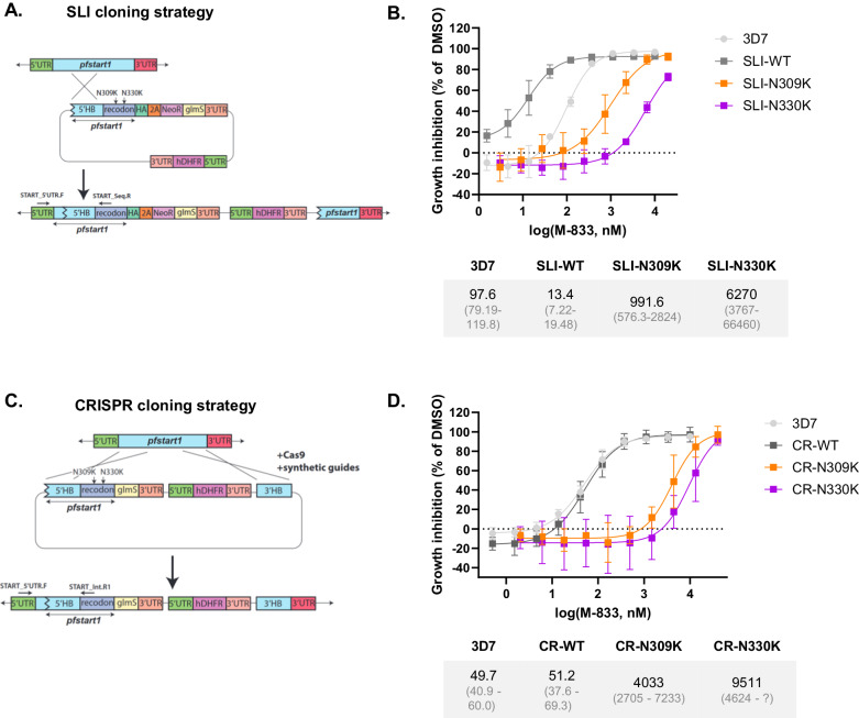
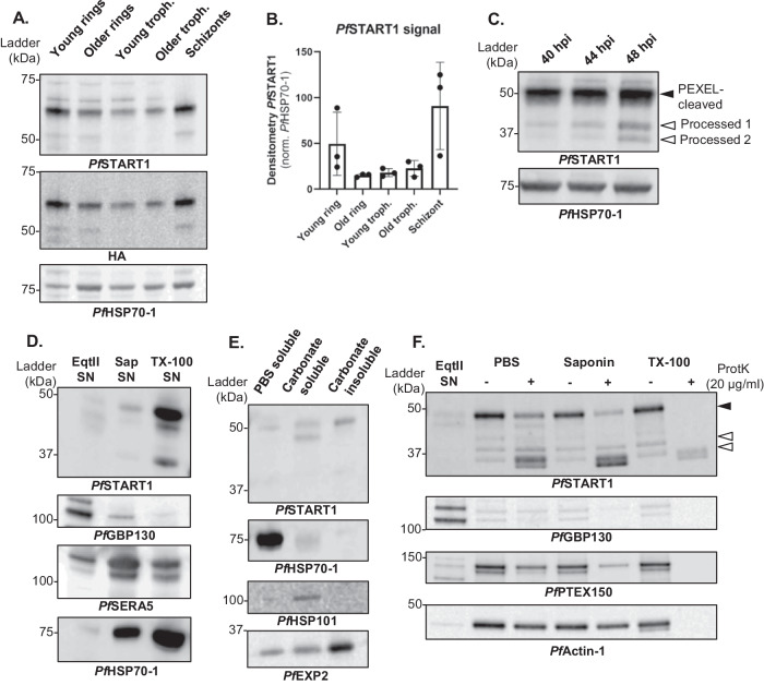
With resistance to most antimalarials increasing, it is imperative that new drugs are developed. We previously identified an aryl acetamide compound, MMV006833 (M-833), that inhibited the ring-stage development of newly invaded merozoites. Here, we select parasites resistant to M-833 and identify mutations in the START lipid transfer protein (PF3D7_0104200, PfSTART1). Introducing PfSTART1 mutations into wildtype parasites reproduces resistance to M-833 as well as to more potent analogues. PfSTART1 binding to the analogues is validated using organic solvent-based Proteome Integral Solubility Alteration (Solvent PISA) assays. Imaging of invading merozoites shows the inhibitors prevent the development of ring-stage parasites potentially by inhibiting the expansion of the encasing parasitophorous vacuole membrane. The PfSTART1-targeting compounds also block transmission to mosquitoes and with multiple stages of the parasite's lifecycle being affected, PfSTART1 represents a drug target with a new mechanism of action.


Conflict of interest statement

The authors declare no competing interests.

Figures

Fig. 1
Fig. 1. M-833 resistant parasites contain mutations in the P. falciparum START1 protein.
A Structure of MMV006833 (M-833). B Parasites populations (Pop) resistant to M-833 were generated by exposing 3D7 P. falciparum parasites to 10 x EC50 of M-833 three times. These parasites were cloned, and growth inhibition assays performed in the presence of 10 µM to 0.04 µM M-833. Growth inhibition was normalised to DMSO controls. Average of EC50 values and 95% confidence intervals are indicated; n = 3 biological replicates. Source data are provided as a Source Data file. C Key domains and cartoon representation of the AlphaFold predicted structure of the StAR-related lipid transfer protein, PfSTART1 (PF3D7_0104200),. Mutations that had been identified in the M-833 resistant parasite populations are shown as sticks (yellow = N309K, N330K; green = I224).
Fig. 2
Fig. 2. Engineering mutations N309K and N330K in PfSTART1 confers resistance to M-833.
Wild-type (WT) or mutant (N309K, N330K) pfstart1 genes were introduced in drug-sensitive 3D7 parasites using a Selection-Linked Integration (SLI) method or using CRISPR-Cas9 (CR). A Schematic of the SLI cloning strategy. The 5’ arm of native pfstart1 was amplified, PCR sewn to a recodonised version of the 3’ arm (either wildtype (WT), N309K or N330K), inserted into a p-HA-2A-Neo-glmS plasmid and transfected into 3D7 parasites, to be integrated into the native gene. B Growth inhibition curves of M-833 treated 3D7 and SLI-parasites. Parasites were incubated in various concentrations of M-833 and growth was assessed 72 h later using a lactate dehydrogenase activity assay. EC50 values and 95% confidence intervals are indicated below the graph; n = 3 biological replicates, mean +/− SD. C Schematic of the CRISPR cloning strategy. The full length 5’ homology blocks for WT, N309K, and N330K were amplified from the SLI plasmids and inserted into the p1.2-glmS plasmid, along with the native 3’ arm of pfstart1. Note that this construct does not include a HA tag. The plasmids were transfected into 3D7 parasites with Cas9 enzyme complexed with a guide RNA, to be integrated into the native gene. (D) Growth inhibition curves of M-833 on 3D7 and CRISPR-parasites (same as B). n = 3 biological replicates, mean +/− SD. Source data are provided as a Source Data file.
Fig. 3
Fig. 3. Knocking-down PfSTART1 sensitises parasites to M-833.
Engineered parasites contain a riboswitch glmS system: when glucosamine (GlcN) is added, PfSTART1 is knocked-down. A 3D7, selection linked integrated (SLI)-wildtype (WT), -N309K or -N330K were exposed for 48 h to 0 or 2.5 mM GlcN, and proteins were extracted from schizonts. Western blots were probed with anti-PfSTART1, anti-HA, and anti-PfEXP2 antibodies. B The densitometry of 3 biological replicates was measured (the PfSTART1 signal was normalised by the corresponding PfEXP2 signal, and then normalised by the 0 mM GlcN condition within each parasite line; mean +/− SD). C EC50 values of M-833 on 3D7 and SLI- parasites in the presence of 0, 0.25, or 2.5 mM glucosamine (GlcN). n = 4 biological replicates, mean +/− SD. Ordinary one-way ANOVA with Dunnett’s multiple comparison test. * adjusted p value = 0.0334. EC50 values and 95% confidence intervals are indicated in the table. D 3D7, CR-WT, -N309K, or -N330K were similarly exposed for 48 h to 0 or 2.5 mM GlcN, and proteins extracted from schizonts. Western blots were probed with anti-PfSTART1 and anti-PfEXP2 antibodies. E The densitometry of 3 biological replicates was measured (PfSTART1 signal was normalised to the corresponding PfEXP2 signal, and to the 0 mM GlcN condition; mean +/− SD). F EC50 values of M-833 on 3D7 and CRISPR- parasites in the presence of 0, 0.25, or 2.5 mM glucosamine (GlcN). n = 3 biological replicates, mean +/− SD. Ordinary one-way ANOVA with Dunnett’s multiple comparison test. *p < 0.05 (p = 0.0137). EC50 values and 95% confidence intervals are indicated in the table. Source data are provided as a Source Data file.
Fig. 4
Fig. 4. Analogues of M-833 are resisted by M-833 resistant lines, SLI- and CRISPR-mutants.
A Optimised analogues of M-833: their structures, EC50 (SD) on 3D7 parasites, toxicity (CC50 (SD)) on HepG2 cells. NT = not tested. (B) EC50 values of M-833 and analogues on 3D7 and M-833 resistant parasites PopD-D7 and PopE-F10, containing the PfSTART1 mutations N309K and N330K, respectively. n = 3 biological replicates, mean +/− SD. EC50 values indicated above the bar, as well as in Supplementary Data 1. Growth curves available in Fig. S3. C EC50 values of W-991 on 3D7, CR-WT, CR-N309K and CR-N330K parasites, in the presence of 0, 0.25 or 2.5 mM glucosamine (GlcN). n = 3 biological replicates, mean +/− SD. Ordinary one-way ANOVA with Dunnett’s multiple comparison test. *p < 0.05. **p < 0.005 (CR-WT, p = 0.0265; CR-N309K, p = 0.0123; CR-N330K, p = 0.008). EC50 values and 95% confidence intervals are indicated in the table. Source data are provided as a Source Data file.
Fig. 5
Fig. 5. The M-833 series demonstrates target engagement for PfSTART1 while the mutant form prevents compound binding.
A Isothermal titration calorimetry (ITC) analysis of M-833 series and recombinant PfSTART(WT) or PfSTART(N330K). Representative thermograms of 90 µM PfSTART1 titrated into 10 µM M-833, W-991, and negative control 4. The bottom panel comprises the data after integration of the peaks and a fitted offset applied. The binding curve shows the fit to a single-site binding model. DP = differential power. The table represents summary of the mean affinity, enthalpy, and entropy obtained for M-833, W-991 and 4 binding to PfSTART1 from n = 2 experiments. Error represents standard deviation. ND = not determined. The second replicate and values for individual parameters are provided in Fig. S4C. B Solvent proteome profiling assays. Parasite lysate was treated with DMSO or 10 µM W-991, challenged with an acetic acid/ethanol/formic acid mixture (AEF) and soluble fractions extracted and analysed via western blots probed with anti-PfSTART1. Western blot replicates are shown in Fig. S4D. C Volcano plots depict differential soluble protein abundance analysis (moderated t-test based on limma package) of parasite lysate treated with W-991 (100 µM) or the DMSO control (n = 3 biological replicates, mean +/− SD) after solvent-induced protein precipitation (7–25% AEF). Non-significant (ns) proteins are plotted in grey, and significant stabilised proteins are in red. Hit selection cut-offs of 0.73 log2 fold-change and p < 0.01 are indicated with dashed lines. Significant stabilisation hits are shown in the bar graphs representing the average of three biological replicates of relative soluble protein abundance in DMSO- or W-991-treated parasite lysate samples after solvent-induced protein precipitation, plotted for Gradient 1 ‘G1’ (7–15% AEF) and Gradient 2 ‘G2’ (17-25% AEF). Error bars represent standard deviation. Source data are provided as a Source Data file.
Fig. 6
Fig. 6. PfSTART1 inhibitors block ring development but their effect is reversible.
A, B Tightly synchronised Hyp1-Nluc schizonts were exposed to the following drugs for 4 h (during the egress/invasion window): M-833 (2 µM), W-991 (60 nM), DMSO (0.02%), ML10 (30 nM) (egress inhibitor), E64 (10 µM) (irreversible egress inhibitor), and heparin (100 µg/mL) (invasion inhibitor). After 4 h, non-egressed schizonts were eliminated with a sorbitol treatment, the drugs washed off, and parasites were followed over 72 h. Smears were taken at each timepoint (A) and growth was assessed with a bioluminescence read-out in relative light units (RLU x 105) (B). n = 3 biological replicates, mean +/− SD. Ordinary one-way ANOVA with Tukey’s multiple comparison test was conducted on the 72 h timepoint. *p < 0.05 (ML10 vs. Heparin; not shown on the graph, p = 0.0484). ***p < 0.005 (DMSO vs. ML10, p = 0.0006; DMSO vs. E64, p = 0.0002). ****p < 0.0001 (DMSO vs. M-833, W-991 and heparin). Highly synchronous ring-stage 3D7 parasites were exposed to M-833 (2 µM), W-991 (60 nM) or DMSO (0.02%). After 48 h, populations of M-833 and W-991-treated parasites were treated with sorbitol, compounds were washed out (w/o) (dotted lines), and morphology visualised by Giemsa-stained thin blood smears (C) and parasitemia was quantified by SYBR Green staining and flow cytometry (D) every 48 h for four following cycles of growth. Error bars indicate the standard deviation of two biological replicates, each made up of three technical replicates. Source data are provided as a Source Data file.
Fig. 7
Fig. 7. W-991 blocks merozoite transition to ameboid ring in RBCs, and blocks transmission in mosquitoes.
A Representative images of 4D Lattice Light Sheet Microscopy of merozoites (MitoTracker; cyan) directly after invasion of RBCs (all membranes are magenta). Treatment with W-991 shows disruption to the formation of the parasitophorous vacuole membrane (magenta) compared to vehicle control. B The mean vacuole sphericity (+/− SD) for W-991 and DMSO treatments across 30 min filming period. Statistical analysis was conducted via a Nested t-test (two-tailed) in GraphPad Prism showing p = 0.716 at T = 0 mins and a p = 0.045 at T = 15 mins (*). Across three independent experiments, there were 20 and 22 events analysed for W-991 (60 nM) and DMSO (0.001%) treatments, respectively (Fig. S6 and Movie S1 and S2). C Stage V gametocytes were quantified on day 17 which demonstrates there was no defect in gametocyte development upon W-991 treatment compared to PMIX/X dual inhibitor WM382. D Oocysts were quantified in mosquito midguts on day 7 after feeding which showed a dose-dependent decrease in oocysts per mosquito with W-991 treatment. Statistical tests (two-way ANOVA) were performed in GraphPad Prism. **** indicates p < 0.0001. Positive control compound, WM832, was used at 50 nM. Error bars indicate SD for n = 30 mosquitoes for each treatment (mean+/−SD). Data are a single representative from 3 independent experiments which can be found in Figure S7C, D. Source data are provided as a Source Data file.
Fig. 8
Fig. 8. PfSTART1 expression and processing.
A Western blot of SLI-WT parasites along the 48 h erythrocytic cycle. A saponin-lysis was performed on tightly synchronised SLI-WT young rings, older rings, young trophozoites, older trophozoites, and schizonts. B Densitometry of PfSTART1 over three biological replicates (normalised by the corresponding PfHSP70-1 signal; mean +/− SD). C Synchronous 3D7 trophozoites were magnet-purified, and whole cells harvested at ~40 hpi (hour post invasion), ~44 hpi and ~48 hpi (this last sample was treated with 10 µM E64 to prevent egress). PEXEL-cleaved PfSTART1 and two further processed forms are indicated by solid and empty arrows respectively. D To localise PfSTART1, Percoll-purified 3D7 schizonts were sequentially lysed with equinatoxin II (EqtII), saponin (Sap) and Triton-X100 (TX-100), and the supernatants (SN) were collected to harvest proteins localising in the red blood cell (RBC) cytosol, the parasitophorous vacuole (PV) and the parasite, respectively. PfGBP130 is a protein known to be exported into the RBC cytosol; PfSERA5 localises to the PV in schizonts; PfHSP70-1 is a parasite cytosolic protein. E To determine the solubility of PfSTART1, saponin-lysed 3D7 schizonts were sequentially lysed in PBS (with five freeze-thaw cycles) and sodium carbonate. The supernatant resulting from the PBS lysis was collected (PBS soluble), as well as the supernatant and the pellet of the carbonate lysis (Carbonate soluble and Carbonate insoluble respectively). Controls are the soluble protein PfHSP70-1, the membrane-associated protein PfHSP101, and the integral transmembrane protein PfEXP2. F Proteinase K protection assay was conducted on Percoll-purified 3D7 schizonts. Schizonts were first lysed in EqtII, the supernatant (SN) of which was collected. The remaining parasite and PV were either incubated in PBS (no lysis), saponin (PVM-lysis), or TX-100 (lysis of all membrane) with or without proteinase K. PfGBP130 is RBC cytosolic protein; PfPTEX150 is a PV protein; PfActin-1 is a parasite cytosolic protein. Note that another replicate for each of these experiments is shown in Fig S8. Source data are provided as a Source Data file.
Fig. 9
Fig. 9. The M-833 series blocks the conversion of newly invaded merozoites into ring-stage parasites by inhibiting the PfSTART1 protein and reducing the expansion of the parasitophorous vacuole membrane (PVM).
(Top) After invasion, the merozoite begins to spin and produce pseudopodia before differentiating into an amoeboid ring-stage parasite. (Bottom) After invasion PfSTART1, is delivered into the vacuole space possibly via secretory organelles fusing with the parasite plasma membrane (PPM). Here PfSTART1 could transfer phospholipids from the PPM to the PVM to allow the latter to expand to accommodate the growing ring-stage parasite. The M-833 compound series tightly binds to PfSTART1, probably displacing the phospholipids and thereby preventing the expansion of the PVM and arresting parasite growth.

References

    1. World Health Organization. World Malaria Report 2023. pp 17–19 (2022).
    1. Bergmann, C. et al. Increase in Kelch 13 Polymorphisms in Plasmodium falciparum, Southern Rwanda. Emerg. Infect. Dis.27, 294–296 (2021). - PMC - PubMed
    1. Dondorp, A. M. et al. Artemisinin resistance in Plasmodium falciparum Malaria. N. Engl. J. Med.361, 455–467 (2009). - PMC - PubMed
    1. Eastman, R. T. & Fidock, D. A. Artemisinin-based combination therapies: a vital tool in efforts to eliminate malaria. Nat. Rev. Microbiol7, 864–874 (2009). - PMC - PubMed
    1. Yoshida, N., Yamauchi, M., Morikawa, R., Hombhanje, F. & Mita, T. Increase in the proportion of Plasmodium falciparum with kelch13 C580Y mutation and decline in pfcrt and pfmdr1 mutant alleles in Papua New Guinea. Malar. J.20, 410 (2021). - PMC - PubMed

MeSH terms