

Use of phase plate cryo-EM reveals conformation diversity of therapeutic IgG with 50 kDa Fab fragment resolved below 6 Å
- PMID: 38890341
- PMCID: PMC11189423
- DOI: 10.1038/s41598-024-62045-8
Use of phase plate cryo-EM reveals conformation diversity of therapeutic IgG with 50 kDa Fab fragment resolved below 6 Å
Abstract
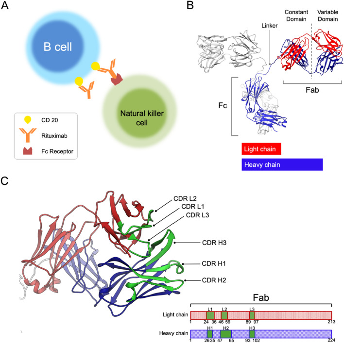
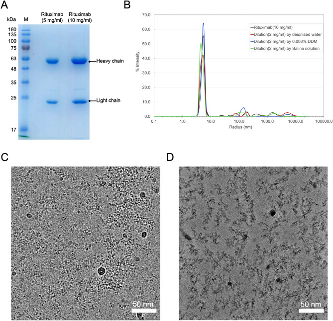
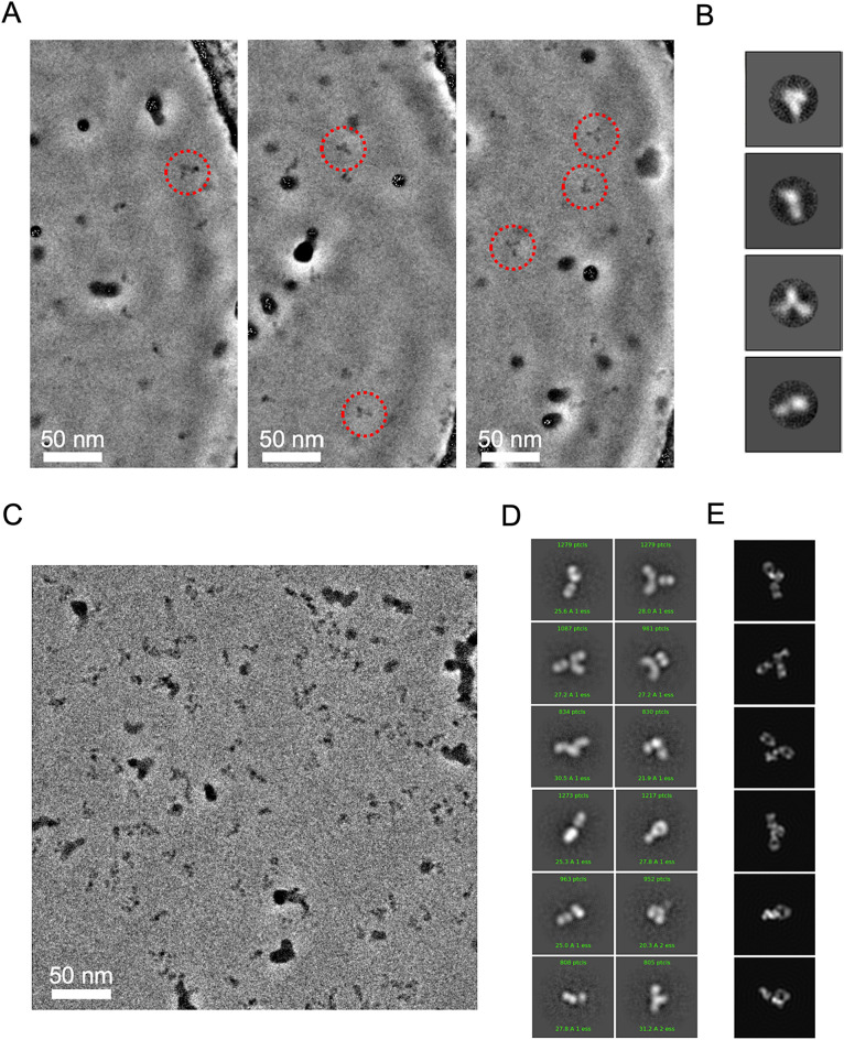
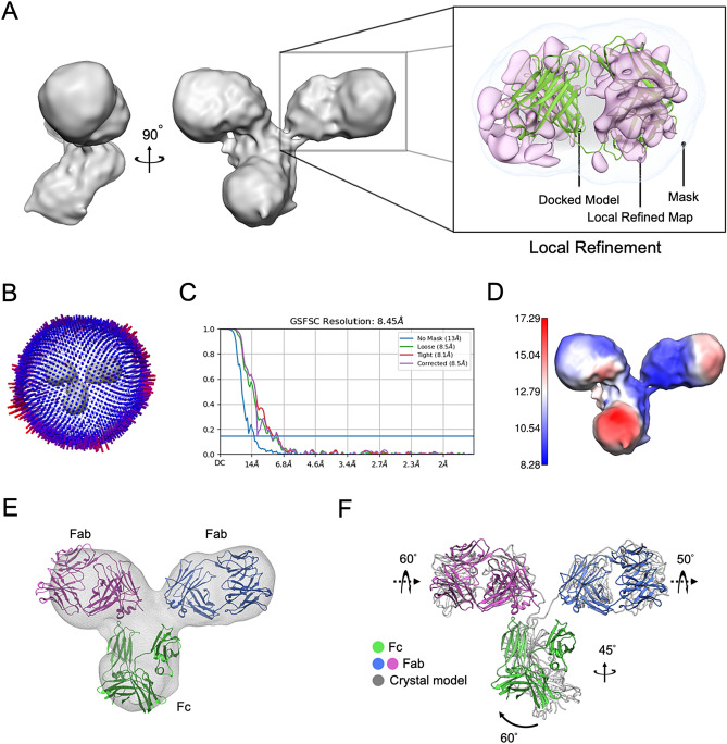
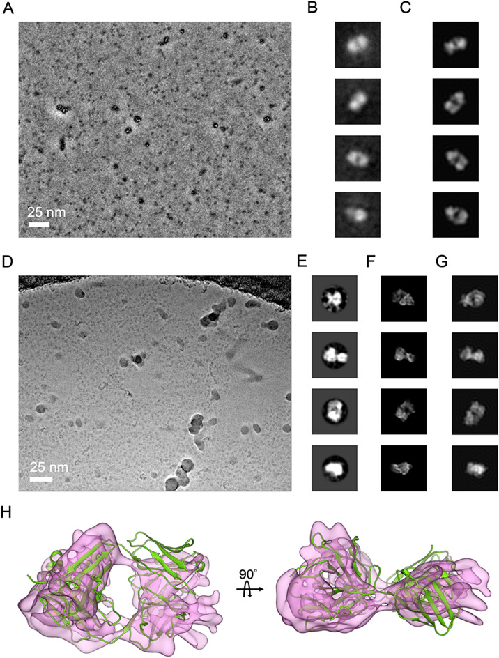
While cryogenic electron microscopy (cryo-EM) is fruitfully used for harvesting high-resolution structures of sizable macromolecules, its application to small or flexible proteins composed of small domains like immunoglobulin (IgG) remain challenging. Here, we applied single particle cryo-EM to Rituximab, a therapeutic IgG mediating anti-tumor toxicity, to explore its solution conformations. We found Rituximab molecules exhibited aggregates in cryo-EM specimens contrary to its solution behavior, and utilized a non-ionic detergent to successfully disperse them as isolated particles amenable to single particle analysis. As the detergent adversely reduced the protein-to-solvent contrast, we employed phase plate contrast to mitigate the impaired protein visibility. Assisted by phase plate imaging, we obtained a canonical three-arm IgG structure with other structures displaying variable arm densities co-existing in solution, affirming high flexibility of arm-connecting linkers. Furthermore, we showed phase plate imaging enables reliable structure determination of Fab to sub-nanometer resolution from ab initio, yielding a characteristic two-lobe structure that could be unambiguously docked with crystal structure. Our findings revealed conformation diversity of IgG and demonstrated phase plate was viable for cryo-EM analysis of small proteins without symmetry. This work helps extend cryo-EM boundaries, providing a valuable imaging and structural analysis framework for macromolecules with similar challenging features.
© 2024. The Author(s).
Conflict of interest statement
The authors declare no competing interests.
Figures

Similar articles
-
Locking the Elbow: Improved Antibody Fab Fragments as Chaperones for Structure Determination.J Mol Biol. 2018 Feb 2;430(3):337-347. doi: 10.1016/j.jmb.2017.12.012. Epub 2017 Dec 19. J Mol Biol. 2018. PMID: 29273204 Free PMC article.
-
High-resolution structure determination of sub-100 kDa complexes using conventional cryo-EM.Nat Commun. 2019 Mar 4;10(1):1032. doi: 10.1038/s41467-019-08991-8. Nat Commun. 2019. PMID: 30833564 Free PMC article.
-
Development of a universal nanobody-binding Fab module for fiducial-assisted cryo-EM studies of membrane proteins.Proc Natl Acad Sci U S A. 2021 Nov 23;118(47):e2115435118. doi: 10.1073/pnas.2115435118. Proc Natl Acad Sci U S A. 2021. PMID: 34782475 Free PMC article.
-
Structure Determination by Single-Particle Cryo-Electron Microscopy: Only the Sky (and Intrinsic Disorder) is the Limit.Int J Mol Sci. 2019 Aug 27;20(17):4186. doi: 10.3390/ijms20174186. Int J Mol Sci. 2019. PMID: 31461845 Free PMC article. Review.
-
Challenges and opportunities in cryo-EM with phase plate.Curr Opin Struct Biol. 2019 Oct;58:175-182. doi: 10.1016/j.sbi.2019.06.013. Epub 2019 Jul 30. Curr Opin Struct Biol. 2019. PMID: 31374473 Review.
Cited by
-
FLEXTAG: A Small and Self-renewable Protein Labeling System for Anti-fading Multicolor Super-resolution Imaging.bioRxiv [Preprint]. 2025 Jul 23:2025.07.19.665678. doi: 10.1101/2025.07.19.665678. bioRxiv. 2025. PMID: 40777492 Free PMC article. Preprint.
References
MeSH terms
Substances
Grants and funding
LinkOut - more resources
Full Text Sources
Research Materials
