. 2024 Aug;38(8):1667-1673.
doi: 10.1038/s41375-024-02300-z. Epub 2024 Jun 18.

Pre-emptive detection and evolution of relapse in acute myeloid leukemia by flow cytometric measurable residual disease surveillance

Affiliations

Pre-emptive detection and evolution of relapse in acute myeloid leukemia by flow cytometric measurable residual disease surveillance

Nicholas McCarthy et al. Leukemia. 2024 Aug.

Abstract

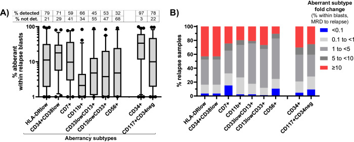
Measurable residual disease (MRD) surveillance in acute myeloid leukemia (AML) may identify patients destined for relapse and thus provide the option of pre-emptive therapy to improve their outcome. Whilst flow cytometric MRD (Flow-MRD) can be applied to high-risk AML/ myelodysplasia patients, its diagnostic performance for detecting impending relapse is unknown. We evaluated this in a cohort comprising 136 true positives (bone marrows preceding relapse by a median of 2.45 months) and 155 true negatives (bone marrows during sustained remission). At an optimal Flow-MRD threshold of 0.040%, clinical sensitivity and specificity for relapse was 74% and 87% respectively (51% and 98% for Flow-MRD ≥ 0.1%) by 'different-from-normal' analysis. Median relapse kinetics were 0.78 log10/month but significantly higher at 0.92 log10/month for FLT3-mutated AML. Computational (unsupervised) Flow-MRD (C-Flow-MRD) generated optimal MRD thresholds of 0.036% and 0.082% with equivalent clinical sensitivity to standard analysis. C-Flow-MRD-identified aberrancies in HLADRlow or CD34+CD38low (LSC-type) subpopulations contributed the greatest clinical accuracy (56% sensitivity, 90% specificity) and notably, by longitudinal profiling expanded rapidly within blasts in > 40% of 86 paired MRD and relapse samples. In conclusion, flow MRD surveillance can detect MRD relapse in high risk AML and its evaluation may be enhanced by computational analysis.


Conflict of interest statement

NM, FD, AP, CR, GA, SG, AA, MJ, and AP, have no conflicts to declare. SDF declares research funding from Jazz and BMS; Speakers Bureau with Jazz, Pfizer and Novartis; advisory committee with MPAACT. CH and GG, The National Heart, Lung, and Blood Institute receives research funding for the laboratory of Dr. Hourigan from the Foundation of the NIH AML MRD Biomarkers Consortium.

Figures

Fig. 1
Fig. 1. MRD relapse detection and kinetics of relapse by flow cytometric MRD monitoring.
A Testing accuracy for pre-emptive detection of relapse by ‘different from normal’ MRD standard analysis. Receiver operating curve (ROC) statistics calculated from the combined cohort (136 true positives [relapse within 4 months of sample] and 121 true negatives [by sustained off-treatment remission after the sample]). Clinical specificity and sensitivity shown for routine assay thresholds (0.05% and ELN 0.10%) and optimum thresholds (derived from Maxstat package). B Relapse kinetics of high-risk AML. Relapse kinetics for 136 paired MRD and relapse bone marrows from 119 high risk AML patients. Relapse leukemic aberrant immunophenotypes were examined in MRD samples in parallel to standard ‘different-from-normal’ MRD analysis and kinetics calculated from the highest MRD values. Summary of relapse kinetics is shown for the overall sample cohort and specific diagnostic genetics subgroups; there was a significant difference (P = 0.026) in relapse kinetics between the FLT3-mutated and adverse cytogenetics /TP53 subgroups by non-parametric Mann-Whitney U testing.
Fig. 2
Fig. 2. Aberrant phenotypes at MRD and Relapse.
Each column represents one relapse. Samples grouped as MRD positive, MRD low positive and MRD negative by C-Flow MRD analysis of MRD timepoint. Results show presence for aberrant phenotype types detected at MRD timepoint (top 3 aberrancies of MRD blasts) and relapse timepoint (subsequent row, red = highest frequency aberrancy where top 3 MRD aberrancies are not detected).
Fig. 3
Fig. 3. Relapse evolution of aberrant progenitor types within blasts.
A Frequency of displayed aberrant phenotypic subtype within relapse sample blasts. Overall frequency for CD34+ and CD34- compartments shown at end of graph. Box plots indicate the median and interquartile range for relapse samples where aberrancy is detectable as assessed objectively by computational unsupervised analysis (whiskers = 5-95th percentiles). Inset table displays detected versus non-detected frequency for each aberrant subtype (% of relapses). B Distribution of fold-changes in specific aberrant subtype frequency within blasts from pre-relapse MRD timepoint to relapse in paired samples (86 MRD samples paired with 76 relapses). Fold changes in total CD34+ and CD34- aberrancies shown at end of graph.

References

    1. Schuurhuis GJ, Heuser M, Freeman S, Bene MC, Buccisano F, Cloos J, et al. Minimal/measurable residual disease in AML: a consensus document from the European LeukemiaNet MRD Working Party. Blood. 2018;131:1275–91. 10.1182/blood-2017-09-801498 - DOI - PMC - PubMed
    1. Heuser M, Freeman SD, Ossenkoppele GJ, Buccisano F, Hourigan CS, Ngai LL, et al. 2021 Update on Measurable Residual Disease (MRD) in Acute Myeloid Leukemia (AML): A Consensus Document from the European LeukemiaNet MRD Working Party. Blood. 2021;138:2753–67. 10.1182/blood.2021013626 - DOI - PMC - PubMed
    1. Ommen HB, Hokland P, Haferlach T, Abildgaard L, Alpermann T, Haferlach C, et al. Relapse kinetics in acute myeloid leukaemias with MLL translocations or partial tandem duplications within the MLL gene. Br J Haematol. 2014;165:618–28. 10.1111/bjh.12792 - DOI - PubMed
    1. Ommen HB, Schnittger S, Jovanovic JV, Ommen IB, Hasle H, Ostergaard M, et al. Strikingly different molecular relapse kinetics in NPM1c, PML-RARA, RUNX1-RUNX1T1, and CBFB-MYH11 acute myeloid leukemias. Blood. 2010;115:198–205. 10.1182/blood-2009-04-212530 - DOI - PubMed
    1. Ommen HB, Touzart A, MacIntyre E, Kern W, Haferlach T, Haferlach C, et al. The kinetics of relapse in DEK-NUP214-positive acute myeloid leukemia patients. Eur J Haematol. 2015;95:436–41. 10.1111/ejh.12511 - DOI - PubMed