. 2024 Jun 19;22(1):345.
doi: 10.1186/s12951-024-02632-z.

Cartilage progenitor cells derived extracellular vesicles-based cell-free strategy for osteoarthritis treatment by efficient inflammation inhibition and extracellular matrix homeostasis restoration

Affiliations

Cartilage progenitor cells derived extracellular vesicles-based cell-free strategy for osteoarthritis treatment by efficient inflammation inhibition and extracellular matrix homeostasis restoration

Kai Feng et al. J Nanobiotechnology. .

Abstract

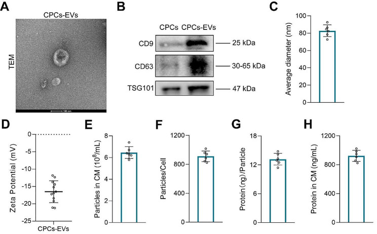
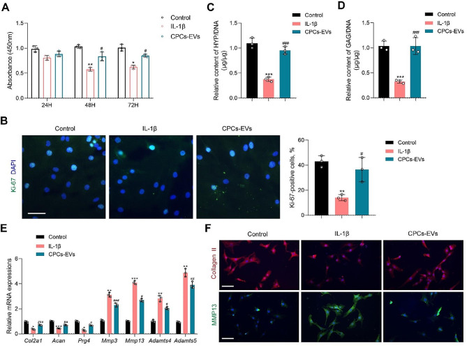
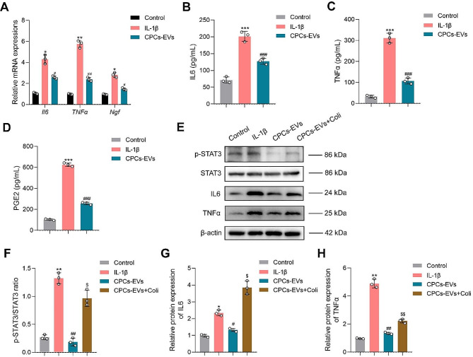
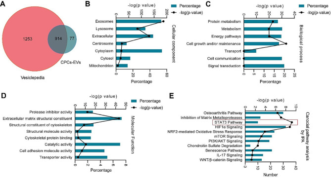
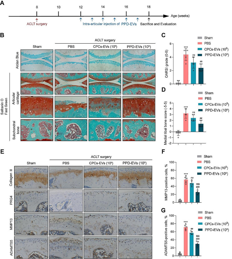
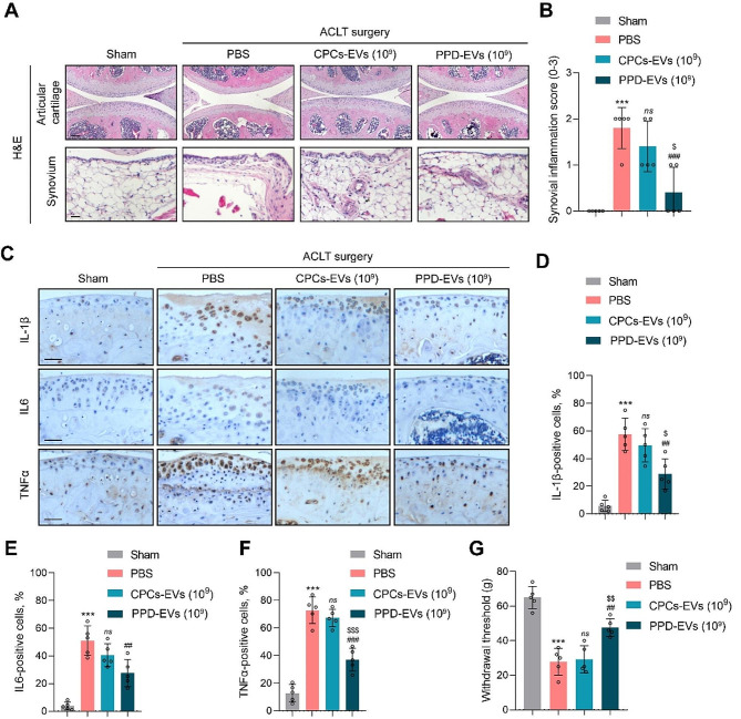
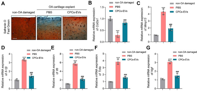
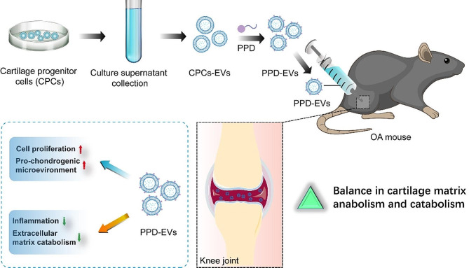
Osteoarthritis (OA) is a common degenerative joint disease which currently lacks of effective agents. It is therefore urgent and necessary to seek an effective approach that can inhibit inflammation and promote cartilage matrix homeostasis. Cartilage progenitor cells (CPCs) are identified as a cell population of superficial zone in articular cartilage which possess strong migration ability, proliferative capacity, and chondrogenic potential. Recently, the application of CPCs may represent a novel cell therapy strategy for OA treatment. There is growing evidence that extracellular vesicles (EVs) are primary mediators of the benefits of stem cell-based therapy. In this study, we explored the protective effects of CPCs-derived EVs (CPCs-EVs) on IL-1β-induced chondrocytes. We found CPCs-EVs exhibited chondro-protective effects in vitro. Furthermore, our study demonstrated that CPCs-EVs promoted matrix anabolism and inhibited inflammatory response at least partially via blocking STAT3 activation. In addition, liquid chromatography-tandem mass spectrometry analysis identified 991 proteins encapsulated in CPCs-EVs. By bioinformatics analysis, we showed that STAT3 regulatory proteins were enriched in CPCs-EVs and could be transported to chondrocytes. To promoting the protective function of CPCs-EVs in vivo, CPCs-EVs were modified with cationic peptide ε-polylysine-polyethylene-distearyl phosphatidylethanolamine (PPD) for surface charge reverse. In posttraumatic OA mice, our results showed PPD modified CPCs-EVs (PPD-EVs) effectively inhibited extracellular matrix catabolism and attenuated cartilage degeneration. Moreover, PPD-EVs down-regulated inflammatory factors expressions and reduced OA-related pain in OA mice. In ex-vivo cultured OA cartilage explants, PPD-EVs successfully promoted matrix anabolism and inhibited inflammation. Collectively, CPCs-EVs-based cell-free therapy is a promising strategy for OA treatment.

Keywords: Cartilage progenitor cells; Engineering modification; Extracellular vesicles; Inflammation; Osteoarthritis.


Conflict of interest statement

The authors declare no competing interests.

Figures

Fig. 1
Fig. 1
Characterization of CPCs-EVs. A Representative morphological image of CPCs-EVs captured by TEM. Scale bar = 100 nm. B The surface markers (CD9, CD63, and TSG101) of CPCs-EVs were detected by western blot analysis. C Average particle diameter of CPCs-EVs was detected by nano-flow cytometry. (n = 6). D The zeta potential of CPCs-EVs was detected by a nanoparticle analyzer. (n = 12). E-H Quantification of CPCs-EVs parameters, including the particle concentration and the protein concentration. (n = 6)
Fig. 2
Fig. 2
The protective effects of CPCs-EVs on IL-1β-induced chondrocytes. A Chondrocyte proliferative ability was detected by CCK-8 assay. (n = 3). B Immunofluorescence staining for Ki-67 (green) and percentage of Ki-67-positive cells in each group. (n = 3). Scale bar = 50 μm. C Relative HYP content was detected after CPCs-EVs treatment in IL-1β-induced chondrocytes. (n = 3). D Relative GAG content was detected after CPCs-EVs treatment in IL-1β-induced chondrocytes. (n = 3). E Relative mRNA expressions of anabolism-related genes (Col2a1, Acan, Prg4) and catabolism-related genes (Mmp3, Mmp13, Adamts4, Adamts5) were detected by PCR analysis. (n = 3). F Immunofluorescence staining for Collagen II (red) and MMP13 (green) in each group. (n = 3). Scale bar = 100 μm. Data represent mean ± S.D. *P < 0.05, **P < 0.01, ***P < 0.001 versus the Control group. #P < 0.05, ##P < 0.01, ###P < 0.001 versus the IL-1β group
Fig. 3
Fig. 3
CPCs-EVs alleviate inflammatory response by inhibiting STAT3 activation in vitro. A Relative mRNA expressions of inflammatory cytokines (Il6, TNFα, Ngf) in mouse chondrocytes were detected by PCR analysis. (n = 3). B The concentration of IL6 was detected by ELISA assay in chondrocytes after different treatments. (n = 3). C The concentration of TNFα was detected by ELISA assay in chondrocytes after different treatments. (n = 3). D The concentration of PGE2 was detected by ELISA assay in chondrocytes after different treatments. (n = 3). E Western blot analysis of p-STAT3, STAT3, IL6, and TNFα expressions in chondrocytes after treatments. (n = 3). F Quantification of p-STAT3/STAT3 ratio in different groups. (n = 3). G Relative protein expression of IL6 in different groups. (n = 3). H Relative protein expression of TNFα in different groups. (n = 3). Data represent mean ± S.D. *P < 0.05, **P < 0.01, ***P < 0.001 versus the Control group. #P < 0.05, ##P < 0.01, ###P < 0.001 versus the IL-1β group. $P < 0.05, $$P < 0.01 versus the CPCs-EVs group
Fig. 4
Fig. 4
Proteomics analysis of CPCs-EVs. A Venn diagram showing the overlapping and unique proteins in CPCs-EVs with Vesiclepedia database. B Gene Ontology Cellular Component analysis of proteins enriched in CPCs-EVs. C Gene Ontology Biological Process analysis of proteins enriched in CPCs-EVs. D Gene Ontology Molecular Function analysis of proteins enriched in CPCs-EVs. E IPA displaying canonical pathway analysis of CPCs-EVs proteins
Fig. 5
Fig. 5
The therapeutic effects of PPD-EVs in ACLT-induced posttraumatic OA mice model. A Schematic of the time course for the experiments in ACLT-induced mice. Mice undergoing ACLT surgery were intra-articularly injected with different treatments and evaluated as indicated. B Light microscopy images of articular cartilage stained by the Alcian Blue and Safranin O-Fast Green in all groups were observed and analyzed. Scale bar = 100 μm and 50 μm. C Cartilage loss and destruction in each group was evaluated scored by the OARSI scoring system. D The score of medial tibial bone in each group was evaluated. E IHC staining for Collagen II, PRG4, MMP13, and ADAMTS5 in mice after treatments. Scale bar = 100 μm. F Quantification of MMP13-positive cells in articular cartilage after different treatments. G Quantification of ADAMTS5-positive cells in each group. Data represent mean ± S.D. ***P < 0.001 versus the Sham group. ##P < 0.01, ###P < 0.001 versus the HA group. $$$P < 0.001 versus the CPCs-EVs (109) group. ns, not significant
Fig. 6
Fig. 6
PPD-EVs ameliorate joint inflammation and OA-related pain in ACLT-induced mice. A Light microscopy images of articular cartilage and synovium by the H&E staining in all groups were observed and analyzed. Scale bar = 100 μm. B The score of synovium inflammation in each group was evaluated. C IHC staining for IL-1β, IL6, and TNFα in articular cartilage of mice after treatments. Scale bar = 100 μm. D Quantification of IL-1β-positive cells in each group. E Quantification of IL6-positive cells in each group. F Quantification of TNFα-positive cells in each group. G Withdrawal threshold in each group was assessed. Data represent mean ± S.D. ***P < 0.001 versus the Sham group. ##P < 0.01, ###P < 0.001 versus the HA group. $P < 0.05, $$P < 0.01, $$$P < 0.001 versus the CPCs-EVs (109) group. ns, not significant
Fig. 7
Fig. 7
PPD-EVs alleviate cartilage destruction and inhibit inflammatory response in ex-vivo cultured OA cartilage explants. A Light microscopy images of articular cartilage stained by the Safranin O-Fast Green in ex-vivo cultured OA cartilage explants after treatment. Scale bar = 150 μm. B Relative mRNA expression of cartilage anabolism-related gene Col2a1 was detected by PCR analysis. C Relative mRNA expression of cartilage catabolism-related gene Mmp13 was detected by PCR analysis. D-G Relative mRNA expressions of inflammatory cytokines (Il-1β, Il6, Tnfα, and Ngf) were detected in OA cartilage explants after treatment by PCR analysis. Data represent mean ± S.D. ***P < 0.001 versus the non-OA damaged group. ###P < 0.001 versus the PBS group
Fig. 8
Fig. 8
Schematic illustration of the chondro-protective effects of positively charged PPD-modified CPCs-EVs (PPD-EVs) in OA mice. Intra-articular injection of PPD-EVs promotes chondrocyte proliferation and extracellular matrix anabolism, inhibits inflammatory response and catabolism, and ultimately alleviates cartilage destruction in OA mice

Similar articles

Cited by

References

    1. Zhu X, Chan Y, Yung P, Tuan R. Jiang YJFic, biology d: Subchondral Bone Remodeling: A Therapeutic Target for Osteoarthritis. 2020, 8:607764. - PMC - PubMed
    1. Mackie S, Dejaco C, Appenzeller S, Camellino D, Duftner C, Gonzalez-Chiappe S, Mahr A, Mukhtyar C, Reynolds G, de Souza A et al. British Society for Rheumatology guideline on diagnosis and treatment of giant cell arteritis: executive summary. 2020, 59:487–94. - PubMed
    1. Belluzzi E, Stocco E, Pozzuoli A, Granzotto M, Porzionato A, Vettor R, De Caro R, Ruggieri P, Ramonda R, Rossato M et al. Contribution of Infrapatellar Fat Pad and Synovial Membrane to Knee Osteoarthritis Pain. 2019, 2019:6390182. - PMC - PubMed
    1. Michela, Battistelli., Marta, Favero., Debora, Burini., Giovanni, Trisolino., Dante, Dallari., Lucia, De Franceschi., Steven R, Goldring., Mary B, Goldring., Elisa, Belluzzi., Giuseppe, Filardo., Brunella, Grigolo., Elisabetta, Falcieri., Eleonora, Olivotto. (2019). Morphological and ultrastructural analysis of normal, injured and osteoarthritic human knee menisci. Eur J Histochem, 63(1), 0. 10.4081/ejh.2019.2998. (PMID: 30739432). - PMC - PubMed
    1. Rice S, Tselepi M, Sorial A, Aubourg G, Shepherd C, Almarza D, Skelton A, Pangou I, Deehan D, Reynard L et al. Prioritization of PLEC and GRINA as Osteoarthritis Risk genes through the identification and characterization of novel methylation quantitative trait loci. 2019, 71:1285–96. - PMC - PubMed

Substances