. 2024 Jun 18;21(1):104.
doi: 10.1186/s12984-024-01405-x.

Automatic gait events detection with inertial measurement units: healthy subjects and moderate to severe impaired patients

Affiliations

Automatic gait events detection with inertial measurement units: healthy subjects and moderate to severe impaired patients

Cyril Voisard et al. J Neuroeng Rehabil. .

Abstract

Background: Recently, the use of inertial measurement units (IMUs) in quantitative gait analysis has been widely developed in clinical practice. Numerous methods have been developed for the automatic detection of gait events (GEs). While many of them have achieved high levels of efficiency in healthy subjects, detecting GEs in highly degraded gait from moderate to severely impaired patients remains a challenge. In this paper, we aim to present a method for improving GE detection from IMU recordings in such cases.

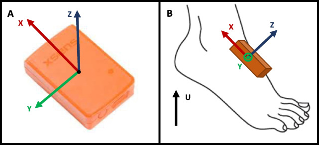
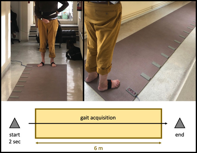
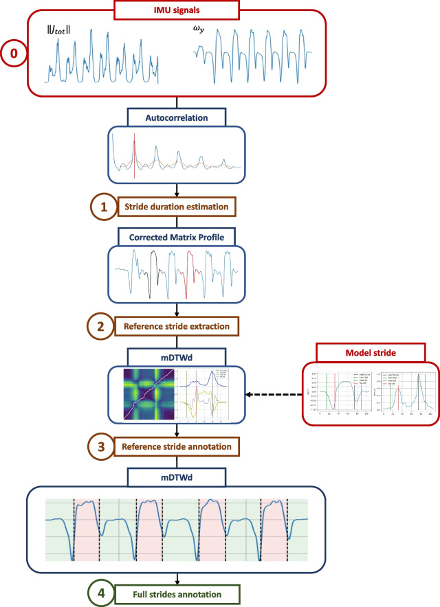
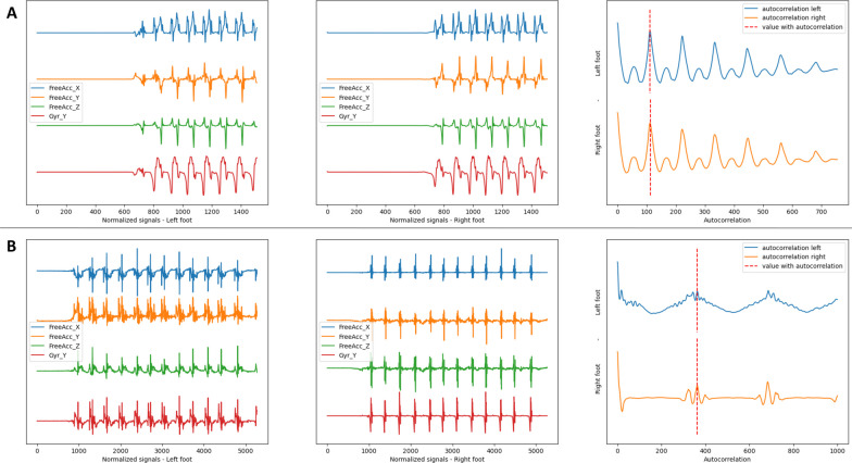
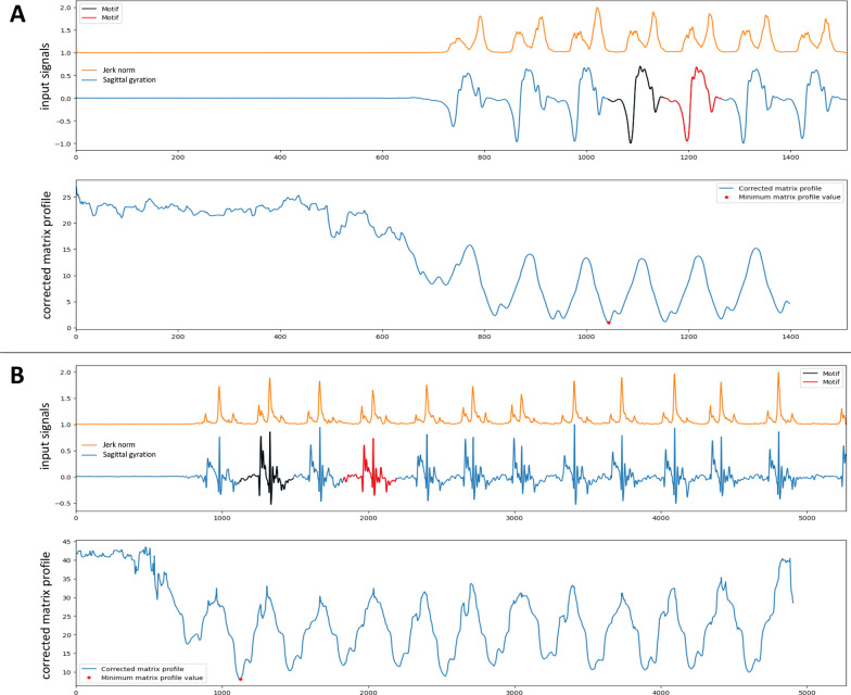
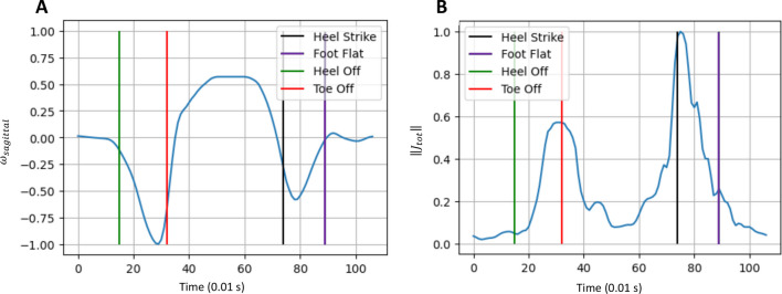
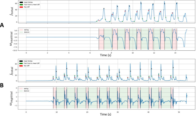
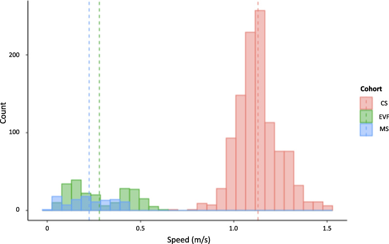
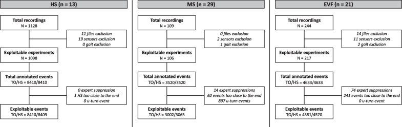
Methods: We recorded 10-meter gait IMU signals from 13 healthy subjects, 29 patients with multiple sclerosis, and 21 patients with post-stroke equino varus foot. An instrumented mat was used as the gold standard. Our method detects GEs from filtered acceleration free from gravity and gyration signals. Firstly, we use autocorrelation and pattern detection techniques to identify a reference stride pattern. Next, we apply multiparametric Dynamic Time Warping to annotate this pattern from a model stride, in order to detect all GEs in the signal.

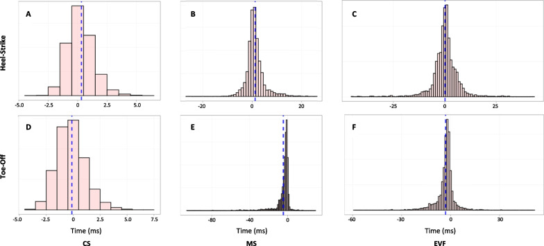
Results: We analyzed 16,819 GEs recorded from healthy subjects and achieved an F1-score of 100%, with a median absolute error of 8 ms (IQR [3-13] ms). In multiple sclerosis and equino varus foot cohorts, we analyzed 6067 and 8951 GEs, respectively, with F1-scores of 99.4% and 96.3%, and median absolute errors of 18 ms (IQR [8-39] ms) and 26 ms (IQR [12-50] ms).

Conclusions: Our results are consistent with the state of the art for healthy subjects and demonstrate a good accuracy in GEs detection for pathological patients. Therefore, our proposed method provides an efficient way to detect GEs from IMU signals, even in degraded gaits. However, it should be evaluated in each cohort before being used to ensure its reliability.

Keywords: Dynamic time warping; Gait analysis; Intertial measurement units; Pathological gaits; Pattern recognition; Step detection.


Conflict of interest statement

The authors declare that they have no competing interest.

Figures

Fig. 1
Fig. 1
Presentation of the sensor. A Tw Awinda XSens® sensor. B Definition of the axis for the sensor on the left foot
Fig. 2
Fig. 2
Gait recording protocol. The patient is equipped with two sensors Mtw Awinda XSens® placed on the dorsal part of each foot using Velcro bands. Walking is initiated upstream of the active surface of the GR
Fig. 3
Fig. 3
Flowchart of the GE detection method. Schematic representation of the 4 parts of the algorithm. The color of the boxes is: red for input data, blue for the corresponding tools and illustrations, brown for intermediate steps, and green for the output result
Fig. 4
Fig. 4
Autocorrelation signal. A Control subject. B Patient from the EVF cohort. The two graphs on the left show the preprocessed signals of interest for both feet. The righthand graph is the resulting multiparametric autocorrelation for each foot and gives the estimated return value of the duration of a stride (dot line)
Fig. 5
Fig. 5
Corrected matrix profile. A Control subject. B Patient from the EVF cohort. Top: gyration signal (blue) and jerk signal (orange). Bottom: CMP, the red star indicates the minimum value of the CMP and allows the detection of the pattern (black) and its nearest neighbor (red)
Fig. 6
Fig. 6
Model stride. Stride from a healthy subject used as a model for all detections. A Gyration (blue) in the sagittal plane. B Total jerk (blue). For each figure, the 4 events of the stride are represented. TO and HS were given by the GR. FF and HO were visually estimated and given as an indication
Fig. 7
Fig. 7
Annotated reference stride. Top: healthy subject from the CS cohort. Bottom: patient from the EVF cohort. A, B mDTWd matrix between model stride (up) and reference stride (left) signals with the corresponding warping path (white line). C, D Annotation of the reference with the 4 GEs. Blue line: jerk. Yellow line: gyration
Fig. 8
Fig. 8
Final gait segmentation. A Healthy subject from the CS cohort. B Patient from the EVF cohort. GEs detected by the algorithm were reported on the jerk signal (top). Gait phases deducted from GEs were reported on the gyration signal (bottom)
Fig. 9
Fig. 9
Walking speed distribution for each cohort. Dot lines represent mean values
Fig. 10
Fig. 10
Flowchart of the data collected and analyzed in the study. Rules for deleting and correcting data are provided in Methods
Fig. 11
Fig. 11
Boxplot of ΔHS and ΔTO. Each dot represents a correctly detected and annotated step
Fig. 12
Fig. 12
Histograms of ΔHS (A–C) and ΔTO(D–F) for each cohort. Dot lines represent the mean error

Similar articles

Cited by

References

    1. Davis RB. Clinical gait analysis. IEEE Eng Med Biol Mag. 1988;7(3):35–40. doi: 10.1109/51.7933. - DOI - PubMed
    1. Davies RJ, Parker J, McCullagh P, Zheng H, Nugent C, Black ND, Mawson S. A personalized self-management rehabilitation system for stroke survivors: a quantitative gait analysis using a smart insole. JMIR Rehabil Assist Technol. 2016;3(2):11. doi: 10.2196/rehab.5449. - DOI - PMC - PubMed
    1. Vienne-Jumeau A, Quijoux F, Vidal P-P, Ricard D. Wearable inertial sensors provide reliable biomarkers of disease severity in multiple sclerosis: a systematic review and meta-analysis. Ann Phys Rehabil Med. 2020;63(2):138–147. doi: 10.1016/j.rehab.2019.07.004. - DOI - PubMed
    1. Takeda R, Tadano S, Todoh M, Morikawa M, Nakayasu M, Yoshinari S. Gait analysis using gravitational acceleration measured by wearable sensors. J Biomech. 2009;42(3):223–233. doi: 10.1016/j.jbiomech.2008.10.027. - DOI - PubMed
    1. Jarchi D, Pope J, Lee TKM, Tamjidi L, Mirzaei A, Sanei S. A review on accelerometry-based gait analysis and emerging clinical applications. IEEE Rev Biomed Eng. 2018;11:177–194. doi: 10.1109/RBME.2018.2807182. - DOI - PubMed

MeSH terms