. 2024 Jun 18;22(1):338.
doi: 10.1186/s12951-024-02579-1.

Targeting SUMOylation with an injectable nanocomposite hydrogel to optimize radiofrequency ablation therapy for hepatocellular carcinoma

Affiliations

Targeting SUMOylation with an injectable nanocomposite hydrogel to optimize radiofrequency ablation therapy for hepatocellular carcinoma

Junfeng Liu et al. J Nanobiotechnology. .

Erratum in

Abstract

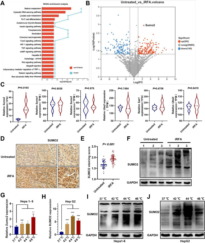
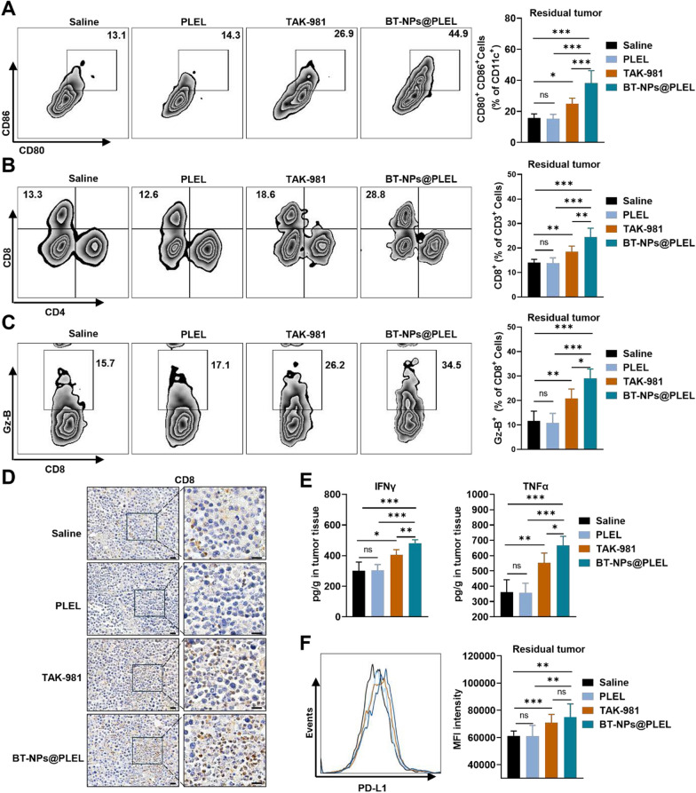
Background: Incomplete radiofrequency ablation (iRFA) in hepatocellular carcinoma (HCC) often leads to local recurrence and distant metastasis of the residual tumor. This is closely linked to the development of a tumor immunosuppressive environment (TIME). In this study, underlying mechanisms and potential therapeutic targets involved in the formation of TIME in residual tumors following iRFA were explored. Then, TAK-981-loaded nanocomposite hydrogel was constructed, and its therapeutic effects on residual tumors were investigated.

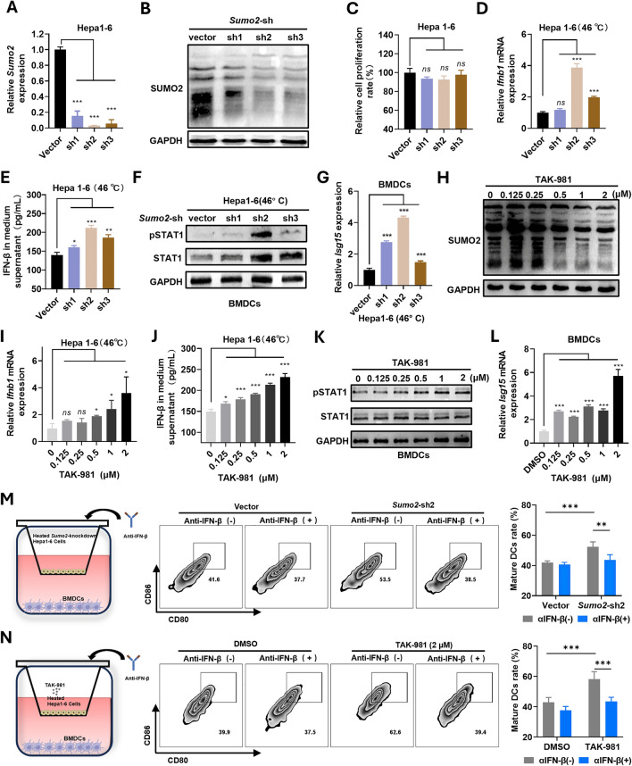
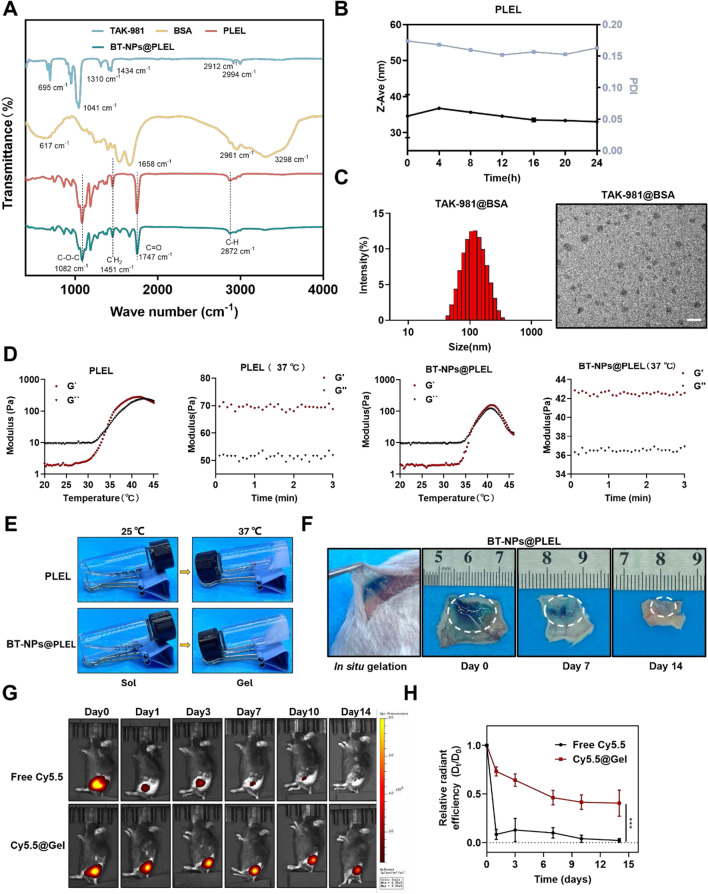
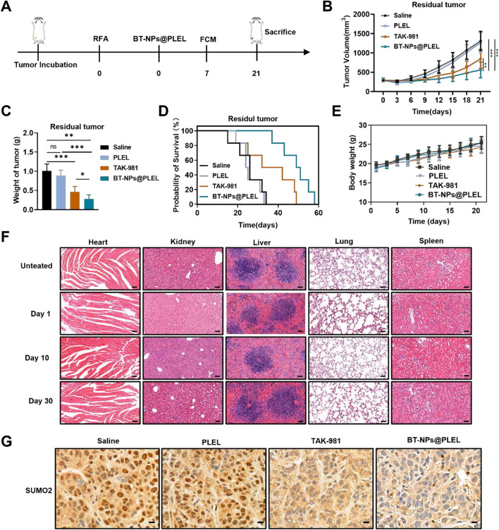
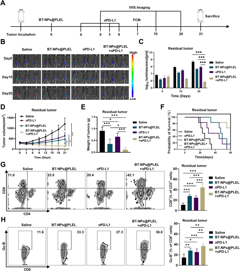
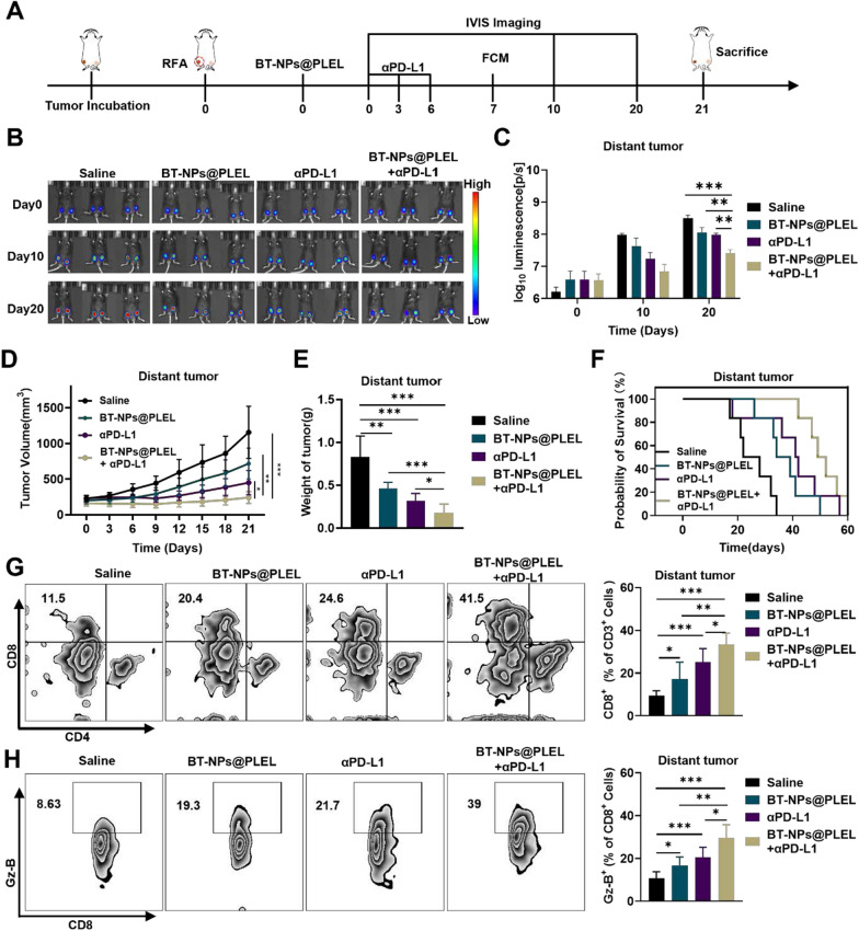
Results: This study reveals that the upregulation of small ubiquitin-like modifier 2 (Sumo2) and activated SUMOylation is intricately tied to immunosuppression in residual tumors post-iRFA. Both knockdown of Sumo2 and inhibiting SUMOylation with TAK-981 activate IFN-1 signaling in HCC cells, thereby promoting dendritic cell maturation. Herein, we propose an injectable PDLLA-PEG-PDLLA (PLEL) nanocomposite hydrogel which incorporates self-assembled TAK-981 and BSA nanoparticles for complementary localized treatment of residual tumor after iRFA. The sustained release of TAK-981 from this hydrogel curbs the expansion of residual tumors and notably stimulates the dendritic cell and cytotoxic lymphocyte-mediated antitumor immune response in residual tumors while maintaining biosafety. Furthermore, the treatment with TAK-981 nanocomposite hydrogel resulted in a widespread elevation in PD-L1 levels. Combining TAK-981 nanocomposite hydrogel with PD-L1 blockade therapy synergistically eradicates residual tumors and suppresses distant tumors.

Conclusions: These findings underscore the potential of the TAK-981-based strategy as an effective therapy to enhance RFA therapy for HCC.

Keywords: Hepatocellular carcinoma; Nanocomposite hydrogel; Radiofrequency ablation; Small ubiquitin-like modifier 2; TAK-981.


Conflict of interest statement

The authors declare that no conflict of interest exists.

Figures

Fig. 1
Fig. 1
Upregulation of Sumo2 and activation of SUMOylation in residual tumors after iRFA. A Significant enrichment of the DEGs of iRFA and untreated group in KEEG terms (top 20). B Volcano plot of DEGs in RNA-seq dataset. C Differences in expressions critical regulators in SUMOylation pathway between iRFA and the untreated group (n = 3). D The IHC staining of SUMO2 in tumor tissues, scale bar = 20 μm. E The IHC staining score of SUMO2 (n = 40). F Western blot analysis of conjugated-SUMO2 in tumor tissues. G The RT-qPCR analysis of the Sumo2 expression in heated Hepa1-6 cells (n=3). H The RT-qPCR analysis of the SUMO2 expression in heated HepG2 cells (n=3). I Western blot analysis of conjugated-SUMO2 in heated Hepa1-6 cells. J Western blot analysis of conjugated-SUMO2 in heated HepG2 cells. ns, not significant, *p < 0.05, **p < 0.01, ***p < 0.001
Fig. 2
Fig. 2
Knockdown of Sumo2 or inhibition of SUMOylation effectively activates the IFN-1 pathway. A The RT-qPCR analysis of Sumo2 in Sumo2-knockdown Hepa1-6 cells (n=3). B Western blot analysis of conjugated- SUMO2 in Sumo2-knockdown Hepa1-6 cells. C The relative proliferation rate in Sumo2-knockdown Hepa1-6 cells (n=3). D The RT-qPCR analysis of the Ifnb1 gene in heated Sumo2-knockdown Hepa1-6 cells (n=3). E ELISA analysis of the IFN-β in the medium supernatant of heated Sumo2-knockdown Hepa1-6 cells (n=3). F Western blot analysis of STAT1/pSTAT1 in BMDCs co-cultured with heated Hepa1-6 cells. G The relative expression of Isg15 in BMDCs co-cultured with heated Hepa1-6 cells (n=3). H Western blot analysis of conjugated-SUMO2 in Hepa1-6 cells treated with TAK-981. I The RT-qPCR analysis of the Ifnb1 gene in Hepa1-6 cells treated with TAK-981 (n=3). J ELISA analysis of the IFN-β in the medium supernatant of Hepa1-6 cells treated with TAK-981 (n=3). K Western blot analysis of STAT1/pSTAT1 in BMDCs co-cultured with heated Hepa1-6 cells treated with TAK-981. L The relative expression of Isg15 in BMDCs co-cultured with heated Hepa1-6 cells treated with TAK-981 (n=3). M The representative flow cytometry plots and statistical analysis of mature DCs rate after co-cultured with heated Sumo2-knockdown Hepa1-6 cells (n=6). N The representative flow cytometry plots and statistical analysis of mature DCs rate after co-cultured with heated Hepa1-6 cells treated with TAK-981. ns, not significant (n=6), *p < 0.05, **p < 0.01, ***p < 0.001
Fig. 3
Fig. 3
Preparation and Characterization of BT-NPs@PLEL. A FTIR spectra analysis of TAK-981, BSA, PLEL, and BT-NPs@PLEL. B The hydrodynamic diameter and polymer dispersity index of PLEL-Sol (1 wt%) within 24 h. C The hydrodynamic diameter and the TEM image of TAK-981@BSA nanoparticles, scale bar = 200 nm. D The rheological behavior of PLEL (25 wt%), BT-NPs@PLEL (25 wt%) in dependent of temperature. G′, storage modulus; G″, loss modulus. E Photographs showing the macroscopic thermo-sensitive sol–gel translation of PLEL and BT-NPs@PLEL (25 wt%). F In vivo gelation and degradation behavior of BT-NPs@PLEL (25 wt%) at different time points. G, H IVIS images and statistical analysis of fluorescence signal recorded at different times after injection of Cy5.5 and Cy5.5@Gel (n=3). ***p < 0.001
Fig. 4
Fig. 4
Inhibition of residual tumor after iRFA in vivo and blocking SUMOylation of the BT-NPs@PLEL. A Schematic representation of treatment of residual tumor after iRFA in C57BL/6 mice. B Tumor volume of residual tumors in different groups (n=6). C The weight of residual tumors post-iRFA on day 21 after different treatments (n=6). D Survival analysis of experimental mice in different groups (n=6). E The body weight changes of mice during treatment. F Images of H&E staining of tissue sections in essential organs after treatment with BT-NPs@PLEL on day 1, day 10, and day 30, scale bar: 50 µm. G Immunohistochemical analysis of SUMO2 in nucleus of residual tumor tissue after different treatments, scale bar: 10 µm. ns, not significant, *p < 0.05, **p < 0.01, ***p < 0.001
Fig. 5
Fig. 5
BT-NPs@PLEL activates anti-tumor immunity of residual tumor after iRFA. A Representative flow cytometry plots and proportions of mature DCs on day 7 (n = 6). B Representative flow cytometry plots and proportions of CD8+ T cells on day 7 (n = 6). C Representative flow cytometry plots and proportions of Granzyme B+ on day 7 (n = 6). D IHC staining of CD8 in residual tumor tissue, scale bar: 20 µm. E ELISA assay to detect the amount of IFN-γ and TNF-α in tumor tissue (n = 6). F The flow cytometry validation of PD-L1 overlay histogram and the mean fluorescence of the cells of tumor tissue after different treatments (n = 6). ns, not significant, *p < 0.05, **p < 0.01, ***p < 0.001
Fig. 6
Fig. 6
Combining BT-NPs@PLEL with anti-PD-L1 treatment for inhibition in residual tumors after iRFA. A Schematic representation of treatment of residual tumor after iRFA in C57/BL6 mice. B Bioluminescence images of mice with residual tumors after iRFA after different treatments on days 0, 10, and 20 (n = 3). C Bioluminescence signals of mice in each group on day 0, 10, and 20 (n = 3). D Tumor volume of residual tumors after iRFA in different groups(n = 6). E The weight of residual tumors on day 21 after different treatments (n = 6). F Survival analysis of experimental mice in different groups (n = 6). G Representative Flow cytometry plots and proportions of CD8+ T cells on day 7 (n = 6). H Representative flow cytometry plots and proportions of Granzyme B+ cells on day 7 (n = 6). ns, not significant *p < 0.05, **p < 0.01, ***p < 0.001
Fig. 7
Fig. 7
Combining BT-NPs@PLEL with anti-PD-L1 treatment for distant tumor inhibition. A Schematic representation of the treatment of residual tumors after iRFA and distant tumors in C57/BL6 mice. B Bioluminescence images of mice with residual tumors after iRFA and distant tumors after different treatments on days 0, 10, and 20 (n = 3). C Bioluminescence signals of mice in each group on day 0, 10, and 20. D Tumor volume of distant tumors in different groups (n = 6). E The weight of distant tumors post-iRFA on day 21 after different treatments (n = 6). F Survival analysis of experimental mice in different groups (n = 6). G Representative flow cytometry plots and proportions of CD8+ T cells in distant tumors on day 7 (n = 6). H Representative flow cytometry plots and proportions of Granzyme B+ cells in distant tumors on day 7 (n = 6). ns, not significant, *p < 0.05, **p < 0.01, ***p < 0.001

Similar articles

Cited by

References

    1. Kim N, Cheng J, Jung I, Liang J, Shih YL, Huang WY, Kimura T, Lee VHF, Zeng ZC, Zhenggan R, et al. Stereotactic body radiation therapy vs. radiofrequency ablation in Asian patients with hepatocellular carcinoma. J Hepatol. 2020;73:121–9. 10.1016/j.jhep.2020.03.005 - DOI - PubMed
    1. Heimbach JK, Kulik LM, Finn RS, Sirlin CB, Abecassis MM, Roberts LR, Zhu AX, Murad MH, Marrero JA. AASLD guidelines for the treatment of hepatocellular carcinoma. Hepatology. 2018;67:358–80. 10.1002/hep.29086 - DOI - PubMed
    1. Cao S, Lyu T, Fan Z, Guan H, Song L, Tong X, Wang J, Zou Y. Long-term outcome of percutaneous radiofrequency ablation for periportal hepatocellular carcinoma: tumor recurrence or progression, survival and clinical significance. Cancer Imag. 2022;22:2.10.1186/s40644-021-00442-2 - DOI - PMC - PubMed
    1. Teng W, Liu KW, Lin CC, Jeng WJ, Chen WT, Sheen IS, Lin CY, Lin SM. Insufficient ablative margin determined by early computed tomography may predict the recurrence of hepatocellular carcinoma after radiofrequency ablation. Liver Cancer. 2015;4:26–38. 10.1159/000343877 - DOI - PMC - PubMed
    1. Shi L, Wang J, Ding N, Zhang Y, Zhu Y, Dong S, Wang X, Peng C, Zhou C, Zhou L, et al. Inflammation induced by incomplete radiofrequency ablation accelerates tumor progression and hinders PD-1 immunotherapy. Nat Commun. 2019;10:5421. 10.1038/s41467-019-13204-3 - DOI - PMC - PubMed