

Activation of kappa opioid receptor suppresses post-traumatic osteoarthritis via sequestering STAT3 on the plasma membrane
- PMID: 38890746
- PMCID: PMC11186255
- DOI: 10.1186/s12964-024-01709-4
Activation of kappa opioid receptor suppresses post-traumatic osteoarthritis via sequestering STAT3 on the plasma membrane
Abstract
Objective: Kappa opioid receptor (KOR) signaling is involved in joint development and inflammation in Osteoarthritis (OA), while the biochemical mechanism remains unclarified. This study aims to investigate downstream molecular events of KOR activation, to provide novel perspectives in OA pathology.
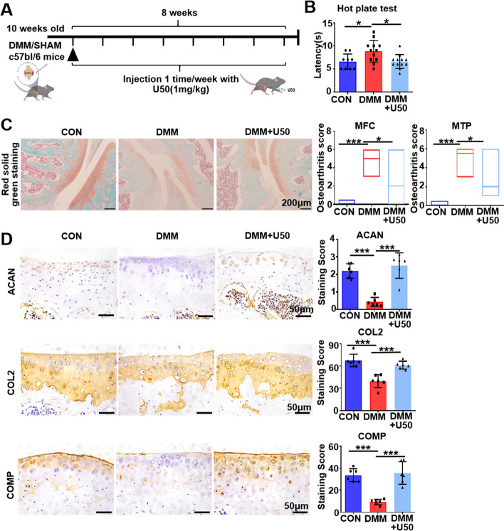
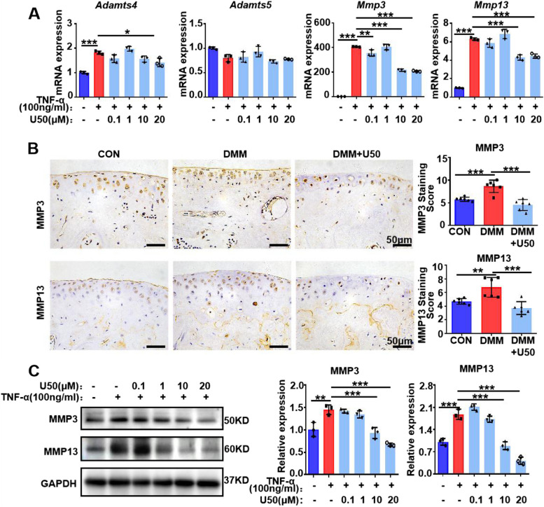
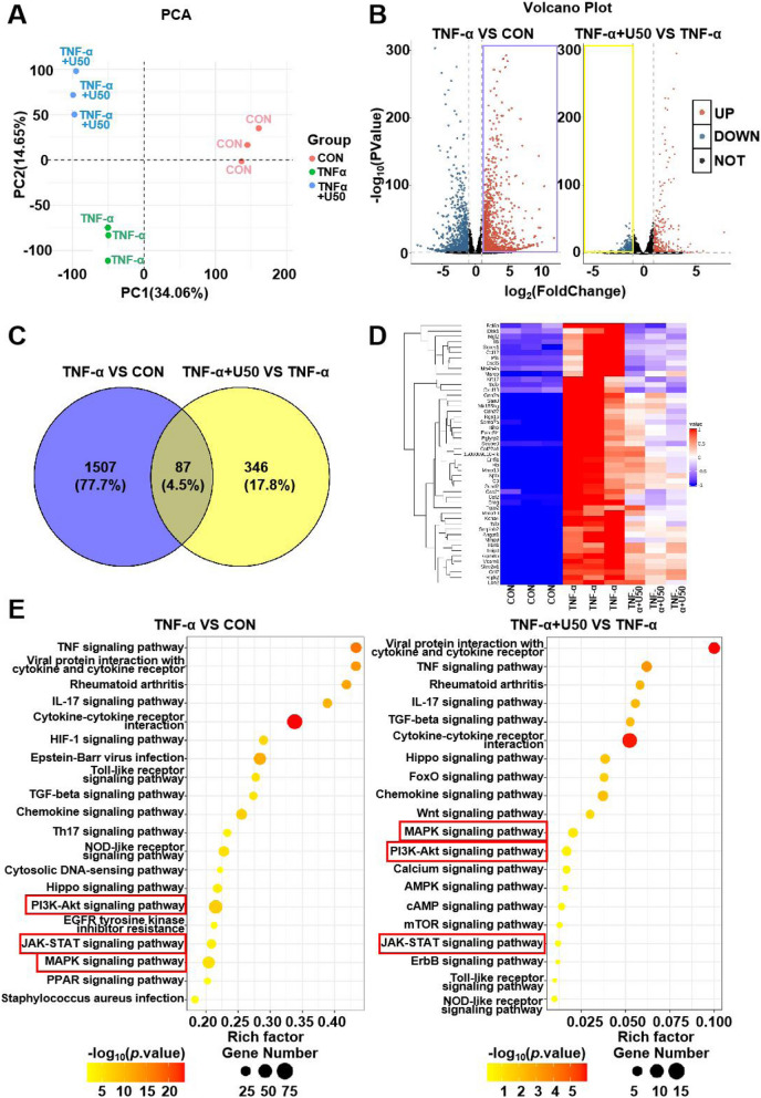
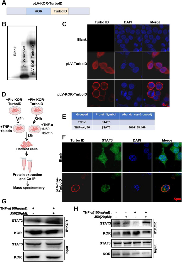
Methods: U50,488H, a selective KOR agonist, was intra-articularly injected in mice upon destabilization of the medial meniscus (DMM) as OA models, with PBS injection as control. The behavioral and histological evaluation was assessed by hot plate test and red solid green staining, respectively. Alterations in mRNA and protein expression were assessed by RNA-seq, RT-qPCR, immunohistochemistry and western blotting (WB) in chondrocytes treated with TNF-α or TNF-α + U50,488H. Proteins interacted with KOR were explored using proximity labeling followed by mass spectrometry and then testified by co-immunoprecipitation (Co-IP) assay and immunofluorescence (IF).
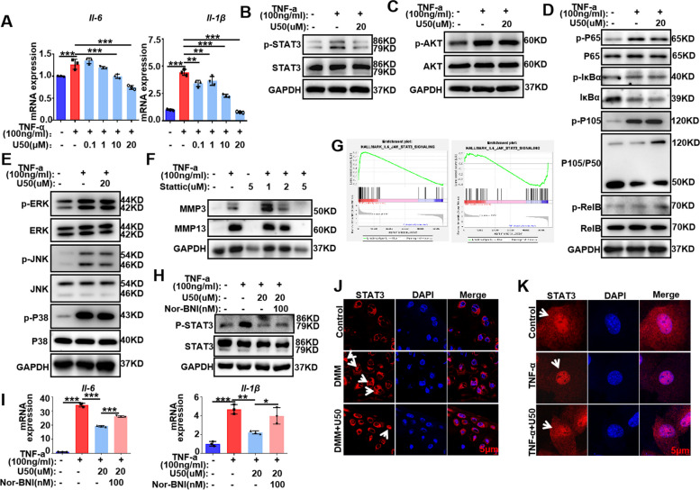
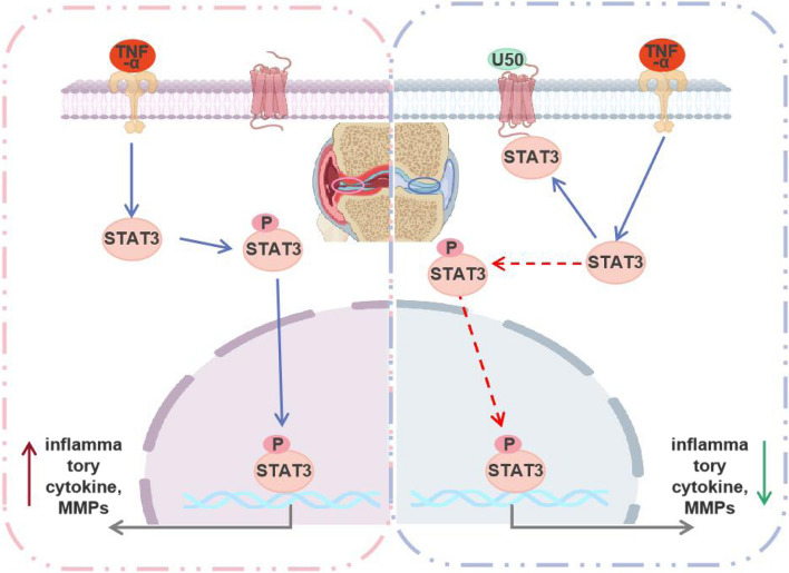
Results: OA-induced pain was reduced and cartilage degeneration was alleviated upon KOR activation in DMM mice. In chondrocytes, activation of KOR reversed the upregulation of MMPs, IL-6, IL-1β and phosphorylated(p-) STAT3, stimulated by TNF-α, while the expression of NF-κB, MAPKs and AKT signaling weren't reversed. RNA-seq and IF results presented that KOR activation evidently reduced STAT3 nuclear translocation in chondrocytes upon TNF-α stimuli. The reduction may be resulted from the binding of KOR and STAT3 in the plasma membrane, revealed by proximity labeling and Co-IP results.
Conclusions: KOR activation protects cartilage from OA, and this protective effect is mainly exerted via sequestering STAT3 on the plasma membrane, resulting in inactivation of STAT3-dependent immune responses which otherwise contributes to OA.
Keywords: Kappa opioid receptor; Osteoarthritis; STAT3; TNF-α.
© 2024. The Author(s).
Conflict of interest statement
The authors declare no competing interests.
Figures
References
Publication types
MeSH terms
Substances
Grants and funding
- 81672214/National Nature Science Foundation of China
- 81672214/National Nature Science Foundation of China
- 81672214/National Nature Science Foundation of China
- 81672214/National Nature Science Foundation of China
- 81672214/National Nature Science Foundation of China
- 81672214/National Nature Science Foundation of China
- 81672214/National Nature Science Foundation of China
- 81672214/National Nature Science Foundation of China
- 81672214/National Nature Science Foundation of China
- 81672214/National Nature Science Foundation of China
- 81672214/National Nature Science Foundation of China
- 202240133/Shanghai Municipal Health Commission Hygiene Industry Clinical Research Project
- 202240133/Shanghai Municipal Health Commission Hygiene Industry Clinical Research Project
- 202240133/Shanghai Municipal Health Commission Hygiene Industry Clinical Research Project
- 202240133/Shanghai Municipal Health Commission Hygiene Industry Clinical Research Project
- 202240133/Shanghai Municipal Health Commission Hygiene Industry Clinical Research Project
- 202240133/Shanghai Municipal Health Commission Hygiene Industry Clinical Research Project
- 202240133/Shanghai Municipal Health Commission Hygiene Industry Clinical Research Project
- 202240133/Shanghai Municipal Health Commission Hygiene Industry Clinical Research Project
- 202240133/Shanghai Municipal Health Commission Hygiene Industry Clinical Research Project
- 202240133/Shanghai Municipal Health Commission Hygiene Industry Clinical Research Project
- 202240133/Shanghai Municipal Health Commission Hygiene Industry Clinical Research Project
- 2023YFC2505903/National Key R&D program of China
- 2023YFC2505903/National Key R&D program of China
- 2023YFC2505903/National Key R&D program of China
- 2023YFC2505903/National Key R&D program of China
- 2023YFC2505903/National Key R&D program of China
- 2023YFC2505903/National Key R&D program of China
- 2023YFC2505903/National Key R&D program of China
- 2023YFC2505903/National Key R&D program of China
- 2023YFC2505903/National Key R&D program of China
- 2023YFC2505903/National Key R&D program of China
- 2023YFC2505903/National Key R&D program of China
LinkOut - more resources
Full Text Sources
Medical
Miscellaneous
