. 2024 May 24;13(11):905.
doi: 10.3390/cells13110905.

Plasma Gelsolin Inhibits Natural Killer Cell Function and Confers Chemoresistance in Epithelial Ovarian Cancer

Affiliations

Plasma Gelsolin Inhibits Natural Killer Cell Function and Confers Chemoresistance in Epithelial Ovarian Cancer

Toshimichi Onuma et al. Cells. .

Abstract

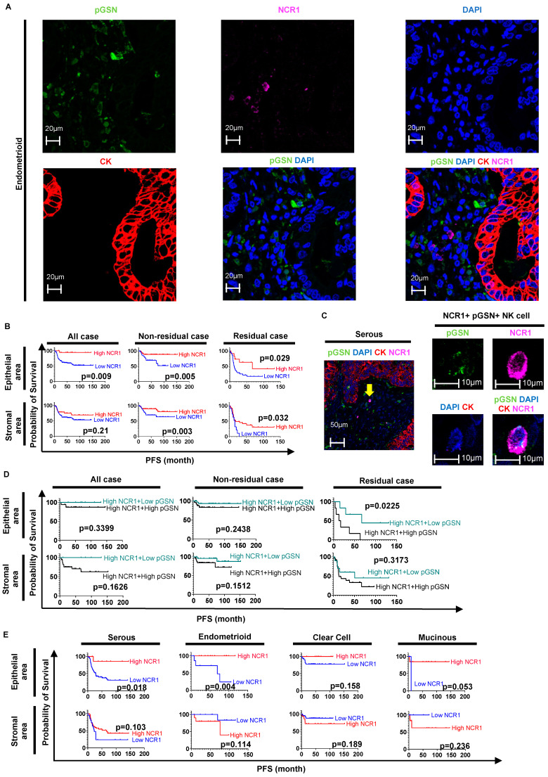
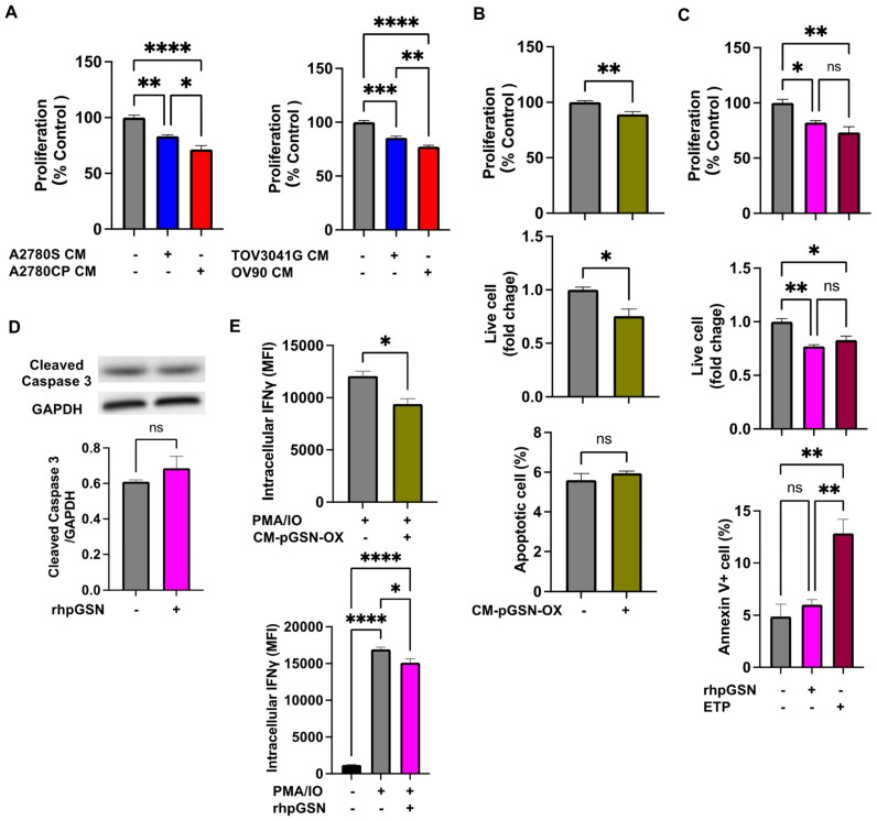
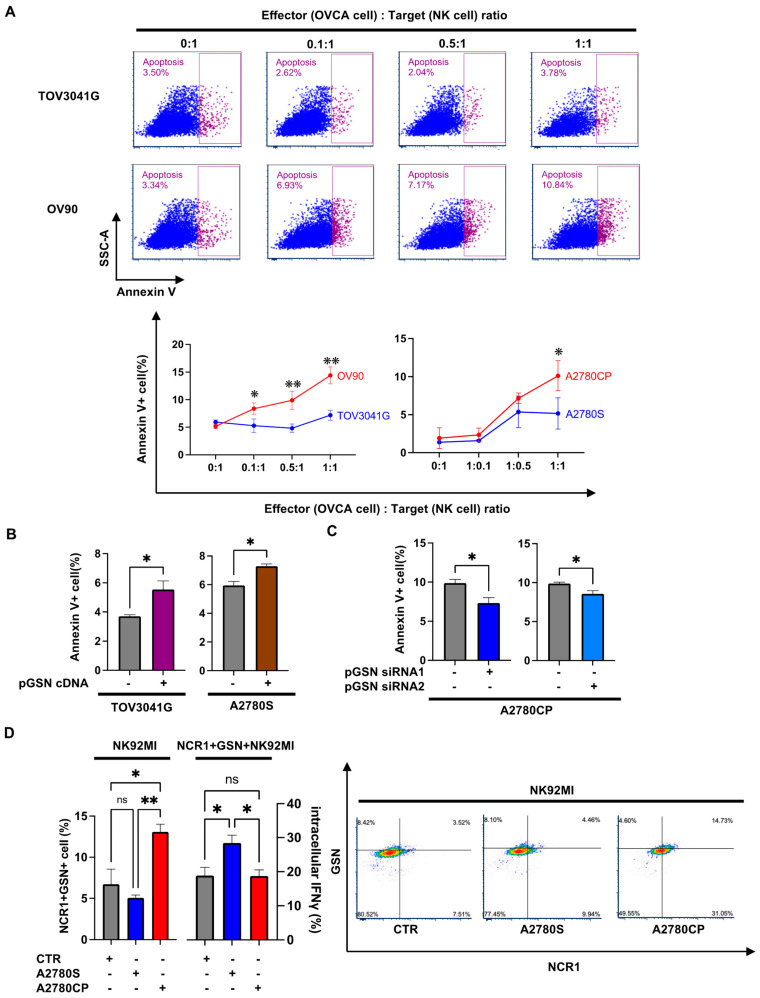
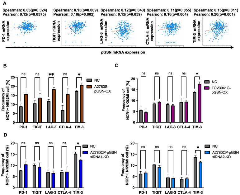
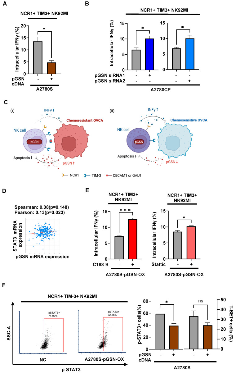
Plasma gelsolin (pGSN) overexpression in ovarian cancer (OVCA) disarms immune function, contributing to chemoresistance. The aim of this study was to investigate the immunoregulatory effects of pGSN expression on natural killer (NK) cell function in OVCA. OVCA tissues from primary surgeries underwent immunofluorescent staining of pGSN and the activated NK cell marker natural cytotoxicity triggering receptor 1 to analyze the prognostic impact of pGSN expression and activated NK cell infiltration. The immunoregulatory effects of pGSN on NK cells were assessed using apoptosis assay, cytokine secretion, immune checkpoint-receptor expression, and phosphorylation of STAT3. In OVCA tissue analyses, activated NK cell infiltration provided survival advantages to patients. However, high pGSN expression attenuated the survival benefits of activated NK cell infiltration. In the in vitro experiment, pGSN in OVCA cells induced NK cell death through cell-to-cell contact. pGSN increased T-cell immunoglobulin and mucin-domain-containing-3 expression (TIM-3) on activated NK cells. Further, it decreased interferon-γ production in activated TIM-3+ NK cells, attenuating their anti-tumor effects. Thus, increased pGSN expression suppresses the anti-tumor functions of NK cells. The study provides insights into why immunotherapy is rarely effective in patients with OVCA and suggests novel treatment strategies.

Keywords: chemoresistance; chemotherapy; gynecological cancers; natural killer cells; ovarian cancer; plasma gelsolin; tumor microenvironment.


Conflict of interest statement

The authors declare no conflict of interest.

Figures

Figure 1
Figure 1
The positive prognostic effect of high NK cell infiltration is suppressed by pGSN overexpression in OVCA tissues. (A) OVCA tissues are stained with anti-pGSN (green), anti-NCR1 (pink), anti-cytokeratin 8/18 (red), and DAPI (blue). The MFI is measured. (B) Patients are divided into high and low NCR1 expression groups using MFI for PFS analysis. (C) Some cells express both pGSN and NCR1 (yellow arrow). (D) Patients are divided into high and low pGSN in high NCR1 expression groups. (E) Patients are categorized into high and low NCR1 expression groups using MFI for PFS analysis for each OVCA histological subtype. The cut-off values separating the high and low NCR1 or pGSN expression groups are provided in Supplementary Tables S5, S7 and S9. OVCA: ovarian cancer; MFI: mean fluorescent intensity; PFS: progression-free survival; NK: natural killer; NCR1: natural cytotoxicity triggering receptor 1; pGSN: plasma gelsolin.
Figure 2
Figure 2
pGSN suppresses cell viability and interferon-γ (IFNγ) production but does not induce NK cell apoptosis without NK cell–OVCA cell contact. (A) NK92MI cells (2.0 × 10⁴ cells) were treated with conditioned media (CM) from chemoresistant (A2780CP and OV90) or chemosensitive (A2780S and TOV3041G) cells for 24 h (n = 4). The results were percent cell viability normalized to control. (B) NK92MI cells were treated with CM from pGSN-overexpressing A2780S cells (A2780S-CM-pGSN-OX) for 24 h (CCK8 2.0 × 10⁴ cells; n = 4, trypan blue 5.0 × 10⁴ cells; n = 4, apoptosis assay 2.0 × 10⁵ cells; n = 3). The results of the CCK8 assay were percent cell viability normalized to control. The results of the trypan blue assay are shown with the control plasmid as the reference value for fold change. (C) NK92MI cells were treated with vehicle, 10 μM rhpGSN, and 6 μM ETP for 12 h (CCK8 2.0 × 10⁴ cells; n = 3, trypan blue 5.0 × 10⁴ cells; n = 3, apoptosis assay 2.0 × 10⁵ cells; n = 3). The results of the CCK8 assay were percent cell viability normalized to control. The results of the trypan blue assay are shown with the vehicle as the reference value for fold change. (D) NK92MI cells (5.0 × 10⁵ cells) were treated with 10 μM rhpGSN and vehicle for 12 h for cleaved caspase-3 expression analysis (n = 3). (E) NK92MI (5.0 × 10⁵ cells) cells were treated with PMA/ionomycin and 10 μM rhpGSN or A2780S-CM-pGSN-OX for 6 h for intracellular IFNγ analysis (n = 3). These experiment results are expressed as means ± SEM from three or four independent experiments. **** p < 0.0001, *** p = 0.0001 to 0.001, ** p = 0.001 to 0.01, * p = 0.01 to 0.05, ns: not significant; NK: natural killer; OVCA: ovarian cancer; ETP: etoposide; pGSN: plasma gelsolin; MFI: median fluorescent intensity.
Figure 3
Figure 3
Increased pGSN induces NK cell dysfunction and apoptosis through cell-to-cell contact (A) CTFR-labeled NK92MI cells were directly co-cultured with chemoresistant (A2780CP and OV90) or chemosensitive (A2780S and TOV3041G) cells for 5 h and cell death examined using apoptosis assay at different effector: target ratios (n = 3). (B) CTFR-labeled NK92MI (2.0 × 10⁵ cells) cells were co-cultured with A2780S cell with pGSN overexpression (A2780S-pGSN-OX) or TOV3041G cell with pGSN overexpression (TOV3041G–pGSN–OX) for 5 h and cell death analyzed using apoptosis assay (n = 3). (C) CTFR-labeled NK92MI (2.0 × 10⁵ cells) cells were co-cultured with A2780CP cell with pGSN knockdown using two siRNA (A2780CP–pGSN siRNA1-KD or A2780CP–pGSN siRNA2-KD) for 5 h for apoptosis assay (n = 3). (D) OVCA cells (2.0 × 10⁵ cells) were seeded and incubated for 24 h. NK92MI (3.0 × 10⁵ cells) cells were co-cultured with chemosensitive (A2780S) and chemoresistant (A2780CP) cells for 6 h. Intracellular pGSN content in activated NK cells (NCR1+) was analyzed, as well as intracellular interferon-γ (IFNγ) expression (n = 3). The results are expressed as mean ± SEM from three independent experiments. ** p = 0.001 to 0.01, * p = 0.01 to 0.05, ns: not significant; CTFR: CellTrace Far Red; NK: natural killer; OVCA: ovarian cancer; NCR1: natural cytotoxicity triggering receptor 1; pGSN: plasma gelsolin.
Figure 4
Figure 4
pGSN increases TIM-3 expression in activated NK cells. (A) The correlation between pGSN mRNA and mRNA expression of immune checkpoint receptors derived from the TCGA dataset was analyzed using cBioPortal (https://www.cbioportal.org/, accessed on 1 September 2022). (BD) NK92MI cells (3.0 × 10⁵ cells) were co-cultured with A2780S cell with pGSN overexpression (A2780S–pGSN–OX), TOV3041G cell with pGSN overexpression (TOV3041G–pGSN–OX), and A2780CP cell with pGSN knockdown using two siRNAs (A2780CP–pGSN siRNA1-KD or A2780CP–pGSN siRNA2-KD) for 6 h. Expression of immune checkpoint receptors (PD-1, TIGIT, LAG-3, CTLA-4, and TIM-3) in activated NK cells (NCR1+) were analyzed using flow cytometry (n = 3). The results are expressed as mean ± SEM from three independent experiments. ** p = 0.001 to 0.01, * p = 0.01 to 0.05, ns: not significant NK: natural killer; NCR1: natural cytotoxicity triggering receptor 1; pGSN: plasma gelsolin; TIGIT: T-cell immunoreceptor with Ig and ITIM domains; PD-1: programmed cell death protein 1; LAG-3: lymphocyte-activation gene 3; CTLA-4: cytotoxic T lymphocyte-associated protein 4; TIM-3: T-cell immunoglobulin and mucin domain 3.
Figure 5
Figure 5
pGSN suppresses activated TIM3+ NK cell function. (A,B) NK92MI cells (3.0 × 10⁵ cells) were co-cultured with A2780S cell with pGSN overexpression (A2780S–pGSN–OX) or A2780CP cell with pGSN knockdown using two siRNA (A2780CP-siRNA1-KD and A2780CP-siRNA2-KD) for 6 h after which activated NK TIM-3+ cells (NCR1+TIM-3+) positive for interferon-γ (IFNγ) were analyzed using flow cytometry (n = 3). (C) In chemoresistant conditions, when NK cells contact with the OVCA, increased pGSN by OVCA cells increased pGSN and TIM-3 content, increased apoptosis, and decreased IFNγ in activated NK cells (i). On the other hand, pGSN levels are relatively lower in chemosensitive conditions and, hence, are unable to suppress activated NK cell function (ii). (D) Correlation between pGSN and STAT3 mRNA expressions derived from the TCGA dataset was analyzed using cBioPortal (https://www.cbioportal.org/, accessed on 1 September 2022). (E) NK92MI cells were treated with STAT3 inhibitor C188-9 or Sttatic 10μM and vehicle for 1 h. Subsequently, NK92MI cells (3.0 × 10⁵ cells) were co-cultured with A2780S–pGSN–OX for 6 h, after which activated NK TIM-3+ cells (NCR1+TIM-3+) positive for IFNγ were analyzed using flow cytometry (n = 3). (F) NK92MI cells (3.0 × 10⁵ cells) were co-cultured with A2780S–pGSN–OX for 6 h, after which activated NK TIM-3+ cells (NCR1+TIM-3+) positive for pSTAT3 and T-BET were analyzed using flow cytometry (n = 3). The results are expressed as mean ± SEM from three independent experiments. *** p = 0.0001 to 0.001, * p = 0.01 to 0.05, ns: not significant; NK: natural killer; NCR1: natural cytotoxicity triggering receptor 1; pGSN: plasma gelsolin; TIM-3: T-cell immunoglobulin and mucin-domain-containing-3 expression; CEACAM-1: carcinoembryonic antigen-related cell adhesion molecules-1; GAL-9: galactine-9; T-BET: T-box expressed in T cells; pSTAT3: phosphorylated signal transducer and activator of transcription 3.

Similar articles

Cited by

References

    1. Kuroki L., Guntupalli S.R. Treatment of Epithelial Ovarian Cancer. BMJ. 2020;371:m3773. doi: 10.1136/bmj.m3773. - DOI - PubMed
    1. Guo C., Song C., Zhang J., Gao Y., Qi Y., Zhao Z., Yuan C. Revisiting Chemoresistance in Ovarian Cancer: Mechanism, Biomarkers, and Precision Medicine. Genes Dis. 2022;9:668–681. doi: 10.1016/j.gendis.2020.11.017. - DOI - PMC - PubMed
    1. Gottesman M.M., Fojo T., Bates S.E. Multidrug Resistance in Cancer: Role of ATP–Dependent Transporters. Nat. Rev. Cancer. 2002;2:48–58. doi: 10.1038/nrc706. - DOI - PubMed
    1. Fletcher J.I., Williams R.T., Henderson M.J., Norris M.D., Haber M. ABC Transporters as Mediators of Drug Resistance and Contributors to Cancer Cell Biology. Drug Resist. Updates. 2016;26:1–9. doi: 10.1016/j.drup.2016.03.001. - DOI - PubMed
    1. Barbato L., Bocchetti M., Di Biase A., Regad T. Cancer Stem Cells and Targeting Strategies. Cells. 2019;8:926. doi: 10.3390/cells8080926. - DOI - PMC - PubMed

Publication types

MeSH terms