. 2024 May 25;13(11):913.
doi: 10.3390/cells13110913.

NLRX1 Mediates the Disruption of Intestinal Mucosal Function Caused by Porcine Astrovirus Infection via the Extracellular Regulated Protein Kinases/Myosin Light-Chain Kinase (ERK/MLCK) Pathway

Affiliations

NLRX1 Mediates the Disruption of Intestinal Mucosal Function Caused by Porcine Astrovirus Infection via the Extracellular Regulated Protein Kinases/Myosin Light-Chain Kinase (ERK/MLCK) Pathway

Jie Tao et al. Cells. .

Abstract

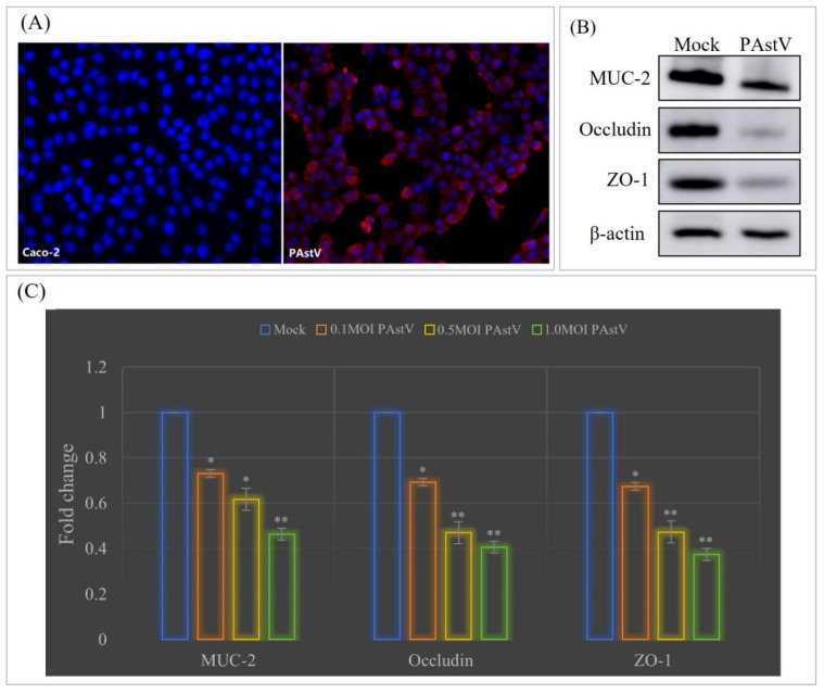
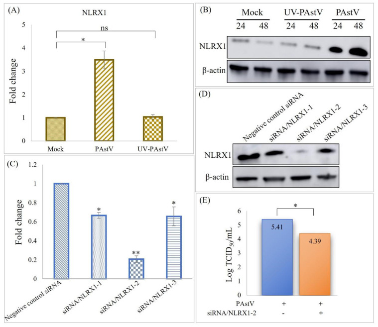
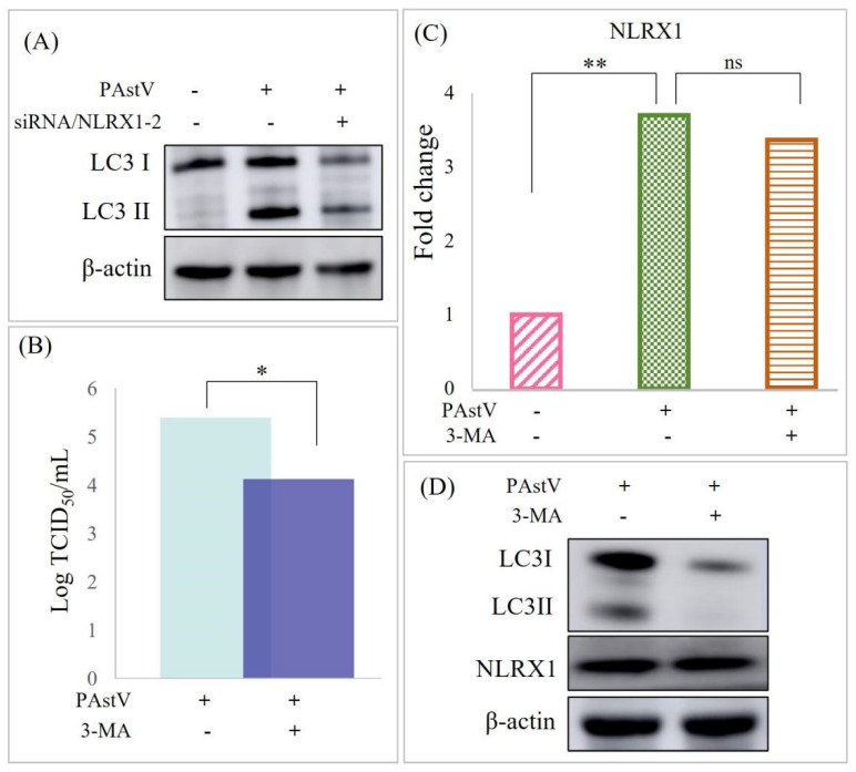
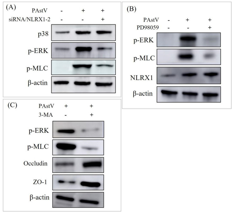
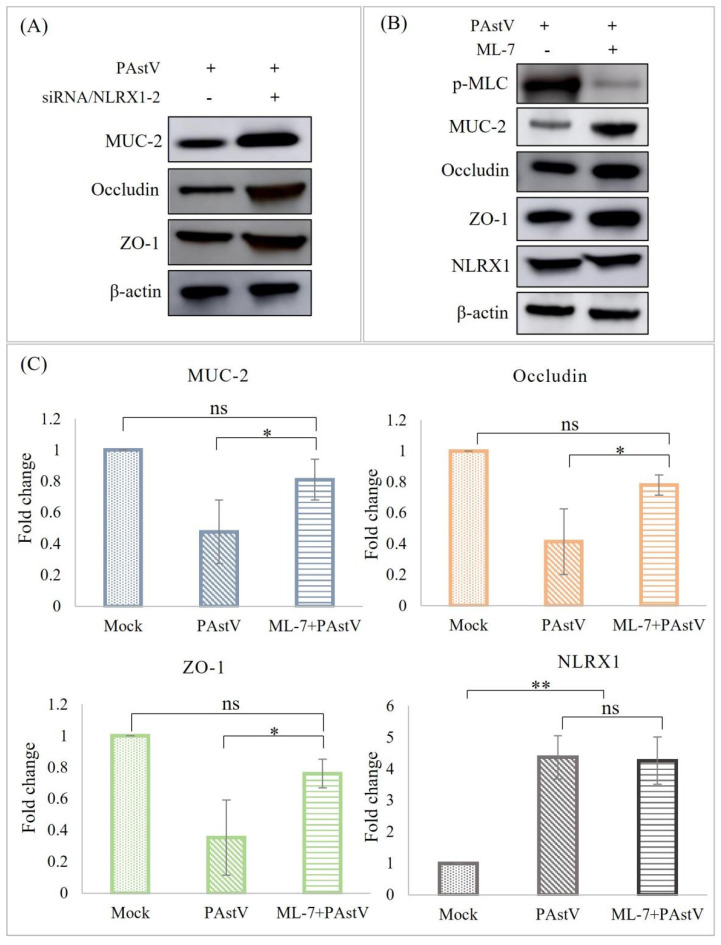
Porcine astrovirus (PAstV) has a potential zoonotic risk, with a high proportion of co-infection occurring with porcine epidemic diarrhea virus (PEDV) and other diarrheal pathogens. Despite its high prevalence, the cellular mechanism of PAstV pathogenesis is ill-defined. Previous proteomics analyses have revealed that the differentially expressed protein NOD-like receptor X1 (NLRX1) located in the mitochondria participates in several important antiviral signaling pathways in PAstV-4 infection, which are closely related to mitophagy. In this study, we confirmed that PAstV-4 infection significantly up-regulated NLRX1 and mitophagy in Caco-2 cells, while the silencing of NLRX1 or the treatment of mitophagy inhibitor 3-MA inhibited PAstV-4 replication. Additionally, PAstV-4 infection triggered the activation of the extracellular regulated protein kinases/ myosin light-chain kinase (ERK/MLCK) pathway, followed by the down-regulation of tight-junction proteins (occludin and ZO-1) as well as MUC-2 expression. The silencing of NLRX1 or the treatment of 3-MA inhibited myosin light-chain (MLC) phosphorylation and up-regulated occludin and ZO-1 proteins. Treatment of the ERK inhibitor PD98059 also inhibited MLC phosphorylation, while MLCK inhibitor ML-7 mitigated the down-regulation of mucosa-related protein expression induced by PAstV-4 infection. Yet, adding PD98059 or ML-7 did not affect NLRX1 expression. In summary, this study preliminarily explains that NLRX1 plays an important role in the disruption of intestinal mucosal function triggered by PAstV-4 infection via the ERK/MLC pathway. It will be helpful for further antiviral drug target screening and disease therapy.

Keywords: ERK/MLCK; NLRX1; intestinal mucosal barrier; mitophagy; porcine astrovirus.


Conflict of interest statement

The authors declare no conflicts of interest.

Figures

Figure 1
Figure 1
Down-regulation of tight junctions in Caco−2 cells upon PAstV infection. (A) PAstV propagation on Caco−2 cells with 15 μg/mL Pancreatin resulted in specific red fluorescence detected in the cytoplasm, as demonstrated by an indirect immunofluorescence assay using a PAstV monoclonal antibody. (B) Following inoculation of Caco-2 cells with 1MOI of PAstV, the expression of tight-junction proteins was detected 24 h later through Western blotting. (C) The influence of PAstV infection at various MOIs (0.1, 0.5, and 1.0) on the transcription of MUC−2, occludin, and ZO−1 was determined by relative fluorescence quantification PCR, utilizing β-actin as the intrinsic reference protein. Fold changes were calculated using the 2−ΔΔCt method. (“**” p < 0.01, ”*” p < 0.05).
Figure 2
Figure 2
Reduction in PAstV replication through NLRX1 knockdown in Caco−2 cells. (A) Caco−2 cells were infected at an MOI of 1.0 with either PAstV or UV−inactivated PAstV for 24 h. Real−time PCR was employed to determine the relative NLRX1 levels, normalized to β−actin. Asterisks denote significant differences from uninfected cells(“**” p < 0.01, ”*” p < 0.05). “ns” indicates no significant difference. (B) Cell lysates were collected at 24 h and 48 h, respectively, following infection, and NLRX1 expression was assessed through Western blotting using the indicated antibodies. (C,D) Caco−2 cells were transfected with negative control siRNA or three different siRNA duplexes targeting NLRX1 for 24 h. Subsequently, the relative NLRX1 and expression levels were detected, with β−actin as an internal control. (E) Cells were transfected with negative control siRNA or siRNA/NLRX1−2 for 24 h and then infected with 1.0 MOI PAstV. Virus titers were determined through a TCID50 assay at 60 h post-infection.
Figure 3
Figure 3
Induction of mitophagy by PAstV infection via up-regulation of NLRX1 protein. (A) Caco-2 cells were transfected with negative control siRNA or siRNA/NLRX1−2 for 24 h, followed by infection with 1MOI PAstV. Cell lysates were blotted with anti-LC3I/II, anti-NLRX1, and anti-β−actin antibodies. (B) Cells treated with the mitophagy inhibitor 3−MA for 6 h were subsequently infected with 1.0MOI PAstV. Virus titers were determined through a TCID50 assay 60 h post-infection. (C,D) show that cells subjected to treatment were infected with 1.0 MOI PAstV, and the relative and expression levels of NLRX1 were assessed 24 h later. (“**” p < 0.01, ”*” p < 0.05). “ns” indicates no significant difference.
Figure 4
Figure 4
Reduction in tight-junction protein expression by PAstV infection via the ERK/MLC pathway. (A) Caco-2 cells were transfected with negative control siRNA or siRNA/NLRX1-2 for 24 h, followed by PAstV infection at an MOI of 1.0. After 24 h, cell lysates were blotted with the indicated antibodies. (B) Cells were treated with 20 μM PD98059 (ERK inhibitor) for 6 h, followed by infection with 1MOI PAstV. Subsequent Western blotting using the indicated antibodies was conducted as described above. (C) Cells were treated with 20 μM 3-MA (mitophagy inhibitor), and the subsequent steps were the same as in (B).
Figure 5
Figure 5
Essential role of NLRX1 in impaired intestinal barrier function induced by PAstV infection. (A) Caco−2 cells were transfected with negative control siRNA or siRNA/NLRX1−2 for 24 h, followed by PAstV infection at an MOI of 1.0. Cell lysates were then blotted with the indicated antibodies 24 h later. Further, cells were treated with 25 μM ML−7 (MLCK inhibitor) for 6 h and infected with 1.0 MOI PAstV. (B) The relative MUC−2, occludin, ZO−1, and NLRX1 levels were detected after treatment with ML−7 (MLCK inhibitor), using β−actin as an internal reference. (C) MUC−2, occludin, ZO−1, and NLRX1 expression levels were assessed after treatment with ML−7 (MLCK inhibitor), with β−actin as an internal control. (“**” p < 0.01, ”*” p < 0.05). “ns” indicates no significant difference.

Similar articles

References

    1. Rawal G., Linhares D.C.L. Scoping review on the epidemiology, diagnostics and clinical significance of porcine astroviruses. Transbound. Emerg. Dis. 2022;69:974–985. doi: 10.1111/tbed.14123. - DOI - PubMed
    1. Nguyen T.V., Piewbang C., Techangamsuwan S. Genetic characterization of canine astrovirus in non-diarrhea dogs and diarrhea dogs in Vietnam and Thailand reveals the presence of a unique lineage. Front. Vet. Sci. 2023;10:1278417. doi: 10.3389/fvets.2023.1278417. - DOI - PMC - PubMed
    1. Chen J., Wan C.M., Gong S.T., Fang F., Sun M., Qian Y., Huang Y., Wang B.X., Xu C.D., Ye L.Y., et al. Chinese clinical practice guidelines for acute infectious diarrhea in children. World J. Pediatr. 2018;14:429–436. doi: 10.1007/s12519-018-0190-2. - DOI - PubMed
    1. Vu D.L., Bosch A., Pinto R.M., Guix S. Epidemiology of Classic and Novel Human Astrovirus: Gastroenteritis and Beyond. Viruses. 2017;9:33. doi: 10.3390/v9020033. - DOI - PMC - PubMed
    1. Bridger J.C. Detection by electron microscopy of caliciviruses, astroviruses and rotavirus–like particles in the faeces of piglets with diarrhoea. Vet. Rec. 1980;107:532–533. - PubMed

Publication types

MeSH terms

Substances

LinkOut - more resources