. 2024 May 30;13(11):953.
doi: 10.3390/cells13110953.

Inhibition of PIKfyve Leads to Lysosomal Disorders via Dysregulation of mTOR Signaling

Affiliations

Inhibition of PIKfyve Leads to Lysosomal Disorders via Dysregulation of mTOR Signaling

Jianhong Xia et al. Cells. .

Abstract

PIKfyve is an endosomal lipid kinase that synthesizes phosphatidylinositol 3,5-biphosphate from phosphatidylinositol 3-phsphate. Inhibition of PIKfyve activity leads to lysosomal enlargement and cytoplasmic vacuolation, attributed to impaired lysosomal fission processes and homeostasis. However, the precise molecular mechanisms underlying these effects remain a topic of debate. In this study, we present findings from PIKfyve-deficient zebrafish embryos, revealing enlarged macrophages with giant vacuoles reminiscent of lysosomal storage disorders. Treatment with mTOR inhibitors or effective knockout of mTOR partially reverses these abnormalities and extend the lifespan of mutant larvae. Further in vivo and in vitro mechanistic investigations provide evidence that PIKfyve activity is essential for mTOR shutdown during early zebrafish development and in cells cultured under serum-deprived conditions. These findings underscore the critical role of PIKfyve activity in regulating mTOR signaling and suggest potential therapeutic applications of PIKfyve inhibitors for the treatment of lysosomal storage disorders.

Keywords: PIKfyve; lysosome; mTOR; macrophage; vacuolation.


Conflict of interest statement

The authors declare no conflicts of interest.

Figures

Figure 1
Figure 1
Defects in macrophages in PIKfyve mutant zebrafish embryos. (A) Representative images of WT and MU embryos at 3 dpf. The arrows show the cells with giant vacuole-like structures in the CHT. Cellular death in the mutant brains was evidenced by grey tissue (asterisk). (B) The relative expression levels of PIKfyve in the WT zebrafish and the homozygous mutants (MU1, MU2) were quantified by RT-qPCR. The data were analyzed by 2−∆∆CT method and ꞵ-actin was used as an internal control (** p < 0.01). (CF) PIKfyve mutation was introduced into transgenic zebrfish lines in which macrophages (mpeg1:EGFP) (C,D) or neutrophils (mpx:EGFP) (E,F) were respectively highlighted with EGFP. In the mutant embryos, the macrophages were enlarged with giant vacuoles, while the neutrophils appeared normal. Scale bar, 20 µm. (D) Quantification of macrophage cell number (left panel) and macrophage diameter (right panel) in the CHT (ns, non-significant; ** p < 0.01). (F) Quantification of neutrophil cell number (left panel) and neutrophil diameter (right panel) in the CHT (ns, non-significant). (G,H) NR staining of microglia in WT siblings and PIKfyve mutants at 3 dpf. (G) Dorsal and lateral view of the microglia in the optic tectum of WT and MU larvae at 3 dpf stained with NR. The yellow arrows show the microglia stained with NR. (H) Quantification of NR-positive microglia cell number in the optic tectum of WT and MU larvae at 3 dpf (ns, non-significant).
Figure 2
Figure 2
PIKfyve deficiency leads to the accumulation of enlarged LAMP1-positive compartments in macrophages. (AD) The giant vacuoles in mutant macrophages were revealed to be fused lysosomes by staining with LysoTracker (A) or anti-LAMP1 antibody (C). (B) and (D) respectively show the quantification of LysoTracker and LAMP1 fluorescence intensity (** p < 0.01, **** p < 0.0001). Scale bar, 20 µm. (E) Representative confocal images and tracks of macrophage movement in the CHT of WT and MU zebrafish larvae at 3 dpf. Tracks are coded for speed. Scale bar, 50 µm. (F) Quantification of migration speed of macrophages in (E) (** p < 0.01).
Figure 3
Figure 3
The defects in PIKfyve mutants are dependent on sustained mTOR activation. (A) mTOR inhibitor (Rapa or Torin-1) treatment or effective mTOR knockout (mTOR_KO) attenuates the developmental defects in PIKfyve mutants. Scale bar, 20 µm. (B) Survival curve showing that the lifespan of MU embryos can be extended when mTOR is inhibited by chemical (Rapa, Torin-1) or genetic (mTOR_KO) approaches (N = 3, n = 30 per group). (** p < 0.01, log-rank test). (C) Quantification of diameter of macrophages in MU zebrafish embryos treated as in (A) (**** p < 0.0001, one-way ANOVA).
Figure 4
Figure 4
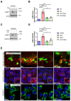
Molecular evidence for the hyperactivation of mTOR signaling in PIKfyve mutant zebrafish. (A) Western blot analysis showing the sustained mTOR activity during early development in PIKfyve mutant zebrafish embryos. (B) Densitometric analysis of phosphorylation status of 4E-BP1 and S6K in (A) (ns, non-significant; * p < 0.05, ** p < 0.01; two-way ANOVA). (C) Western blot analysis of mTOR activity in WT and MU zebrafish embryos at 5 dpf with or without rapamycin treatment. (D) Densitometric analysis of phosphorylation status of 4E-BP1 and S6K in (C) (ns, non-significant; ** p < 0.01, *** p < 0.001, **** p < 0.0001; two-way ANOVA). (E) WT and MU zebrafish embryos with or without rapamycin treatment were fixed at 5 dpf, permeabilized, and immuno-stained with phospho-S6K antibody. Arrows denote the macrophages with intense phospho-S6K staining. Scale bar, 20 µm.
Figure 5
Figure 5
Rapamycin treatment ameliorates the lysosomal defects induced by PIKfyve deficiency in vivo and in vitro. (AD) Western blot analysis showing the rapamycin treatment restored the elevated LAMP1 level induced by PIKfyve deficiency in zebrafish (A,B) and RAW264.7 cells (C,D). (B) and (D) are densitometric analysis of phosphorylation status of 4E-BP1 and S6K in (A) and (C), respectively (ns, non-significant; * p < 0.05, ** p < 0.01, *** p < 0.001; one-way ANOVA). (E) Lysotracker and LAMP1 staining in zebrafish and RAW264.7 cells. The Lysotracker/LAMP1-positive vacuoles were significantly enlarged in PIKfyve mutant cells and in Apm-treated cells, which were ameliorated by rapamycin treatment.
Figure 6
Figure 6
PIKfyve is required for starvation-induced mTOR shutdown. (A) HeLa cells were pretreated with DMSO vehicle control or 200 nM Apm for 3 h, followed by starvation in HBSS in the absence or in the presence of 200 nM Apm (HBSS + Apm) for the indicated time. Lysates were probed with the indicated antibodies. (B) Densitometric analysis of phosphorylation status of 4E-BP1 and S6K in (A). Data are represented as mean ± SD of 3 independent experiments (ns, non-significant; ** p < 0.01, *** p < 0.001, **** p < 0.0001; two-way ANOVA). (C) HeLa cells were treated as in (A), fixed at the selected time point 0 (Fed) and 120 min (HBSS 2 h). Cells were stained with the indicated antibodies and visualized by confocal microscopy. Scale bar, 20 µm. (D) Quantification of TSC2/LAMP1 colocalization from (C) (**** p < 0.0001, one-way ANOVA). (E) A working model for PIKfyve-dependent regulation of mTOR activity. The red arrow pointing upward indicates hyperactivation of mTOR activity. The prohibition sign indicates mTOR inhibition.

Similar articles

References

    1. Huang P.-T., Einav S., Asquith C.R.M. PIKfyve: A Lipid Kinase Target for COVID-19, Cancer and Neurodegenerative Disorders. Nat. Rev. Drug Discov. 2021;20:730. doi: 10.1038/d41573-021-00158-9. - DOI - PMC - PubMed
    1. Zolov S.N., Bridges D., Zhang Y., Lee W.-W., Riehle E., Verma R., Lenk G.M., Converso-Baran K., Weide T., Albin R.L., et al. In Vivo, Pikfyve Generates PI(3,5)P2, Which Serves as Both a Signaling Lipid and the Major Precursor for PI5P. Proc. Natl. Acad. Sci. USA. 2012;109:17472–17477. doi: 10.1073/pnas.1203106109. - DOI - PMC - PubMed
    1. Kim J., Jahng W.J., Di Vizio D., Lee J.S., Jhaveri R., Rubin M.A., Shisheva A., Freeman M.R. The Phosphoinositide Kinase PIKfyve Mediates Epidermal Growth Factor Receptor Trafficking to the Nucleus. Cancer Res. 2007;67:9229–9237. doi: 10.1158/0008-5472.CAN-07-1333. - DOI - PMC - PubMed
    1. de Lartigue J., Polson H., Feldman M., Shokat K., Tooze S.A., Urbé S., Clague M.J. PIKfyve Regulation of Endosome-Linked Pathways. Traffic. 2009;10:883–893. doi: 10.1111/j.1600-0854.2009.00915.x. - DOI - PMC - PubMed
    1. Soares A.C., Ferreira A., Mariën J., Delay C., Lee E., Trojanowski J.Q., Moechars D., Annaert W., De Muynck L. PIKfyve Activity Is Required for Lysosomal Trafficking of Tau Aggregates and Tau Seeding. J. Biol. Chem. 2021;296:100636. doi: 10.1016/j.jbc.2021.100636. - DOI - PMC - PubMed

Publication types

MeSH terms

Substances