. 2024 Jun 3;13(11):968.
doi: 10.3390/cells13110968.

Interleukin-8/CXCR1 Signaling Contributes to the Progression of Pulmonary Adenocarcinoma Resulting in Malignant Pleural Effusion

Affiliations

Interleukin-8/CXCR1 Signaling Contributes to the Progression of Pulmonary Adenocarcinoma Resulting in Malignant Pleural Effusion

Yi-Ming Chang et al. Cells. .

Abstract

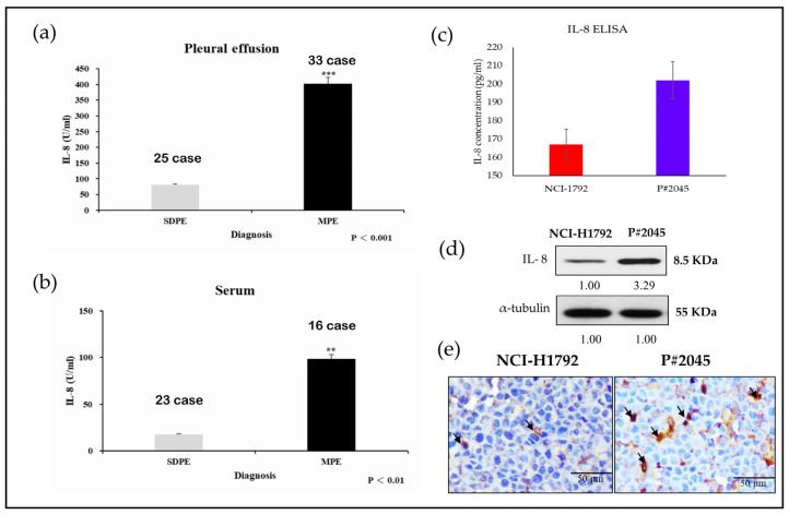
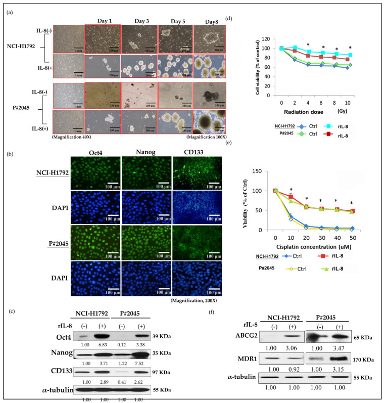
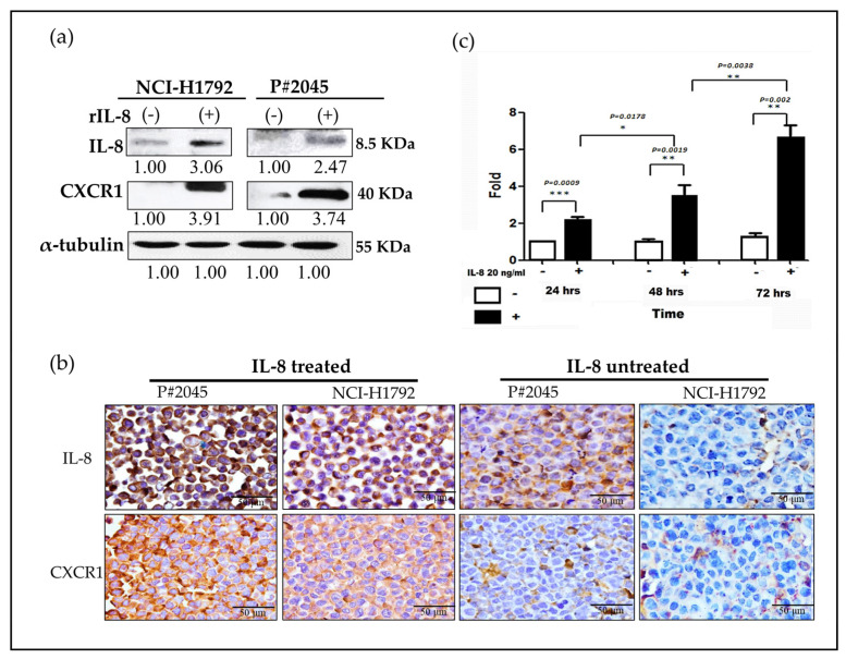
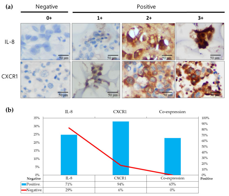
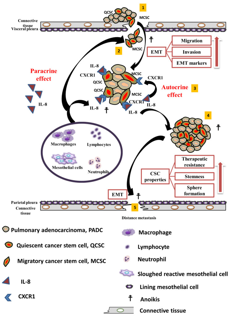
Pulmonary adenocarcinoma (PADC) treatment limited efficacy in preventing tumor progression, often resulting in malignant pleural effusion (MPE). MPE is filled with various mediators, especially interleukin-8 (IL-8). However, the role of IL-8 and its signaling mechanism within the fluid microenvironment (FME) implicated in tumor progression warrants further investigation. Primary cultured cells from samples of patients with MPE from PADC, along with a commonly utilized lung cancer cell line, were employed to examine the role of IL-8 and its receptor, CXCR1, through comparative analysis. Our study primarily assessed migration and invasion capabilities, epithelial-mesenchymal transition (EMT), and cancer stem cell (CSC) properties. Additionally, IL-8 levels in MPE fluid versus serum, along with immunohistochemical expression of IL-8/CXCR1 signaling in tumor tissue and cell blocks were analyzed. IL-8/CXCR1 overexpression enhanced EMT and CSC properties. Furthermore, the immunocytochemical examination of 17 cell blocks from patients with PADC and MPE corroborated the significant correlation between upregulated IL-8 and CXCR1 expression and the co-expression of IL-8 and CXCR1 in MPE with distant metastasis. In summary, the IL-8/ CXCR1 axis in FME is pivotal to tumor promotion via paracrine and autocrine signaling. Our study provides a therapeutic avenue for improving the prognosis of PADC patients with MPE.

Keywords: CXCR1; cancer stem cell; epithelial-mesenchymal transition; fluid microenvironment; interleukin-8; interleukin-8/CXCR1 signaling.


Conflict of interest statement

The authors declare no conflicts of interest.

Figures

Figure 1
Figure 1
Comparative analysis of IL-8 levels in pleural effusion and serum, with further comparisons of IL-8 in two representative cell lines. (a) High levels of IL-8 were observed in PADC with MPE which were statistically higher than those in SDPE (*** p < 0.001, Student’s t-test) (Experiments were conducted in triplicate). (b) Similar results were found with higher IL-8 levels in the serum of patients with PADC and MPE, compared to that in patients without MPE (** p < 0.01, Student’s t-test). (Experiments were conducted in triplicate). Interestingly, higher IL-8 levels were found in pleural effusion compared to serum. (c) Comparison of IL-8 levels in the culture media of P#2045 and NCI-1792 showed different levels, with a higher level of IL-8 noted in MPE (P#2045) by ELISA (Experiments were conducted in triplicate). (d) IL-8 protein was observed in both cell lines, with higher levels noted in MPE (P#2045) by Western blotting (Experiments were conducted in triplicate). (e) Stronger expression of IL-8 was found in MPE (P#2045) compared to PADC (NCI-1792) by ICC (arrows indicate positive IL-8 findings) (magnification, ×400, Scale bar: 50 μm).
Figure 2
Figure 2
Paracrine effect of IL-8 via the administration of rIL-8 enhanced migration and invasion activities, and increased EMT properties. (a) Migration assays showed that rIL-8 significantly enhanced migration properties in NCI-H1792 and P#2045 cell lines (** p < 0.01 and * p < 0.05, respectively, Student’s t-test) (Experiments were conducted in triplicate) (The red arrows indicate tumor cells). (b) Invasion assays revealed that rIL-8 significantly enhanced invasion properties in NCI-H1792 and P#2045 cell lines (* p < 0.05 and * p < 0.05, respectively, Student’s t-test) (Experiments were conducted in triplicate) (The red arrows indicate tumor cells). (c) Treatment with rIL-8 resulted in enhanced EMT properties in both cell lines, including decreased expression of E-cadherin, increased expression of N-cadherin, vimentin, and transcription factors Twist-1 and Snail (Experiments were conducted in triplicate). (d) The wound-healing assay experiment at different time points, 0, 12, and 24 h in both NCI-H1792 and P#2045 cell lines. The results indicate that compared to the cell line NCI-H1792, the distance between P#2045 cells decreased by 50% within 12 h, and by 24 h, the cells on both sides had already connected. This wound-healing assay resulted in the significant finding that P#2045 cells were easily migrated to the scratched center, indicating an increase of migratory activities compared to NCI-H1792 cells (magnification, ×400, Scale bar = 100 μm) (Experiments were conducted in triplicate). (e) In the wound-healing assay, after exposure to rIL8 for 12 and 24 h, cell migration in the NCI-H1792 cell line increased by 1.58-fold and 1.66-fold, respectively. In the P#2045 cell line, the migration increased by 1.55-fold at 12 h and 1.96-fold at 24 h. (f) In the wound-healing assay, the P#2045 cell line treated with rIL8, the increase in the invasive front was observed to be 3.2-fold at 12 h and 169.2-fold at 24 h (*** p < 0.001, Student’s t-test).
Figure 3
Figure 3
IL-8 promotion and maintenance of CSC properties: sphere formation, CSC markers, radioresistance, chemoresistance, and drug resistance. (a) rIL-8 enhanced sphere size and increased sphere number on different days (day 1, day 3, day 5, and day 9) in both NCI-H1792 and P#2045 (magnification, ×100). (b) Expression of CSC markers, including OCT4, Nanog, and CD133 in both NCI-H1792 and P#2045 was verified by immunofluorescence (magnification, ×200). (c) rIL-8 promoted the expression of CSC markers, including Oct4, Nanog, and CD133, in both NCI-H1792 and P#2045 by western blotting (Experiments were conducted in triplicate). (d) Both cell lines treated with rIL-8 showed a good survival rate in radiosensitivity assays (* p < 0.05, ANOVA) (Experiments were conducted in triplicate). (e) Both cell lines treated with rIL-8 also exhibited a good survival rate in chemosensitivity assays (* p < 0.05, ANOVA) (Experiments were conducted in triplicate). (f) Both cell lines treated with rIL-8 increased expression of ABCG2 and MDR1 (Experiments were conducted in triplicate).
Figure 4
Figure 4
Demonstration and verification of paracrine and autocrine effects of IL-8/CXCR1. (a) Two representative cell lines, NCI-H1792 and P#2045, were treated with exogenous IL-8 for 24 h. Subsequent analyses were conducted using western blot and ICC to assess the effects of the treatment. Increased CXCR1 expression in both cell lines (NCI-H1792 and P#2045) following the addition of rIL-8, as shown by western blotting (Experiments were conducted in triplicate). (b) A marked increase in the expression of CXCR1 upon the addition of rIL-8 in both cell lines (NCI-H1792 and P#2045), was demonstrated by ICC. (magnification, ×400, Scale bar: 50 μm). Enhanced IL-8 expression was also observed in both cells using ICC (magnification, ×400, Scale bar: 50 μm). However, stronger expressions of IL-8/CXCR1 were seen in P#2045 compared to NCI-H1792 due to the long-term influence of FME, implicating the importance of FME. (c) A time-dependent trend showing progressively increasing levels of IL-8 was observed in P#2045 cells, implying an autocrine mechanism, which demonstrated a systematic escalation in IL-8 concentration over the course of the experiment at 24, 48, and 72 h. (Experiments were conducted in triplicate) (* p < 0.05, ** p < 0.01, *** p < 0.001, respectively, Student’s t-test).
Figure 5
Figure 5
Validation of IL-8 and CXCR1 expression in MPE (a) Representative images of IL-8 and CXCR1 expression on cytology are shown, indicating absent (0), weak (1+), moderate (2+), and strong (3+) staining intensity (Scale bar: 50 μm). (b) The proportion of increased IL-8 expression in cell blocks of PADC with MPE was estimated to be 71%, whereas the corresponding CXCR1 expression in cell blocks of PADC with MPE was measured up to 94%. Moreover, the majority (11/17) of MPE cases showing co-expression of IL-8/CXCR1 accounted for 65% of the total.
Figure 6
Figure 6
Diagrammatic illustration demonstrates a model depicting the paracrine and autocrine effects of IL8/CXCR1 signaling in MPE, which were divided into five sequential steps in the metastatic cascade. (1) PADCs are usually found in the peripheral areas at the edges of each lobe. PADC cells, consisting of a small portion of subset CSCs within the tumor, display traits akin to inactive or dormant cancer stem cells (CSCs), known as “quiescent CSCs, (QCSC)” when along with signs of EMT, a few of them become as active form also known as migratory CSCs (MCSCs), which show more aggressive behavior. These cells actively move and penetrate the connective tissue and visceral pleura, eventually leading to the pleural space and causing MPE. (2) Isolated or small clusters of PADC cells in MPE sometimes display micropapillary patterns or become smaller spheroids. To avoid the threat of anoikis, not only managing survival but also showing continuous proliferation which is due to the unique FME, particularly enriched with interleukin-8 (IL-8) produced by inflammatory cells such as lymphocytes, neutrophils, macrophages, and possibly mesothelial cells via paracrine effects. (3) Following the paracrine effect of IL-8 in conjunction with the CXCR1 receptor near a specialized premetastatic area on either MCSC or QCSC cell surfaces, self-stimulation, and proliferation by the IL-8/CXCR1 axis maintains cancerous tissue and ensures tumor cell survival in an autocrine manner. (4) Then to preserve and increase CSC properties, tumor nests become larger, and round clusters proliferate rapidly in IL-8 nutrient-rich effusion, leading to the development of more spheroids with increased cancer stemness and therapeutic resistance to chemotherapy, radiation, and drugs. (5) Finally, specific cells within these spheroids are stimulated by environmental signals, revive EMT and CSC characteristics, and evolve into metastatic CSCs. They then invade the nearby parietal pleura with rich angiolymphatic vessels, paving the way for further spreading to distant organs.

Similar articles

Cited by

References

    1. Chen S.F., Lin Y.S., Jao S.W., Chang Y.C., Liu C.L., Lin Y.J., Nieh S. Pulmonary Adenocarcinoma in Malignant Pleural Effusion Enriches Cancer Stem Cell Properties during Metastatic Cascade. PLoS ONE. 2013;8:e54659. doi: 10.1371/journal.pone.0054659. - DOI - PMC - PubMed
    1. Fousek K., Horn L.A., Palena C. Interleukin-8: A chemokine at the intersection of cancer plasticity, angiogenesis, and immune suppression. Pharmacol. Ther. 2021;219:107692. doi: 10.1016/j.pharmthera.2020.107692. - DOI - PMC - PubMed
    1. Sainz B., Jr., Carron E., Vallespinos M., Machado H.L. Cancer Stem Cells and Macrophages: Implications in Tumor Biology and Therapeutic Strategies. Mediat. Inflamm. 2016;2016:9012369. doi: 10.1155/2016/9012369. - DOI - PMC - PubMed
    1. Waugh D.J., Wilson C. The interleukin-8 pathway in cancer. Clin. Cancer Res. 2008;14:6735–6741. doi: 10.1158/1078-0432.CCR-07-4843. - DOI - PubMed
    1. Nagarsheth N., Wicha M.S., Zou W. Chemokines in the cancer microenvironment and their relevance in cancer immunotherapy. Nat. Rev. Immunol. 2017;17:559–572. doi: 10.1038/nri.2017.49. - DOI - PMC - PubMed

Publication types

MeSH terms

LinkOut - more resources