. 2024 Jun 4;13(11):972.
doi: 10.3390/cells13110972.

A Novel CRISPR-Cas9 Strategy to Target DYSTROPHIN Mutations Downstream of Exon 44 in Patient-Specific DMD iPSCs

Affiliations

A Novel CRISPR-Cas9 Strategy to Target DYSTROPHIN Mutations Downstream of Exon 44 in Patient-Specific DMD iPSCs

Neha R Dhoke et al. Cells. .

Abstract

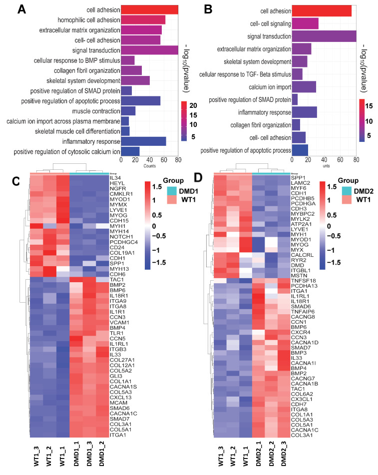
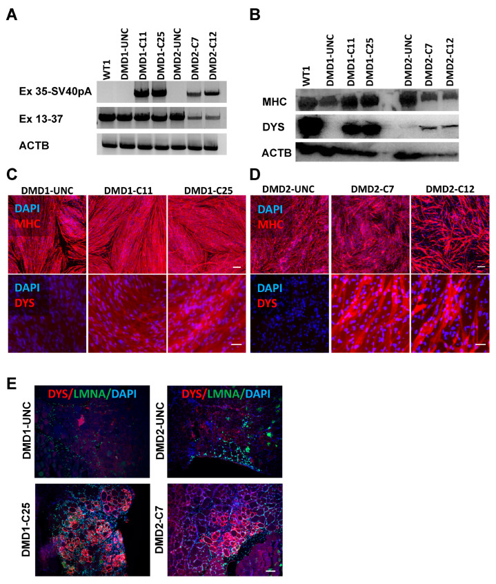
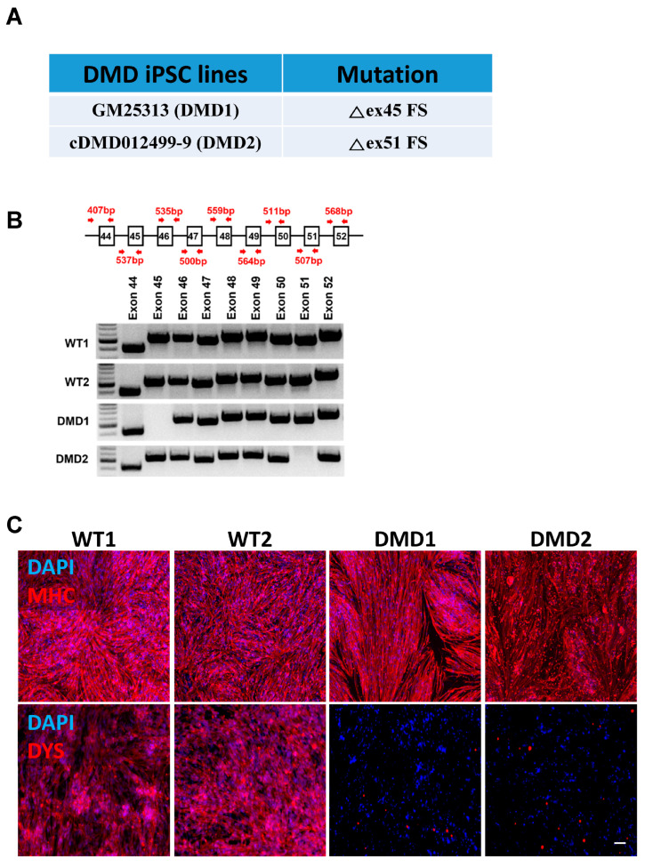
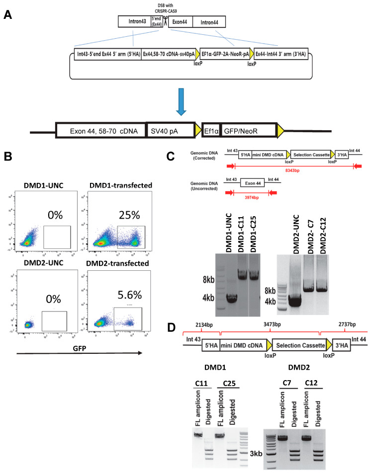
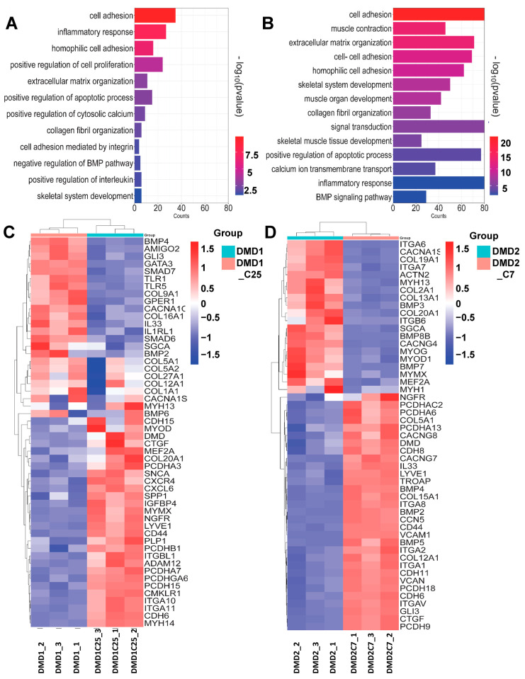
Mutations in the DMD gene cause fatal Duchenne Muscular Dystrophy (DMD). An attractive therapeutic approach is autologous cell transplantation utilizing myogenic progenitors derived from induced pluripotent stem cells (iPSCs). Given that a significant number of DMD mutations occur between exons 45 and 55, we developed a gene knock-in approach to correct any mutations downstream of exon 44. We applied this approach to two DMD patient-specific iPSC lines carrying mutations in exons 45 and 51 and confirmed mini-DYSTROPHIN (mini-DYS) protein expression in corrected myotubes by western blot and immunofluorescence staining. Transplantation of gene-edited DMD iPSC-derived myogenic progenitors into NSG/mdx4Cv mice produced donor-derived myofibers, as shown by the dual expression of human DYSTROPHIN and LAMIN A/C. These findings further provide proof-of-concept for the use of programmable nucleases for the development of autologous iPSC-based therapy for muscular dystrophies.

Keywords: CRISPR-Cas9; DMD; dystrophin; gene editing; patient-specific iPS cells; transplantation.


Conflict of interest statement

R.C.R.P. is cofounder and holds equity in Myogenica. All other authors have no competing financial interests.

Figures

Figure 2
Figure 2
Transcriptional profile of DMD iPSC-derived myotubes. (A,B) Functional classification based on gene ontology (GO) analysis using DAVID. Significantly enriched GO terms in DEGs from Biological Process in DMD1 and DMD2 iPSC-derived myotubes compared to WT counterparts, displayed according to −log(adj p-value). (C,D) Heatmaps indicate genes involved in enriched biological processes between WT1 vs. DMD1 (C) and WT1 vs. DMD2 (D) iPSC-derived myotubes.
Figure 4
Figure 4
Detection of DYSTROPHIN expression in vitro and in vivo. (A) RT-PCR analysis of corrected and uncorrected DMD1 iPSC-derived myotubes. Top panel shows the amplification of DMD exon 35 to SV40pA sequences where the amplicons are specific to gene-corrected DMD1 myotubes. Middle panel shows the amplification of DMD exon 13 to 37 sequences, where the amplicons are present in all samples, as expected. Lower panel shows the ACTB amplification that serves as loading control. (B) Western blot shows the rescue of mini-DYS expression, with a molecular weight of 314.42 kDa, in clones of gene-corrected DMD1 (C11 and C25) and DMD2 (C7 and C12) iPSC-derived myotubes, respectively. Uncorrected DMD1 and DMD2 myotubes show the absence of DYS expression. Control WT1 iPSC-derived myotubes show full-length dystrophin with a molecular weight of 427 kDa. ACTB (43 kDa) and MCH (223 kDa) served as loading and differentiation controls, respectively. (C,D) Representative images show immunostaining for MHC (upper) and DYS (lower) in myotubes derived from gene-corrected and uncorrected counterparts. DAPI stains nuclei (in blue). Scale bar, 100 μm. (E) Representative images show the engraftment of gene-corrected DMD1 (C25) and DMD2 (C7) iPSC-derived myogenic progenitors following their transplantation into TA muscles of mdx-NSG mice. Myogenic progenitors from uncorrected iPSCs served as negative controls. Panel shows immunostaining for human DYS (in red) and human LMNA (in green). DAPI stains nuclei (in blue). Scale bar, 100 µm.
Figure 1
Figure 1
Characterization of patient-specific DMD iPSCs. (A) Information of patient-specific DMD iPSC lines. (B) Validation of patient-specific mutations by PCR amplifications for each exon. DMD1 is absent of exon 45 while DMD2 is absent of exon 51. (C) Representative images show immunostaining for MHC (upper panel) and DYSTROPHIN (DYS, lower panel) in WT and DMD iPSC-derived myotubes. DYSTROPHIN is absent in DMD samples. DAPI stains nuclei (in blue). Scale bar, 100 μm.
Figure 3
Figure 3
Gene correction of patient-specific DMD iPSCs. (A) Scheme outlining the gene editing strategy for correction of DMD mutations through HDR-based gene knock-in. (B) FACS plots show GFP expression in gene-edited (bulk) DMD iPSCs. WT iPSCs served as negative control. (C) PCR shows validation of mini-DYS cDNA knock-in. Gene-edited DMD iPSC clones show amplicons of approximately 8.3 kb while uncorrected iPSC counterparts display 4 kb amplicons. Gel images are cropped only to show relevant lanes. (D) Digestion analysis of PCR amplicons shows the expected sizes of bands of 2.1 kb, 2.7 kb, and 3.5 kb.
Figure 5
Figure 5
Transcriptomic profile of DMD iPSC patient-derived myotubes and their corrected counterparts. (A,B) Functional classification based on gene ontology (GO) analysis using DAVID. Significantly enriched GO terms from biological process in DMD1 vs. DMD1-C25 and DMD 2 vs. DMD2-C7 mutant lines compared to the WT iPSC derived myotubes displayed according to −log(adj p-value). (C,D) Heat map showing genes enriched in biological process between uncorrected DMD1 iPSC-derived myotubes and its corrected counterpart DMD1-C25 iPSC-derived myotubes as well as DMD 2 iPSC-derived myotubes and its corrected counterpart DMD2-C7, respectively.

Similar articles

Cited by

References

    1. Olson E.N. Toward the correction of muscular dystrophy by gene editing. Proc. Natl. Acad. Sci. USA. 2021;118:e2004840117. doi: 10.1073/pnas.2004840117. - DOI - PMC - PubMed
    1. Schultz T.I., Raucci F.J., Jr., Salloum F.N. Cardiovascular Disease in Duchenne Muscular Dystrophy: Overview and Insight into Novel Therapeutic Targets. JACC Basic Transl. Sci. 2022;7:608–625. doi: 10.1016/j.jacbts.2021.11.004. - DOI - PMC - PubMed
    1. Duan D., Goemans N., Takeda S.I., Mercuri E., Aartsma-Rus A. Duchenne muscular dystrophy. Nat. Rev. Dis. Primers. 2021;7:13. doi: 10.1038/s41572-021-00248-3. - DOI - PMC - PubMed
    1. Gao Q.Q., McNally E.M. The Dystrophin Complex: Structure, Function, and Implications for Therapy. Compr. Physiol. 2015;5:1223–1239. doi: 10.1002/cphy.c140048. - DOI - PMC - PubMed
    1. Mias-Lucquin D., Dos Santos Morais R., Chéron A., Lagarrigue M., Winder S.J., Chenuel T., Pérez J., Appavou M.-S., Martel A., Alviset G., et al. How the central domain of dystrophin acts to bridge F-actin to sarcolemmal lipids. J. Struct. Biol. 2020;209:107411. doi: 10.1016/j.jsb.2019.107411. - DOI - PubMed

Publication types

LinkOut - more resources