. 2024 Jun 5;13(11):981.
doi: 10.3390/cells13110981.

Conditional Knockout of Neurexins Alters the Contribution of Calcium Channel Subtypes to Presynaptic Ca2+ Influx

Affiliations

Conditional Knockout of Neurexins Alters the Contribution of Calcium Channel Subtypes to Presynaptic Ca2+ Influx

Johannes Brockhaus et al. Cells. .

Abstract

Presynaptic Ca2+ influx through voltage-gated Ca2+ channels (VGCCs) is a key signal for synaptic vesicle release. Synaptic neurexins can partially determine the strength of transmission by regulating VGCCs. However, it is unknown whether neurexins modulate Ca2+ influx via all VGCC subtypes similarly. Here, we performed live cell imaging of synaptic boutons from primary hippocampal neurons with a Ca2+ indicator. We used the expression of inactive and active Cre recombinase to compare control to conditional knockout neurons lacking either all or selected neurexin variants. We found that reduced total presynaptic Ca2+ transients caused by the deletion of all neurexins were primarily due to the reduced contribution of P/Q-type VGCCs. The deletion of neurexin1α alone also reduced the total presynaptic Ca2+ influx but increased Ca2+ influx via N-type VGCCs. Moreover, we tested whether the decrease in Ca2+ influx induced by activation of cannabinoid receptor 1 (CB1-receptor) is modulated by neurexins. Unlike earlier observations emphasizing a role for β-neurexins, we found that the decrease in presynaptic Ca2+ transients induced by CB1-receptor activation depended more strongly on the presence of α-neurexins in hippocampal neurons. Together, our results suggest that neurexins have unique roles in the modulation of presynaptic Ca2+ influx through VGCC subtypes and that different neurexin variants may affect specific VGCCs.

Keywords: calcium channel subtypes; endocannabinoid system; genetically encoded calcium indicator; neurexin; presynapse.


Conflict of interest statement

The authors declare no conflicts of interest.

Figures

Figure 2
Figure 2
Presynaptic Ca2+ transients recorded from individual active boutons with synGCaMP7b. (A) Example picture of fluorescence intensity of synGCaMP7b before stimulation (left, F0, shown in magenta), representing the baseline fluorescence; fluorescence intensity changes after stimulation with 3 APs, isolated by subtraction (middle, ΔF, shown in green). The green fluorescence dots lighting up indicate active boutons. Both images merged represent the effect image (right) that allows the identification of active boutons that are not disturbed by high baseline fluorescence of other sources like Cre-EGFP-fluorescent cell nuclei (asterisk). (B) Enlarged perspective (yellow box in A), showing the change in fluorescence (ΔF, green) as well as the cell process morphology indicated by co-transfected RFP (red). (C) ROIs (red circles) were placed on active boutons for the quantification of presynaptic Ca2+ transients. (D) Averaged synGCaMP7b fluorescence changes from Nx123 cKO neurons with Nxs (Cremut, n = 14 cells/1045 boutons) or without all Nx variants (dashed line, Cre, 13/916) show Ca2+ transients following a single-AP stimulation. (E) Neurons lacking only Nx1α (Cre, 8/681) and equivalent controls (Cremut, 12/1074) showed comparable fluorescence alterations as those seen in N123 cKO. (F) Comparing peak values of Ca2+ transients (mean ± SEM) in Cremut and Cre cells from both mouse lines in response to a single-AP stimulation. Nx123 cKO: Cremut 48 cells/3964 boutons, Cre 58/4582; Nx1α cKO: Cremut 21/1672, Cre 25/2051. The mean values of all boutons of a single cell are shown as dots and used for statistics. Columns were compared with an unpaired t-test. * p < 0.05, **** p < 0.0001.
Figure 1
Figure 1
Conditional deletion of the single Nx1α variant. (A) Immunoblots of Nxs in Nx123 cKO cells tested with 3 different antibodies: pan-Nx1 (A1, Millipore #AB161-I), Nx1α (A2, Frontier Institute #AB_2571817), and Nx123 (A3, SySy #175003). Con, mouse line without floxed α-Nxs. (B) Wild-type allele of the 5′ end of the Nx1α gene including the first coding exon (indicated in red) is illustrated. After successful homologous recombination of the wild-type allele with the targeting vector (not depicted), the knock-in allele that resulted is indicated. The 5′ loxP site is introduced via the BamH1 (‘B’) site upstream of the first coding exon. Downstream of the first coding exon and at the EcoR1 site (‘E’), the 3′ loxP site and NeoR (Neomycin resistance) gene are inserted (blunt-end cloning). Via the addition of a Cre-recombinase, the knock-in allele is converted into the knockout allele. The region between the loxP sites of the Nx1α gene including the first coding exon is excised. Further restriction sites: S = Spel, N = Nhel. (C) Immunoblots of control neurons (C1) without floxed α-Nx and Nx1α cKO neurons (C2) were probed with anti-Nx123 (SySy #175003). (D) Quantification of αNx normalized to ΔCre condition (100%) for control neurons, Nx123 cKO neurons, and Nx1α cKO neurons. Data are based on n independent immunoblot experiments like in A3 and C (control: 3, 2; Nx123: 4, 4; Nx1α: 3, 3); columns were compared with an unpaired t-test. n.s. = non-significant: p > 0.05, ** p < 0.01, *** p < 0.001.
Figure 3
Figure 3
Deletion of all Nx variants decreased presynaptic Ca2+ influx primarily via P/Q-type VGCCs. Pharmacologically isolated VGCC subtype contribution to the Ca2+ influx measured during single-AP stimulation in Nx123 cKO neurons with synGCaMP7b by sequential addition of specific blockers: ω-agatoxin IVA (AgTX, 0.1 μM; P/Q-type); ω-conotoxin GVIA (CTX, 2 μM; N-type); nifedipine (Nif, 20 μM; L-type); SNX-482 (SNX, 0.5 μM; R-type). (A) Averaged traces of control neurons (Cremut, 12 cells/869 boutons, left) and neurons lacking all neurexin variants (Cre, 13/916, right); colors indicate traces after subsequent application of subtype-specific blockers as depicted. Thus, the area in dimmed colors above the traces indicates the amount of Ca2+ influx sensitive to the given blocker. (B) Ca2+ transients that reflect Ca2+ influx through the given VGCC subtypes are isolated by subtraction from the traces in A, comparing Nx123 cKO Cremut (continuous lines) and Nx123 cKO Cre (dashed lines). (C) Mean ± SEM of relative VGCC subtype contribution (% of control) calculated for each bouton (ROI, relative to total Ca2+ influx) in Nx123 cKO neurons. The number of examined boutons/cells is shown in the P/Q columns and applies to all VGCC subtypes. Columns were compared with Kruskal-Wallis test, n.s.: p > 0.05, **: p < 0.01, ****: p < 0.0001. (D) Each presynaptic bouton’s P/Q-type contribution was determined, and the spreading is depicted in a histogram that contrasts the distribution of Nx123 cKO neurons with and without Nxs. The data show that without Nxs, the number of boutons with more than 50% P/Q-type Ca2+ influx is almost lost (Cremut, 584/12; Cre, 501/13). The same analysis is shown in (E) for the N-type and (F) for the L-type portion in individual boutons.
Figure 4
Figure 4
Single Nx1α deletion changed the VGCC subtype contribution to presynaptic Ca2+ influx. VGCC subtype contribution to Ca2+ influx after single-AP stimulation was measured in Nx1α cKO neurons with synGCaMP7b by sequential addition of specific blockers as described in Figure 3. (A) Averaged traces of control neurons (Nx1α cKO Cremut, 13 cells/1154 boutons; left) and neurons lacking only Nx1α (Cre, 11/956, right); colors indicate traces after subsequent application of subtype-specific blockers as depicted. Thus, the area in dimmed colors above the traces indicates the amount of Ca2+ influx sensitive to the given blocker. (B) Ca2+ transients that specifically reflect Ca2+ influx through the sequentially blocked VGCC subtypes were isolated by subtraction from the traces in A and compared between Nx1α cKO Cremut (continuous lines) and Nx1α cKO Cre (dashed lines). (C) Mean ± SEM of relative Ca2+ contribution (% of control) per VGCC subtype calculated for individual boutons (ROIs) in Nx1α cKO neurons. The number of examined boutons/cells is shown in the P/Q columns and applies to all VGCC subtypes. Columns were compared with Kruskal-Wallis test, n.s.: p > 0.05, ****: p < 0.0001. (D) The P/Q-type portion of Ca2+ transients was calculated for each synaptic bouton, and the spreading is shown in a histogram comparing the variability in neurons with and without Nx1α (Cremut, 755/13; Cre, 552/11), in (E) for the N-type and (F) for the L-type.
Figure 5
Figure 5
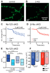
Endocannabinoid-evoked CB1-receptor activation reduces presynaptic Ca2+ transients in an Nx-dependent manner. (A) Several presynaptic boutons of a synGCaMP7b-transfected Nx123 cKO Cre neuron are shown in an exemplary ΔF image during 3-AP stimulation. (B) The identical presynaptic boutons to those in A after 5 min of CB1-receptor activation with 2 µM 2-AG, again during a 3-AP stimulation. (C) Repetitive stimulation (1 AP every 30 s) shows a reduction in Ca2+ transients in response to the application of 2-AG, averaged (mean ± SEM) from neurons of Nx123 Cremut (13 neurons, continuous line) and Nx123 Cre (16, dotted line), displayed as relative changes normalized to the mean of four stimulations before 2-AG application. (D) Similar recordings as in C for β-Nx cKO Cremut (10, continuous line) and β-Nx cKO Cre (9, dotted line). (E) Boxplot (quartiles and median) of Ca2+ ΔF/F0 for Nx123 Cremut (960 ROIs/13 cells) and Nx123 Cre (1168/16) before and after 2-AG application. (F) Relative change (%) in presynaptic Ca2+ transients by activation of CB1-receptor with 2-AG. Cells were measured under both conditions (control and 5 min of 2-AG), and reduction was calculated for each bouton separately, plotted as mean ± SEM in Nx123 cKO (blue) and β-Nx cKO (red) neurons (Cremut and Cre). Outliers were detected and removed with the ROUT method (Q = 1), and columns were compared with an unpaired t-test; * p < 0.05; **** p < 0.0001; numbers (included ROIs/cells) are given in the columns.

References

    1. Catterall W.A. Structure and Regulation of Voltage-Gated Ca2+-Channels. Annu. Rev. Cell Dev. Biol. 2000;16:521–555. doi: 10.1146/annurev.cellbio.16.1.521. - DOI - PubMed
    1. Dolphin A.C., Lee A. Presynaptic calcium channels: Specialized control of synaptic neurotransmitter release. Nat. Rev. Neurosci. 2020;21:213–229. doi: 10.1038/s41583-020-0278-2. - DOI - PMC - PubMed
    1. Sheng J., He L., Zheng H., Xue L., Luo F., Shin W., Sun T., Kuner T., Yue D.T., Wu L.G. Calcium-channel number critically influences synaptic strength and plasticity at the active zone. Nat. Neurosci. 2012;15:998–1006. doi: 10.1038/nn.3129. - DOI - PMC - PubMed
    1. Nakamura Y., Harada H., Kamasawa N., Matsui K., Rothman J.S., Shigemoto R., Silver R.A., DiGregorio D.A., Takahashi T. Nanoscale distribution of presynaptic Ca2+ channels and its impact on vesicular release during development. Neuron. 2015;85:145–158. doi: 10.1016/j.neuron.2014.11.019. - DOI - PMC - PubMed
    1. Li L., Bischofberger J., Jonas P. Differential gating and recruitment of P/Q-, N-, and R-type Ca2+ channels in hippocampal mossy fiber boutons. J. Neurosci. 2007;27:13420–13429. doi: 10.1523/JNEUROSCI.1709-07.2007. - DOI - PMC - PubMed

Publication types

LinkOut - more resources