. 2024 Jun 6;13(11):988.
doi: 10.3390/cells13110988.

Mecp2 Deficiency in Peripheral Sensory Neuron Improves Cognitive Function by Enhancing Hippocampal Dendritic Spine Densities in Mice

Affiliations

Mecp2 Deficiency in Peripheral Sensory Neuron Improves Cognitive Function by Enhancing Hippocampal Dendritic Spine Densities in Mice

Yuting Feng et al. Cells. .

Abstract

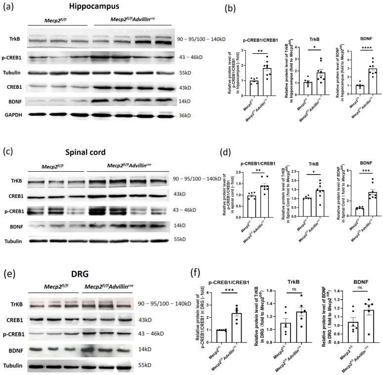
Methyl-CpG-binding protein 2 (Mecp2) is an epigenetic modulator and numerous studies have explored its impact on the central nervous system manifestations. However, little attention has been given to its potential contributions to the peripheral nervous system (PNS). To investigate the regulation of Mecp2 in the PNS on specific central regions, we generated Mecp2fl/flAdvillincre mice with the sensory-neuron-specific deletion of the Mecp2 gene and found the mutant mice had a heightened sensitivity to temperature, which, however, did not affect the sense of motion, social behaviors, and anxiety-like behavior. Notably, in comparison to Mecp2fl/fl mice, Mecp2fl/flAdvillincre mice exhibited improved learning and memory abilities. The levels of hippocampal synaptophysin and PSD95 proteins were higher in Mecp2fl/flAdvillincre mice than in Mecp2fl/fl mice. Golgi staining revealed a significant increase in total spine density, and dendritic arborization in the hippocampal pyramidal neurons of Mecp2fl/flAdvillincre mice compared to Mecp2fl/fl mice. In addition, the activation of the BDNF-TrkB-CREB1 pathway was observed in the hippocampus and spinal cord of Mecp2fl/flAdvillincre mice. Intriguingly, the hippocampal BDNF/CREB1 signaling pathway in mutant mice was initiated within 5 days after birth. Our findings suggest a potential therapeutic strategy targeting the BDNF-TrkB-CREB1 signaling pathway and peripheral somasensory neurons to treat learning and cognitive deficits associated with Mecp2 disorders.

Keywords: BDNF/CREB1 pathway; cognitive function; methyl-CpG-binding protein 2; peripheral sensory neuron.


Conflict of interest statement

The authors have no relevant financial or non-financial interests to disclose. The authors have no conflicts of interest to declare that are relevant to the content of this article. All authors certify that they have no affiliations with or involvement in any organization or entity with any financial interest or non-financial interest in the subject matter or materials discussed in this manuscript. The authors have no financial or proprietary interests in any material discussed in this article.

Figures

Figure 1
Figure 1
Generation of Mecp2fl/fl Advillincre mice. Female Mecp2 floxed mice were bred with male Advillin-Cre mice to generate Mecp2fl/flAdvillincre mice. (a) Analysis of the PCR products of tail DNA from pups that were produced from crossings between Mecp2fl/flAdvillincre males and females carrying both Mecp2 floxed and Advillin-Cre transgene by agarose gel electrophoresis. Mu: mutant (Mecp2 280 bp, Advillincre 150 bp); WT: wild-type (Mecp2 180 bp, Advillincre 530 bp); NC: negative control; He: heterozygous; M: DNA ladder DL2000; (b) western blot analysis of protein samples prepared from DRG and hippocampus of adult Mecp2fl/fl and Mecp2fl/flAdvillincre mice. Mecp2fl/fl n = 3, Mecp2fl/flAdvillincre n = 6. (c) Mecp2 immunohistochemical analysis of DRG sections from adult Mecp2fl/fl and Mecp2fl/flAdvillincre mice. The brownish yellow particles represent positive protein expression. Scale bar: 200 µm. AOD: Average optical density. **** p < 0.0001.
Figure 2
Figure 2
Mecp2 deficiency in sensory neuron leads to increased sensitivity to temperature and pain. Somatic sensations of Mecp2 fl/fl and Mecp2fl/fl Advillincre mice were measured by hot-plate test (a) and cold tail-flick latency reflex test (b). The sense of motion and position were measured by pole-climbing test (c), and forelimb and hindlimb grip strength (d,e). Mecp2fl/fl n = 10, Mecp2fl/flAdvillincre n = 11. ** p < 0.01.
Figure 3
Figure 3
Mecp2 in peripheral sensory neuron is not associated with compulsive-like behaviors and sociability of mice. The compulsive-like behaviors of Mecp2fl/fl and Mecp2fl/flAdvillincre mice were measured by marble-burying test (a) and nestlet-shredding test (b). The sociability (c) and social novelty preference (d) of Mecp2fl/fl and Mecp2fl/fl Advillincre mice were evaluated by 3-chamber social interaction assay. Mecp2fl/fl n = 10, Mecp2fl/flAdvillincre n = 11, CSDS n = 9. * p < 0.05, ** p < 0.01, *** p < 0.001.
Figure 4
Figure 4
Mecp2 in peripheral sensory neuron did not affect the anxiety-like behavior of mice. In open field test, the time spent in the central square (a) and the distance traveled in the arena (b) were measured. In elevated plus maze, the time of mice spent in the open (c) and closed (d) arms were recorded, and their ratio was calculated (e). Mecp2fl/fl n = 10, Mecp2fl/fl Advillincre n = 15.
Figure 5
Figure 5
Peripheral sensory neuron deletion of Mecp2 led to improved cognitive function in mice. Spatial learning and memory of mice were assessed by Morris water maze and object recognition test. During the training days of Morris water maze, the time it took each mouse to reach the platform (latency) was recorded (a). Twenty-four hours after the last training day, the probe trials of Morris water maze were performed. The activity traces (b), the frequency of the platform entries (c), the distance spent in the target quadrant (d), and the mean speed in the pool (e) were recorded. In object recognition test, during 5 min, the exploration time of one of the objects and the exploration time of the novel object were recorded and the discrimination index of objects was calculated (f). Mecp2fl/fl n = 10, Mecp2fl/flAdvillincre n = 15. The inverse relationship between the exploration time of the novel object and the tail-flick latency (g) or paw-lick latency (h) were analyzed including Mecp2fl/fl and Mecp2fl/flAdvillincre mice. Mecp2fl/fl n = 10, Mecp2fl/flAdvillincre n = 11. ns: no significance, * p < 0.05, ** p < 0.01.
Figure 6
Figure 6
Peripheral sensory neuron deletion of Mecp2 induced modification of synaptic phenotypes in hippocampus. Mice brains were staining by HE and toluidine blue. Images were captured under a light microscope. Scale bar: 600 µm (HE), 2 mm, and 60 µm (Nissl) (a). The hippocampal cell count of three mice in each group were analyzed. For each mouse, three microscope fields were randomly selected to calculate the average number of cells per square millimeter in each area of the hippocampus, including CA1, CA2, CA3, and DG (b). Golgi staining was performed to measure the spine density of hippocampal pyramidal neurons (c) and the dendritic arborization (d) in mice. Protein levels of PSD95 and synaptophysin were determined by western blot (e). The protein levels of target genes were normalized to β actin. The values for Mecp2fl/fl were set at 100% as control. Mecp2fl/fl n = 4, Mecp2fl/flAdvillincre n = 6. ns: no significance, * p < 0.05, *** p < 0.001, **** p < 0.0001.
Figure 7
Figure 7
Peripheral sensory neuron deletion of Mecp2 activates BDNF-TrkB-CREB1 signaling pathway. Protein levels of BDNF, p-CREB1, CREB1, and TrkB in hippocampus (a), spinal cord (c), and DRG (e) were determined by western blot. The semi-quantitative protein levels of target genes were normalized to Tubulin or GAPDH (b,d,f). Mecp2fl/fl n = 6, Mecp2fl/flAdvillincre n = 8. p value was determined by t-test. ns: no significance, * p < 0.05, ** p < 0.01, *** p < 0.001, **** p < 0.0001.
Figure 8
Figure 8
The BDNF-TrkB-CREB1 signaling pathway was activated in forebrain of Mecp2fl/flAdvillincre mice after birth. Protein levels of BDNF, p-CREB1, CREB1, and TrkB in forebrain (a) were determined by western blot. The semi-quantitative protein levels of target genes were normalized to Tubulin (b). Mecp2fl/fl n = 6, Mecp2fl/flAdvillincre n = 8. p value was determined by two-way ANOVA. * p < 0.05, ** p < 0.01, *** p < 0.001, **** p < 0.0001.

Similar articles

References

    1. Mattei A.L., Bailly N., Meissner A. DNA methylation: A historical perspective. Trends Genet. 2022;38:676–707. doi: 10.1016/j.tig.2022.03.010. - DOI - PubMed
    1. Ibrahim A., Papin C., Mohideen-Abdul K., Le Gras S., Stoll I., Bronner C., Dimitrov S., Klaholz B.P., Hamiche A. MeCP2 is a microsatellite binding protein that protects CA repeats from nucleosome invasion. Science. 2021;372:eabd5581. doi: 10.1126/science.abd5581. - DOI - PubMed
    1. Wang J., Xiao Y., Liu C., Huang Y., Petersen R.B., Zheng L., Huang K. Emerging physiological and pathological roles of MeCP2 in non-neurological systems. Arch. Biochem. Biophys. 2021;700:108768. doi: 10.1016/j.abb.2021.108768. - DOI - PubMed
    1. Ramocki M.B., Tavyev Y.J., Peters S.U. The MECP2 duplication syndrome. Am. J. Med. Genet. A. 2010;152A:1079–1088. doi: 10.1002/ajmg.a.33184. - DOI - PMC - PubMed
    1. Ip J.P.K., Mellios N., Sur M. Rett syndrome: Insights into genetic, molecular and circuit mechanisms. Nat. Rev. Neurosci. 2018;19:368–382. doi: 10.1038/s41583-018-0006-3. - DOI - PMC - PubMed

Publication types

MeSH terms

Substances