. 2024 Jun 1;13(11):1536.
doi: 10.3390/plants13111536.

UV-B Radiation Disrupts Membrane Lipid Organization and Suppresses Protein Mobility of GmNARK in Arabidopsis

Affiliations

UV-B Radiation Disrupts Membrane Lipid Organization and Suppresses Protein Mobility of GmNARK in Arabidopsis

Qiulin Liu et al. Plants (Basel). .

Abstract

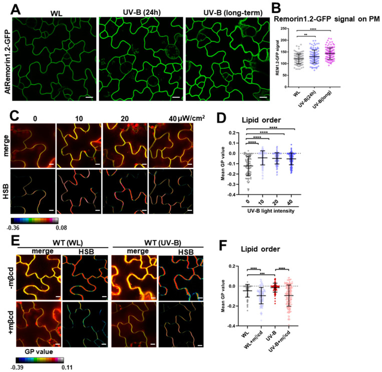
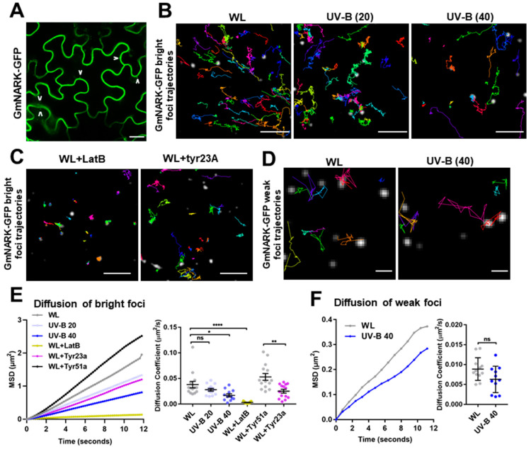
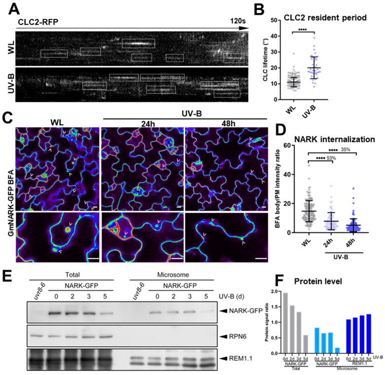
While it is well known that plants interpret UV-B as an environmental cue and a potential stressor influencing their growth and development, the specific effects of UV-B-induced oxidative stress on the dynamics of membrane lipids and proteins remain underexplored. Here, we demonstrate that UV-B exposure notably increases the formation of ordered lipid domains on the plasma membrane (PM) and significantly alters the behavior of the Glycine max nodule autoregulation receptor kinase (GmNARK) protein in Arabidopsis leaves. The GmNARK protein was located on the PM and accumulated as small particles in the cytoplasm. We found that UV-B irradiation interrupted the lateral diffusion of GmNARK proteins on the PM. Furthermore, UV-B light decreases the efficiency of surface molecule internalization by clathrin-mediated endocytosis (CME). In brief, UV-B irradiation increased the proportion of the ordered lipid phase and disrupted clathrin-dependent endocytosis; thus, the endocytic trafficking and lateral mobility of GmNARK protein on the plasma membrane are crucial for nodule formation tuning. Our results revealed a novel role of low-intensity UV-B stress in altering the organization of the plasma membrane and the dynamics of membrane-associated proteins.

Keywords: GmNARK; UV-B; endocytosis; lipid organization.


Conflict of interest statement

The authors declare no conflicts of interest.

Figures

Figure 1
Figure 1
UV-B light increases the proportion of ordered lipid phase and disrupts clathrin-mediated endocytosis. (A,B) WL-grown pAtRemorin1.2:AtRemorin1.2-GFP (in Arabidopsis) seedlings were exposed to UV-B light (40 μW/cm2) for 24 h or in long-term (4 d) treatment or continuously grown under WL. The Remorin1.2-GFP fluorescence signal on the PM was calculated (from left to right: n = 118, 128, and 123 cells (B)). (CF) PM lipid order was visualized in Arabidopsis leaf epidermal cells in a series of UV-B treatments (C,D). The seedlings were treated with WL or UV-B (40 μW/cm2, 24 h) in the absence or presence of 10 mM of mβcd (24 h) (E,F). Then, the treated seedlings were stained with di-4-ANEPPDHQ. Radiometric color-coded GP images were generated in HSB pictures (C,E), and mean GP value was calculated (from left to right: n = 88, 147, 165, and 156 images (D); n = 85, 110, 88, and 148 images (F)). Scale bars, 10 μm (A,C,E). Error bar = S.D. p-values were determined using two-tailed Student’s t-test, assuming equal variances (** p < 0.01; *** p < 0.001; **** p < 0.0001).
Figure 2
Figure 2
UV-B disrupts the internalization and lateral diffusion of GmNARK proteins. (A) GmNARK-GFP distribution of WL-grown 35s:GmNARK-GFP (in Arabidopsis) was visualized. The internalized GmNARK-GFP proteins within the cytosol are highlighted by arrows. (BF) GmNARK-GFP foci on the PM were recorded using VA-TIRFM in 5-day-old 35s:GmNARK-GFP (in Arabidopsis) seedlings under WL, UV-B light (20 or 40 μW/cm2 for 48 h), WL supplemented with LatB (10 μM, 1 h), or WL supplemented with Tyr23A/51A (30 μM, 1 h). Cotyledon epidermal cells were observed. Trajectories of GmNARK-GFP bright foci (B,C,E) and weak foci (D,F) were individually tracked. Mean-squared displacement (MSD) and diffusion coefficient of GmNARK-GFP particle are plotted and quantified in (E) (left panel: n = 60 images; right panel: n = 14, 14, 12, 11, 14, and 14 images), (F) (left panel: n = 15 images; right panel: n = 12 and 11 images). Scale bars, 10 µm (A), 5 µm (B,C), and 1 µm (D). Error bar = S.D. p-values were determined using two-tailed Student’s t-test, assuming equal variances (* p < 0.05; ** p < 0.01; **** p < 0.0001; ns, not significant).
Figure 3
Figure 3
UV-B impairs mobility of GmNARK proteins and attenuation of GmNARK protein levels. (A,B) CLC2-RFP lifetime on PM was quantitatively measured in WT Arabidopsis leaf epidermal cells. 5-day-old WT seedlings were treated with WL or UV-B (40 μW/cm2) for 48 h before being imaged using VA-TIRFM. The kymographs of the 120 s time course showed the residence of CLC2-RFP particles on the PM, indicated by white boxes (A). Quantitative analyses of CLC2-RFP particle lifetimes are shown (B: n = 241 and 39 images for each treatment). (C,D) UV-B-treated (40 μW/cm2, 24 h or 48 h pretreatment) and WL-grown 5-day-old 35s:GmNARK-GFP (in Arabidopsis) specimens were incubated in 200 µM of BFA for 2 h. Internalization signal of GmNARK-GFP-termed BFA body was quantified as a ratio comparing each GmNARK-GFP signal on the PM (D: n = 142, 74, and 109 cells from left to right). The comparison ratio of the internalization signal under UV-B relative to WL is shown as percentage (D). (E,F) Four-day-old 35s:GmNARK-GFP Arabidopsis seedlings were subjected to UV-B-treatment (10 μW/cm−2, 0, 2 d, 3 d, or 5 d pretreatment). Total GmNARK-GFP protein and AtRemorin1.1 levels and microsome protein were tested using Western blot. Proteins were analyzed via immunoblotting with anti-GFP, anti-Remorin1.1, and anti-RPN6 antibodies. RPN6 was used as a loading control. uvr8-6 is Atuvr8 mutant Arabidopsis (E). Quantification of GmNARK-GFP and REM1.1 protein levels was conducted. Protein signal ratios were used to indicate the signal strength of GmNARK-GFP and REM1.1 relative to each RPN6 signal on the protein blot, measured in arbitrary units via densitometry (F). Scale bars, 10 µm (C); error bar = S.D. p-values were determined using two-tailed Student’s t-test assuming equal variances; **** p < 0.0001).

Similar articles

References

    1. Madronich S., McKenzie R.L., Björn L.O., Caldwell M.M. Changes in biologically active ultraviolet radiation reaching the Earth’s surface. J. Photochem. Photobiol. B. 1998;46:5–19. doi: 10.1016/S1011-1344(98)00182-1. - DOI - PubMed
    1. Frohnmeyer H., Staiger D. Ultraviolet-B radiation-mediated responses in plants. Balancing damage and protection. Plant Physiol. 2003;133:1420–1428. doi: 10.1104/pp.103.030049. - DOI - PMC - PubMed
    1. Nawkar G.M., Maibam P., Park J.H., Sahi V.P., Lee S.Y., Kang C.H. UV-Induced cell death in plants. Int. J. Mol. Sci. 2013;14:1608–1628. doi: 10.3390/ijms14011608. - DOI - PMC - PubMed
    1. Turunen M., Latola K. UV-B radiation and acclimation in timberline plants. Environ. Pollut. 2005;137:390–403. doi: 10.1016/j.envpol.2005.01.030. - DOI - PubMed
    1. Hideg E., Jansen M.A., Strid A. UV-B exposure, ROS, and stress: Inseparable companions or loosely linked associates? Trends Plant Sci. 2013;18:107–115. doi: 10.1016/j.tplants.2012.09.003. - DOI - PubMed

LinkOut - more resources