Review
. 2024 May 21;25(11):5572.
doi: 10.3390/ijms25115572.

Limb Girdle Muscular Dystrophy Type 2B (LGMD2B): Diagnosis and Therapeutic Possibilities

Affiliations
Review

Limb Girdle Muscular Dystrophy Type 2B (LGMD2B): Diagnosis and Therapeutic Possibilities

Bal Hari Poudel et al. Int J Mol Sci. .

Abstract

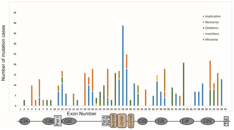
Dysferlin is a large transmembrane protein involved in critical cellular processes including membrane repair and vesicle fusion. Mutations in the dysferlin gene (DYSF) can result in rare forms of muscular dystrophy; Miyoshi myopathy; limb girdle muscular dystrophy type 2B (LGMD2B); and distal myopathy. These conditions are collectively known as dysferlinopathies and are caused by more than 600 mutations that have been identified across the DYSF gene to date. In this review, we discuss the key molecular and clinical features of LGMD2B, the causative gene DYSF, and the associated dysferlin protein structure. We also provide an update on current approaches to LGMD2B diagnosis and advances in drug development, including splice switching antisense oligonucleotides. We give a brief update on clinical trials involving adeno-associated viral gene therapy and the current progress on CRISPR/Cas9 mediated therapy for LGMD2B, and then conclude by discussing the prospects of antisense oligomer-based intervention to treat selected mutations causing dysferlinopathies.

Keywords: LGMD2B; antisense oligonucleotides; dysferlinopathies; gene therapy; readthrough therapy.


Conflict of interest statement

The authors declare no conflicts of interest.

Figures

Figure 1
Figure 1
Architecture of the human dysferlin protein. C2A–C2G; Calcium-binding C2 domains, TM; transmembrane domain, Fer; Ferlin and dysF; dysferlin domains. The amino acid residue positions are indicated above the domain structure, with the encoding exons below. The dysferlin exon map with a reading frame is shown below the domain structure, with the rectangular blocks indicating exons with splice junctions occurring between codons, whereas exons with chevron sides indicate splice junctions that occur within a codon <1:2< or >2:1>. Adapted from Abdullah et al., 2014 [17], Sula et al., 2014 [20], and NM_003494.4.
Figure 2
Figure 2
The role of dysferlin in membrane repair in a calcium-dependent manner. (a) Normal sarcolemmal membrane showing the location of dysferlin (green), annexin (blue), MG53 (pink), and caveolin (grey). (b) Membrane damage causes high influx of calcium into the cytosol and membrane repair vesicles loaded with mini-dysferlin (dysferlin are cleaved by calpain to form mini-dysferlin) are brought near the site of injury by the interaction of multiple proteins as shown. (c) The membrane is repaired by sealing the lesion by patch formation. Modified from Renzhi Han 2011 and Lek et al., 2013 [24,25].
Figure 3
Figure 3
A schematic showing the protein structure of dysferlin and the mutation spectrum. The positions of each of the seven C2 domains (C2A–C2G), the FerA/B, DYSF, and transmembrane domains, and the exon arrangement are shown on the y-axis. The histogram represents the incidence of pathogenic mutations within each of the 55 exons encoding the canonical skeletal muscle isoform of dysferlin (data derived from the Leiden muscular dystrophy database) [8].
Figure 4
Figure 4
Some possible therapeutic strategies at a glance. CRISPR/Cas9 mediated correction of mutations, antisense oligomer (AO) therapies (exon skipping, exon retention, splice correction), gene therapy, readthrough (red X is blocking of stop codon by Ataluren) and small molecule-based therapies have shown some potential to restore either fully or partially functional dysferlin expression.

References

    1. Liu J., Aoki M., Illa I., Wu C., Fardeau M., Angelini C., Serrano C., Urtizberea J.A., Hentati F., Hamida M.B., et al. Dysferlin, a novel skeletal muscle gene, is mutated in Miyoshi myopathy and limb girdle muscular dystrophy. Nat. Genet. 1998;20:31–36. doi: 10.1038/1682. - DOI - PubMed
    1. Lennon N.J., Kho A., Bacskai B.J., Perlmutter S.L., Hyman B.T., Brown R.H. Dysferlin interacts with Annexins A1 and A2 and mediates sarcolemmal wound-healing. J. Biol. Chem. 2003;278:50466–50473. doi: 10.1074/jbc.M307247200. - DOI - PubMed
    1. Britton S., Freeman T., Vafiadaki E., Keers S., Harrison R., Bushby K., Bashir R. The third human FER-1-like protein is highly similar to dysferlin. Genomics. 2000;68:313–321. doi: 10.1006/geno.2000.6290. - DOI - PubMed
    1. Bulankina A.V., Thoms S. Functions of Vertebrate Ferlins. Cells. 2020;9:534. doi: 10.3390/cells9030534. - DOI - PMC - PubMed
    1. karishma Dhuri C.B., Quijano E., Pham H., Gupta A., Bikram A., Bahal R. Antisense Oligonucleotides: An Emerging Area in Drug Discovery and Development. Clin. Med. 2020;9:2004. - PMC - PubMed

MeSH terms

Supplementary concepts

Grants and funding