Review
. 2024 May 21;25(11):5587.
doi: 10.3390/ijms25115587.

Revisiting Glutamate Excitotoxicity in Amyotrophic Lateral Sclerosis and Age-Related Neurodegeneration

Affiliations
Review

Revisiting Glutamate Excitotoxicity in Amyotrophic Lateral Sclerosis and Age-Related Neurodegeneration

Frederick J Arnold et al. Int J Mol Sci. .

Abstract

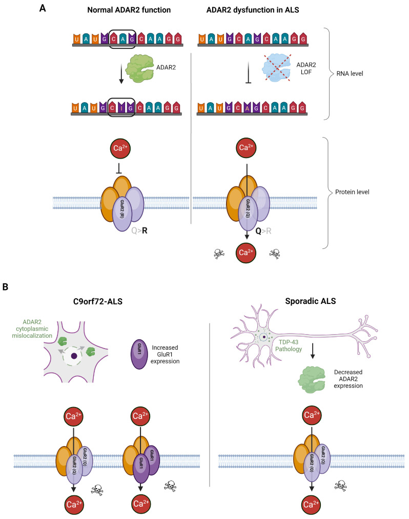
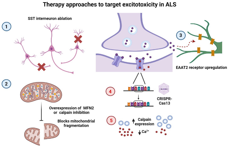
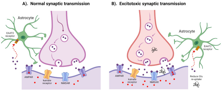
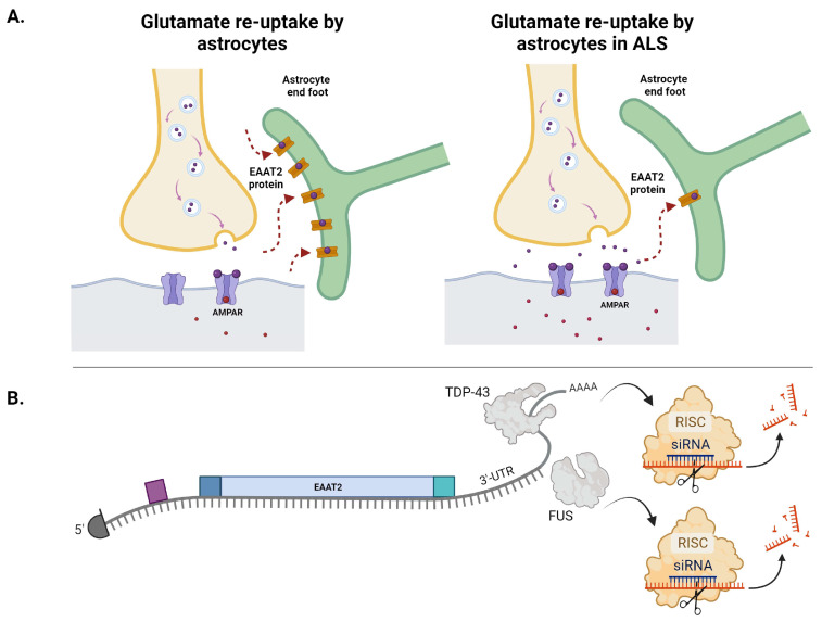
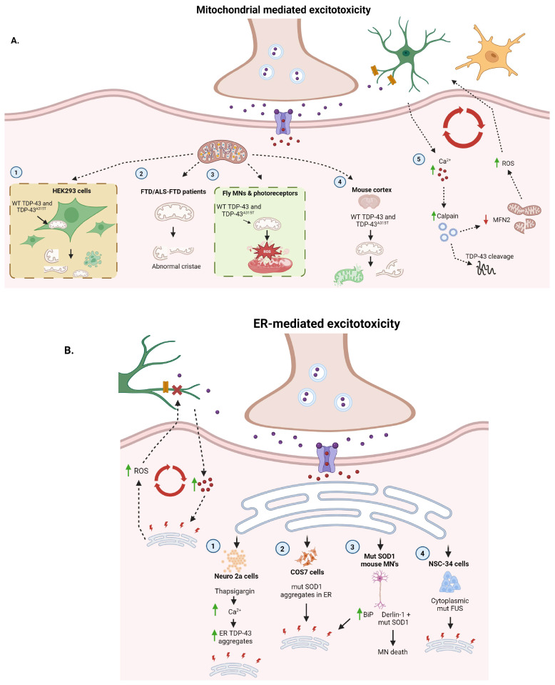
Amyotrophic lateral sclerosis (ALS) is the most common motor neuron disorder. While there are five FDA-approved drugs for treating this disease, each has only modest benefits. To design new and more effective therapies for ALS, particularly for sporadic ALS of unknown and diverse etiologies, we must identify key, convergent mechanisms of disease pathogenesis. This review focuses on the origin and effects of glutamate-mediated excitotoxicity in ALS (the cortical hyperexcitability hypothesis), in which increased glutamatergic signaling causes motor neurons to become hyperexcitable and eventually die. We characterize both primary and secondary contributions to excitotoxicity, referring to processes taking place at the synapse and within the cell, respectively. 'Primary pathways' include upregulation of calcium-permeable AMPA receptors, dysfunction of the EAAT2 astrocytic glutamate transporter, increased release of glutamate from the presynaptic terminal, and reduced inhibition by cortical interneurons-all of which have been observed in ALS patients and model systems. 'Secondary pathways' include changes to mitochondrial morphology and function, increased production of reactive oxygen species, and endoplasmic reticulum (ER) stress. By identifying key targets in the excitotoxicity cascade, we emphasize the importance of this pathway in the pathogenesis of ALS and suggest that intervening in this pathway could be effective for developing therapies for this disease.

Keywords: AMPA receptors; GluR2 editing; NMDA receptors; astrocytes; glutamate excitotoxicity.


Conflict of interest statement

We declare no financial, personal, or professional conflicts of interest relating to this manuscript/subject matter.

Figures

Figure 2
Figure 2
GluR2 editing by the ADAR2 enzyme and AMPAR composition in ALS. (A) The ADAR2 enzyme converts the GluR2 mRNA CAG codon to a CIG codon at the RNA level (left panel). This A-to-I RNA editing changes the gene-encoded glutamine to a positively charged arginine residue at the protein level. The incorporation of ADAR2-edited GluR2 results in a calcium-impermeable AMPAR (left panel). This RNA editing dysfunction allows calcium permeability of the AMPAR (right panel) and downstream excitotoxicity [10]. (B) In both FALS and SALS, multiple mechanisms have been proposed that ultimately result in increased calcium permeability of AMPARs. In C9orf72-ALS (left panel), nucleocytoplasmic mislocalization of ADAR2 has been observed in multiple model systems as well as in postmortem patient spinal cord tissue. As described, this leads to reduced editing of the GluR2 mRNA and an increase in AMPAR calcium permeability [44]. Additionally, increased expression of calcium-permeable GluR1 was reported in C9orf72-ALS iPSC-derived motor neurons. In SALS (right panel), TDP-43 pathology corresponds with decreased ADAR2 expression, which also results in decreased editing of the GluR2 mRNA and increased calcium permeability via AMPARs.
Figure 5
Figure 5
Existing and potential therapeutic strategies to target excitotoxicity in ALS. Examples of therapy interventions, previously proposed (blue circles, 1–3) or untested (red circles, 4–5), to target the excitotoxicity pathway: (1) SST interneuron ablation (red cross) restores normal excitability of layer-5 pyramidal neurons [91]. (2) Overexpression of MFN2 or inhibition of calpain activation blocks calcium-induced mitochondrial fragmentation [108]. (3) Upregulation of astrocytic EAAT2 receptors reduces excess glutamate at the synapse [135]. (4) AAV delivery of CRISPR-Cas13 machinery to replace ADAR2 function in editing the GluR2 pre-mRNA. (5) Upregulation of calpains or other neuronal calcium-buffering proteins to decrease cytosolic Ca2+ concentration.
Figure 1
Figure 1
Physiologic versus excitotoxic synaptic glutamate transmission and re-uptake. (A) In typical synaptic transmission, physiological levels of glutamate (purple circles) are trafficked within vesicles to the synapse. Synaptic glutamate activates three ionotropic glutamate receptors expressed on the postsynaptic neuron: N-methyl-D-aspartate receptor (NMDAR), α-amino-3-hydroxy-5-methyl-4-isoxazolepropionic acid receptor (AMPAR), and kainate receptors. Regulated activation of calcium-permeable vs. calcium-impermeable AMPARs by glutamate facilitates the entry of calcium ions (red dots) into the postsynaptic cell. Balanced re-uptake of glutamate from the synapse is predominantly undertaken by EAAT2 receptors on the astrocytes (red arrow). (B) Prominent mechanisms underlying excitotoxic transmission include excessive glutamate release at the synapse, inefficient glutamate re-uptake via astrocytic EAAT2 receptors [10], and increased calcium permeability of AMPARs.
Figure 3
Figure 3
EAAT2 re-uptake of synaptic glutamate and EAAT2 mRNA dysregulation in ALS. (A) Astrocytes play an important role in the re-uptake of excess glutamate from the synaptic cleft. While the precise etiologic mechanism remains unknown, EAAT2 protein levels are significantly reduced in the motor cortex and spinal cord of ALS patients [68]. Such EAAT2 protein loss has been suggested to result from factors such as oxidative stressors, aberrant splicing mechanisms, and toxic DPRs generated from the C9orf72 expansion in FALS. (B) The ALS-associated RNA binding proteins (RBPs) TDP-43 and FUS are known to bind within the 3′UTR of EAAT2 mRNA [79]. Altered RBP interactions with the EAAT2 3′UTR may change cis-regulatory element regulation of the EAAT2 transcript, such as increased miRNA binding, as one possible mechanism contributing to EAAT2 loss in ALS.
Figure 4
Figure 4
Mitochondrial and ER involvement in secondary excitotoxicity processes. (A) (1) In HEK293 cells, mitochondrial localization of WT TDP-43 or TDP-43A315T inhibits mitochondrial complex 1, resulting in reduced mitochondrial size, abnormal/loss of cristae, and cell death. (2) Electron microscopy of mitochondria within multiple ALS/FTLD-TDP brain regions shows altered mitochondrial morphology, such as abnormal or loss of cristae. (3) In Drosophila photoreceptors, transgenic expression of WT TDP-43 and TDP-43A315T alters mitochondrial morphology and increases intracellular ROS. (4) Similarly, Lentiviral overexpression of WT TDP-43 or TDP-43A315T in mouse cortices leads to fragmentation and dysfunction of mitochondria and cell death. (5) Through various mechanisms discussed here, excess synaptic glutamate leads to increased intracellular calcium in MNs which are sensitive to calcium dysregulation. This activates calpain which cleaves TDP-43 and generates aggregation-prone TDP-43 fragments. Calpain also degrades MFN2, leading to impaired mitochondrial function, induction of cell death, and increased ROS. Elevated ROS can disrupt astrocyte (green) and microglia (yellow) function, further contributing to elevated synaptic glutamate and intracellular calcium. (B) Proteins implicated in ALS, such as SOD1, TDP-43, and FUS, are all linked to ER stress and UPR induction. (1) Treating Neuro2a cells with Thapsigargin, which increases intracellular Ca2+, causes cytoplasmic TDP-43 aggregation, including C-terminal TDP-43 fragments that colocalize with the ER. (2) Cos7 cells transfected with mutant (mut) SOD1 (but not WT) exhibit SOD1 aggregates localized to the ER and increased ER stress. (3) Similarly, spinal cord MNs of mutant SOD1 transgenic mice display an increase in ER chaperone proteins, such as BiP, indicative of ER stress. Furthermore, an interaction between SOD1 and Derlin-1, a member of the ER-associated degradation (ERAD) machinery, induces ER stress via activation of ASK1, resulting in MN death. (4) Like TDP-43, mutant FUS exhibits nuclear clearance and cytoplasmic aggregation in ALS. In mouse NSC-34 cells, redistribution of mutant FUS to the cytoplasm triggers ER stress. Together, these pathways support a feedback loop that can be initiated via either increased intracellular Ca2+ or ER stress.

References

    1. Brown R.H., Al-Chalabi A. Amyotrophic Lateral Sclerosis. N. Engl. J. Med. 2017;377:162–172. doi: 10.1056/NEJMra1603471. - DOI - PubMed
    1. King A.E., Woodhouse A., Kirkcaldie M.T.K., Vickers J.C. Excitotoxicity in ALS: Overstimulation, or overreaction? Exp. Neurol. 2016;275:162–171. doi: 10.1016/j.expneurol.2015.09.019. - DOI - PubMed
    1. Sun Y., Li X., Bedlack R. An evaluation of the combination of sodium phenylbutyrate and taurursodiol for the treatment of amyotrophic lateral sclerosis. Expert Rev. Neurother. 2023;23:1–7. doi: 10.1080/14737175.2023.2174018. - DOI - PubMed
    1. Bellingham M.C. A Review of the Neural Mechanisms of Action and Clinical Efficiency of Riluzole in Treating Amyotrophic Lateral Sclerosis: What have we Learned in the Last Decade? CNS Neurosci. Ther. 2011;17:4–31. doi: 10.1111/j.1755-5949.2009.00116.x. - DOI - PMC - PubMed
    1. Takei K., Watanabe K., Yuki S., Akimoto M., Sakata T., Palumbo J. Edaravone and its clinical development for amyotrophic lateral sclerosis. Amyotroph. Lateral Scler. Front. Degener. 2017;18:5–10. doi: 10.1080/21678421.2017.1353101. - DOI - PubMed

MeSH terms