. 2024 May 21;25(11):5613.
doi: 10.3390/ijms25115613.

Minimal Change Disease: Pathogenetic Insights from Glomerular Proteomics

Affiliations

Minimal Change Disease: Pathogenetic Insights from Glomerular Proteomics

Andrada Alina Bărar et al. Int J Mol Sci. .

Abstract

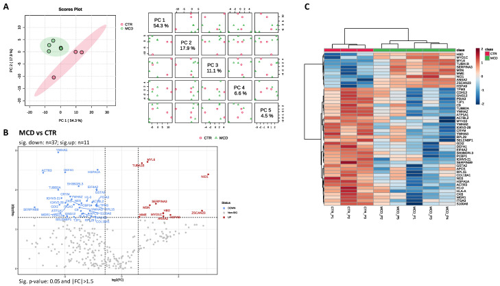
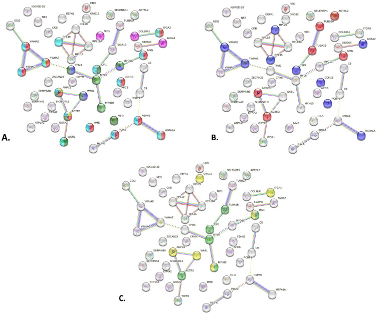
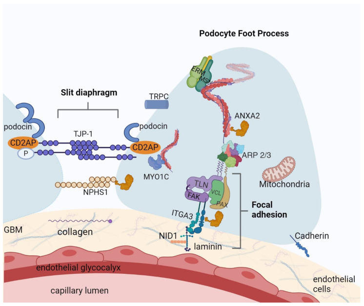
The mechanism underlying podocyte dysfunction in minimal change disease (MCD) remains unknown. This study aimed to shed light on the potential pathophysiology of MCD using glomerular proteomic analysis. Shotgun proteomics using label-free quantitative mass spectrometry was performed on formalin-fixed, paraffin-embedded (FFPE) renal biopsies from two groups of samples: control (CTR) and MCD. Glomeruli were excised from FFPE renal biopsies using laser capture microdissection (LCM), and a single-pot solid-phase-enhanced sample preparation (SP3) digestion method was used to improve yield and protein identifications. Principal component analysis (PCA) revealed a distinct separation between the CTR and MCD groups. Forty-eight proteins with different abundance between the two groups (p-value ≤ 0.05 and |FC| ≥ 1.5) were identified. These may represent differences in podocyte structure, as well as changes in endothelial or mesangial cells and extracellular matrix, and some were indeed found in several of these structures. However, most differentially expressed proteins were linked to the podocyte cytoskeleton and its dynamics. Some of these proteins are known to be involved in focal adhesion (NID1 and ITGA3) or slit diaphragm signaling (ANXA2, TJP1 and MYO1C), while others are structural components of the actin and microtubule cytoskeleton of podocytes (ACTR3 and NES). This study suggests the potential of mass spectrometry-based shotgun proteomic analysis with LCM glomeruli to yield valuable insights into the pathogenesis of podocytopathies like MCD. The most significantly dysregulated proteins in MCD could be attributable to cytoskeleton dysfunction or may be a compensatory response to cytoskeleton malfunction caused by various triggers.

Keywords: laser capture microdissection; minimal change disease; podocyte cytoskeleton; proteomics; tandem mass spectrometry.


Conflict of interest statement

The authors declare no conflicts of interest.

Figures

Figure 1
Figure 1
Differential expression analysis results. (A) Principal component analysis (PCA): (left) score plot between the selected PCs; (right) pairwise score plots between the first five PCs; the explained variance of each PC is shown in the corresponding diagonal cell; (B) volcano plot of differentially expressed proteins (t-test, independent unequal variance, p ≤ 0.05 and |FC| > 1.5) in MCD group vs. CTR; Red dots represent differentially expressed proteins with higher abundance in MCD group, while the blue dots correspond to with higher abundance in CTR group; black proteins showed no significant difference between groups; (C) proteins differentially expressed between MCD and CTR samples depicted as a heatmap of hierarchical cluster analysis results (t-test, independent unequal variance, p ≤ 0.05; Euclidean distance and clustering algorithm using averages).
Figure 2
Figure 2
STRING network enrichment analysis using the proteins differentially expressed between MCD and CTR samples. (A) Enriched terms using the Compartments subcellular localization database. (B) Enriched terms using Gene Ontology Molecular Function database. (C) Enriched pathways highlighted by KEGG database.
Figure 3
Figure 3
Proteins differentiated in MCD: an illustration of podocyte foot process with focal adhesion and slit diaphragm structure. Some of the proteins with statistically significant upregulation (ANXA2, NID1, MSN, MYO1C) or downregulation (ARP3, ITGA3, TJP-1, COL18A1 component of collagen, CDH13 member of cadherin) in MCD versus CTR are represented at the podocyte level. For understanding, we also incorporated proteins that are presented in the podocytes, but did not exhibit statistical significance in our study (NPHS1, podocin, CD2AP, TRPC, ERM, TLN, FAK, VCL, PAX, laminin network). This figure was created with BioRender.com.
Figure 4
Figure 4
Immunohistochemical annexin staining of one of the kidney biopsies. (A) ANXA2 staining, 10× magnification, showing positive staining at the luminal border of the tubules and intense staining in the glomerulus. (B) ANXA2, 40× magnification showing intense staining in one of the positive glomeruli.

References

    1. Barisoni L., Schnaper H.W., Kopp J.B. A Proposed Taxonomy for the Podocytopathies. Clin. J. Am. Soc. Nephrol. 2007;2:529–542. doi: 10.2215/CJN.04121206. - DOI - PubMed
    1. Purohit S., Piani F., Ordoñez F.A., de Lucas-Collantes C., Bauer C., Cara-Fuentes G. Molecular Mechanisms of Proteinuria in Minimal Change Disease. Front. Med. 2021;8:761600. doi: 10.3389/fmed.2021.761600. - DOI - PMC - PubMed
    1. Bertelli R., Bonanni A., Caridi G., Canepa A., Ghiggeri G.M. Molecular and Cellular Mechanisms for Proteinuria in Minimal Change Disease. Front. Med. 2018;5:170. doi: 10.3389/fmed.2018.00170. - DOI - PMC - PubMed
    1. New L.A., Martin C.E., Scott R.P., Platt M.J., Keyvani Chahi A., Stringer C.D., Lu P., Samborska B., Eremina V., Takano T., et al. Nephrin Tyrosine Phosphorylation Is Required to Stabilize and Restore Podocyte Foot Process Architecture. J. Am. Soc. Nephrol. 2016;27:2422–2435. doi: 10.1681/ASN.2015091048. - DOI - PMC - PubMed
    1. Verma R., Venkatareddy M., Kalinowski A., Li T., Kukla J., Mollin A., Cara-Fuentes G., Patel S.R., Garg P. Nephrin Is Necessary for Podocyte Recovery Following Injury in an Adult Mature Glomerulus. PLoS ONE. 2018;13:e0198013. doi: 10.1371/journal.pone.0198013. - DOI - PMC - PubMed

LinkOut - more resources