

Localization of Catecholaminergic Neurofibers in Pregnant Cervix as a Possible Myometrial Pacemaker
- PMID: 38891818
- PMCID: PMC11171499
- DOI: 10.3390/ijms25115630
Localization of Catecholaminergic Neurofibers in Pregnant Cervix as a Possible Myometrial Pacemaker
Abstract
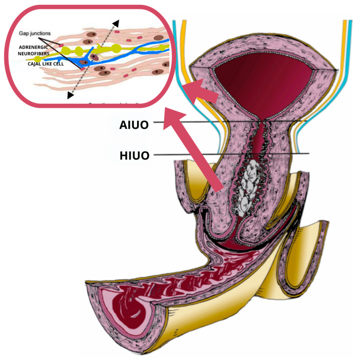
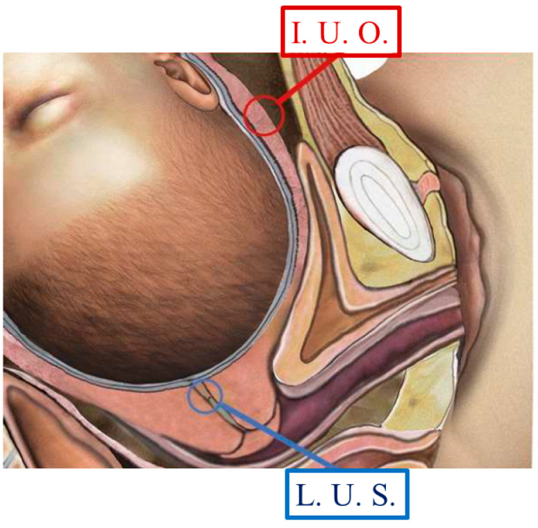
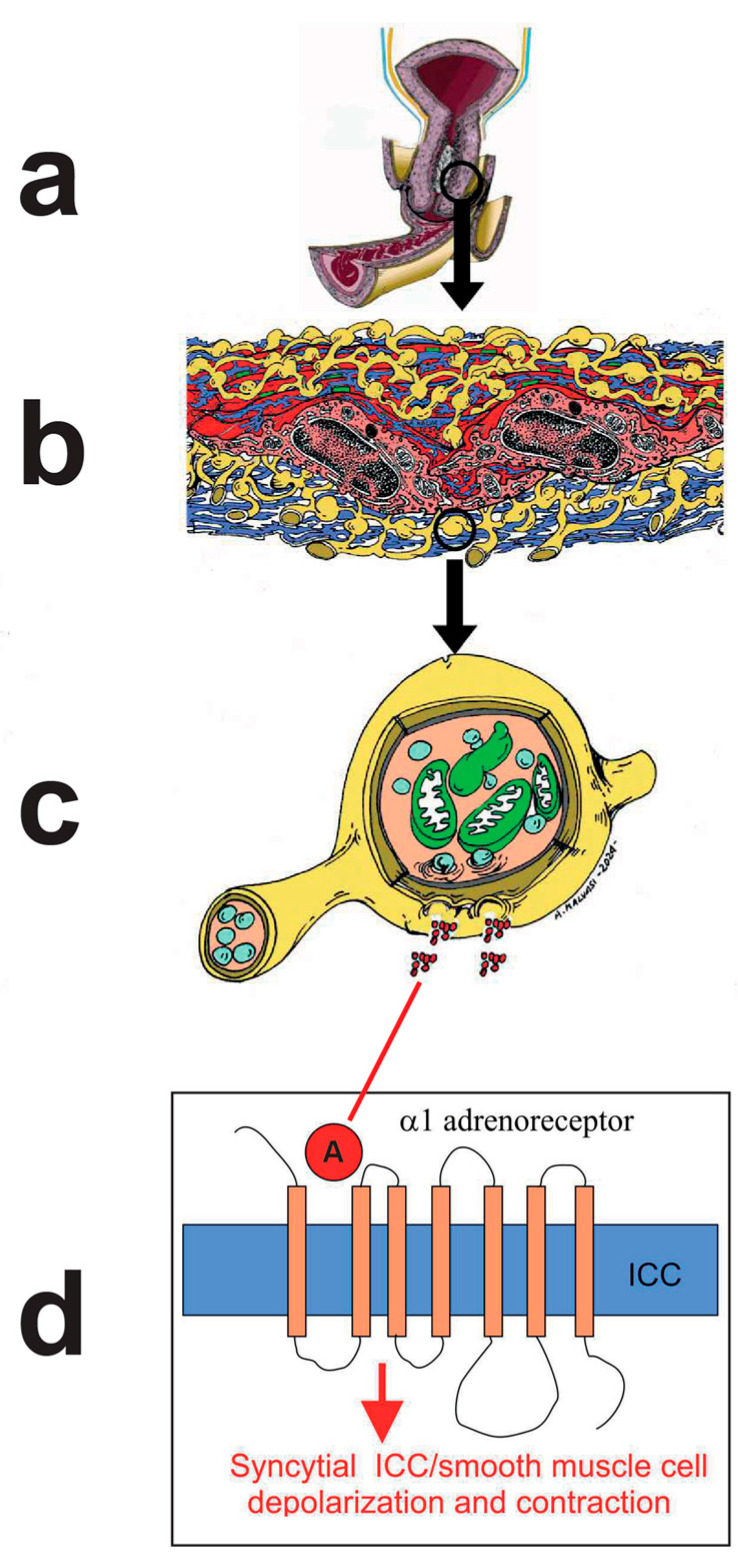
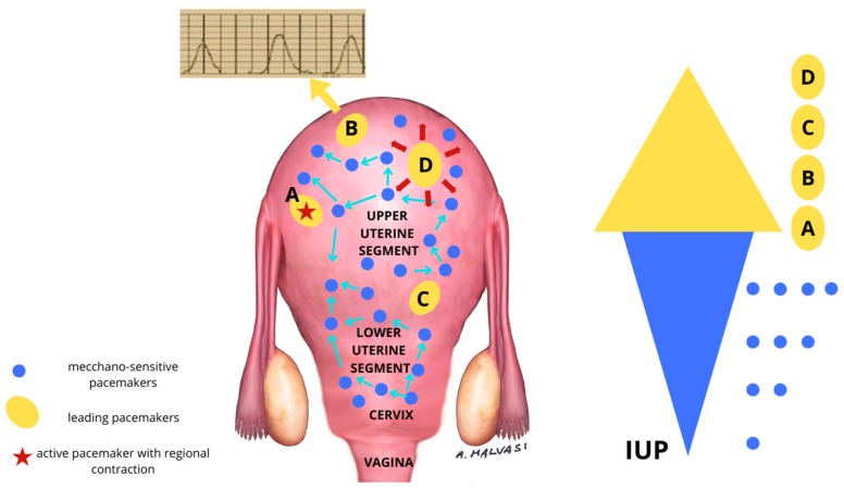
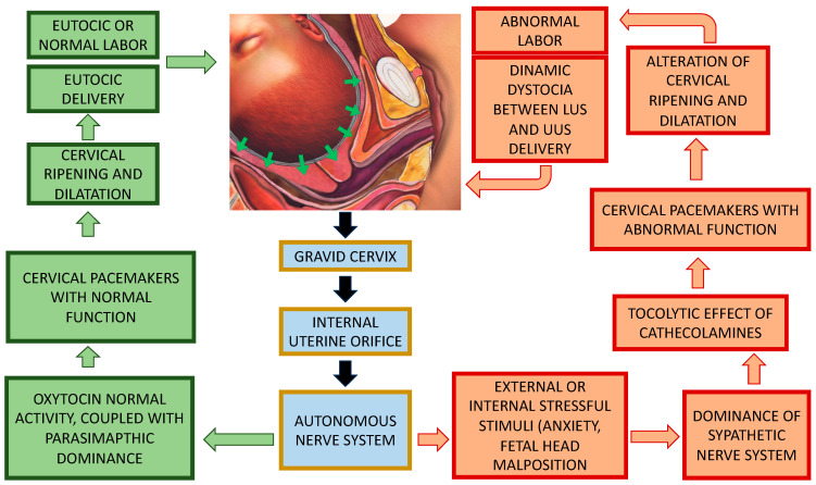
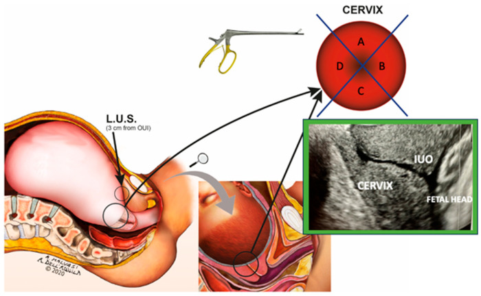
In eutocic labor, the autonomic nervous system is dominated by the parasympathetic system, which ensures optimal blood flow to the uterus and placenta. This study is focused on the detection of the quantitative presence of catecholamine (C) neurofibers in the internal uterine orifice (IUO) and in the lower uterine segment (LUS) of the pregnant uterus, which could play a role in labor and delivery. A total of 102 women were enrolled before their submission to a scheduled cesarean section (CS); patients showed a singleton fetus in a cephalic presentation outside labor. During CS, surgeons sampled two serial consecutive full-thickness sections 5 mm in depth (including the myometrial layer) on the LUS and two randomly selected samples of 5 mm depth from the IUO of the cervix. All histological samples were studied to quantify the distribution of A nerve fibers. The authors demonstrated a significant and notably higher concentration of A fibers in the IUO (46 ± 4.8) than in the LUS (21 ± 2.6), showing that the pregnant cervix has a greater concentration of A neurofibers than the at-term LUS. Pregnant women's mechanosensitive pacemakers can operate normally when the body is in a physiological state, which permits normal uterine contractions and eutocic delivery. The increased frequency of C neurofibers in the cervix may influence the smooth muscle cell bundles' activation, which could cause an aberrant mechano-sensitive pacemaker activation-deactivation cycle. Stressful circumstances (anxiety, tension, fetal head position) cause the sympathetic nervous system to become more active, working through these nerve fibers in the gravid cervix. They might interfere with the mechano-sensitive pacemakers, slowing down the uterine contractions and cervix ripening, which could result in dystocic labor.
Keywords: catecholamine; cervix; cesarean section; delivery; labor; lower uterine segment (LUS); neurotransmitters; pregnancy; uterine pacemaker.
Conflict of interest statement
Dr. Lorenzo Malgieri was Chief Innovation Officer in CLE, 70124 Bari, Italy. The remaining authors declare that the research was conducted in the absence of any commercial or financial relationships that could be construed as a potential conflict of interest. The funder was not involved in the study design, collection, analysis, interpretation of data, the writing of this article, or the decision to submit it for publication.
Figures
Similar articles
-
A Qualitative and Quantitative Study of the Innervation of the Human Non Pregnant Uterus.Curr Protein Pept Sci. 2017;18(2):140-148. doi: 10.2174/1389203717666160330105341. Curr Protein Pept Sci. 2017. PMID: 27063643
-
Laminin and Collagen IV: Two Polypeptides as Marker of Dystocic Labor.Curr Protein Pept Sci. 2017;18(2):149-154. doi: 10.2174/1389203717666160322150125. Curr Protein Pept Sci. 2017. PMID: 27001062
-
Dystocic Labor and Adrenergic and Noradrenergic Neurotransmitters: A Morphological Experimental Study.Int J Mol Sci. 2022 Sep 27;23(19):11379. doi: 10.3390/ijms231911379. Int J Mol Sci. 2022. PMID: 36232680 Free PMC article.
-
Adrenergic and Cholinergic Uterine Innervation and the Impact on Reproduction in Aged Women.Curr Pharm Des. 2020;26(3):358-362. doi: 10.2174/1381612826666200128092256. Curr Pharm Des. 2020. PMID: 32003664 Review.
-
Review and Study of Uterine Bioelectrical Waveforms and Vector Analysis to Identify Electrical and Mechanosensitive Transduction Control Mechanisms During Labor in Pregnant Patients.Reprod Sci. 2021 Mar;28(3):838-856. doi: 10.1007/s43032-020-00358-5. Epub 2020 Oct 22. Reprod Sci. 2021. PMID: 33090378 Review.
Cited by
-
Dystocia, Delivery, and Artificial Intelligence in Labor Management: Perspectives and Future Directions.J Clin Med. 2024 Oct 25;13(21):6410. doi: 10.3390/jcm13216410. J Clin Med. 2024. PMID: 39518549 Free PMC article.
References
-
- Ameer M.A., Fagan S.E., Sosa-Stanley J.N., Peterson D.C. StatPearls. StatPearls Publishing; Treasure Island, FL, USA: 2023. Anatomy, Abdomen and Pelvis: Uterus. 6 December 2022. - PubMed
-
- Malvasi A., Tinelli A., Cavallotti C., Stark M. Der Kaiserschnitt. Elsevier; Amsterdam, The Netherlands: 2009. Anatomische Aspekte von Schwangerschaft und Kaiserschnitt; pp. 39–66.
-
- Miftahof R.N., Akhmadeev N.R. Soft Biological Shells in Bioengineering. IOP Publishing Ltd.; Bristol, UK: 2019. The gravid uterus, Chapter 13; pp. 13–59.
MeSH terms
Substances
LinkOut - more resources
Full Text Sources
