

Dentin Mechanobiology: Bridging the Gap between Architecture and Function
- PMID: 38891829
- PMCID: PMC11171917
- DOI: 10.3390/ijms25115642
Dentin Mechanobiology: Bridging the Gap between Architecture and Function
Abstract
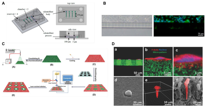
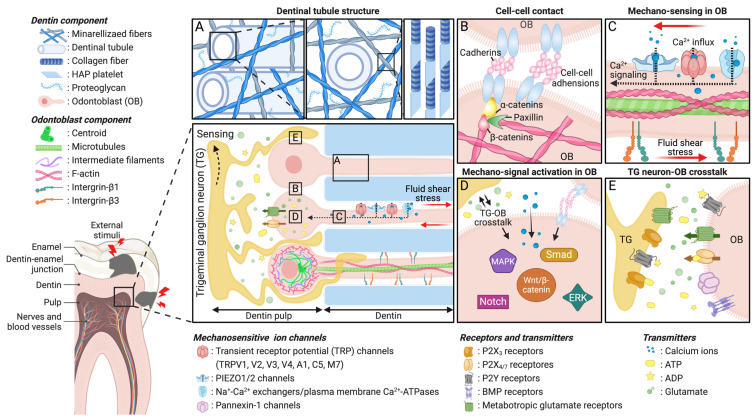
It is remarkable how teeth maintain their healthy condition under exceptionally high levels of mechanical loading. This suggests the presence of inherent mechanical adaptation mechanisms within their structure to counter constant stress. Dentin, situated between enamel and pulp, plays a crucial role in mechanically supporting tooth function. Its intermediate stiffness and viscoelastic properties, attributed to its mineralized, nanofibrous extracellular matrix, provide flexibility, strength, and rigidity, enabling it to withstand mechanical loading without fracturing. Moreover, dentin's unique architectural features, such as odontoblast processes within dentinal tubules and spatial compartmentalization between odontoblasts in dentin and sensory neurons in pulp, contribute to a distinctive sensory perception of external stimuli while acting as a defensive barrier for the dentin-pulp complex. Since dentin's architecture governs its functions in nociception and repair in response to mechanical stimuli, understanding dentin mechanobiology is crucial for developing treatments for pain management in dentin-associated diseases and dentin-pulp regeneration. This review discusses how dentin's physical features regulate mechano-sensing, focusing on mechano-sensitive ion channels. Additionally, we explore advanced in vitro platforms that mimic dentin's physical features, providing deeper insights into fundamental mechanobiological phenomena and laying the groundwork for effective mechano-therapeutic strategies for dentinal diseases.
Keywords: dentin; dentin-mimicking in vitro platforms; mechanobiology; mechanosensing; mechanotransduction; viscoelastic properties.
Conflict of interest statement
The authors declare no competing interests.
Figures
Similar articles
-
Odontoblasts: Specialized hard-tissue-forming cells in the dentin-pulp complex.Congenit Anom (Kyoto). 2016 Jul;56(4):144-53. doi: 10.1111/cga.12169. Congenit Anom (Kyoto). 2016. PMID: 27131345 Review.
-
Topical review. Dental pain and odontoblasts: facts and hypotheses.J Orofac Pain. 2010 Fall;24(4):335-49. J Orofac Pain. 2010. PMID: 21197505 Review.
-
Odontoblasts in odontogenic tumors.Med Hypotheses. 2013 Sep;81(3):371-3. doi: 10.1016/j.mehy.2013.05.015. Epub 2013 Jun 17. Med Hypotheses. 2013. PMID: 23786903
-
PIEZO1 Ion Channels Mediate Mechanotransduction in Odontoblasts.J Endod. 2022 Jun;48(6):749-758. doi: 10.1016/j.joen.2022.02.005. Epub 2022 Feb 25. J Endod. 2022. PMID: 35219748
-
The extent of odontoblast processes in the dentin is distinct between cusp and cervical regions during development and aging.Arch Histol Cytol. 2002 Jun;65(2):179-88. doi: 10.1679/aohc.65.179. Arch Histol Cytol. 2002. PMID: 12164341
Cited by
-
The Hippo-YAP/β-catenin signaling axis coordinates odontogenic differentiation in dental pulp stem cells: Implications for dentin-pulp regeneration.PLoS One. 2025 Jun 26;20(6):e0326978. doi: 10.1371/journal.pone.0326978. eCollection 2025. PLoS One. 2025. PMID: 40570025 Free PMC article.
-
Cellular porosity in dentin exhibits complex network characteristics with spatio-temporal fluctuations.PLoS One. 2025 Jul 16;20(7):e0327030. doi: 10.1371/journal.pone.0327030. eCollection 2025. PLoS One. 2025. PMID: 40668788 Free PMC article.
-
Regenerative Endodontic Therapies: Harnessing Stem Cells, Scaffolds, and Growth Factors.Polymers (Basel). 2025 May 26;17(11):1475. doi: 10.3390/polym17111475. Polymers (Basel). 2025. PMID: 40508718 Free PMC article. Review.
-
Biomineralization of dental tissues with natural drugs: a comprehensive review.Saudi Dent J. 2025 Jul 15;37(4-6):29. doi: 10.1007/s44445-025-00036-9. Saudi Dent J. 2025. PMID: 40663199 Free PMC article. Review.
References
-
- Jansen van Vuuren L., Broadbent J.M., Duncan W.J., Waddell J.N. Maximum voluntary bite force, occlusal contact points and associated stresses on posterior teeth. J. R. Soc. N. Z. 2020;50:132–143. doi: 10.1080/03036758.2019.1691612. - DOI
Publication types
MeSH terms
Grants and funding
LinkOut - more resources
Full Text Sources
Miscellaneous
