

Neurotoxicity and Developmental Neurotoxicity of Copper Sulfide Nanoparticles on a Human Neuronal In-Vitro Test System
- PMID: 38891838
- PMCID: PMC11172337
- DOI: 10.3390/ijms25115650
Neurotoxicity and Developmental Neurotoxicity of Copper Sulfide Nanoparticles on a Human Neuronal In-Vitro Test System
Abstract
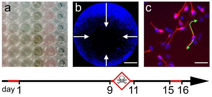
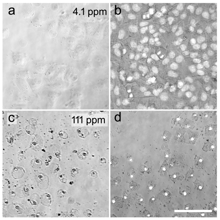
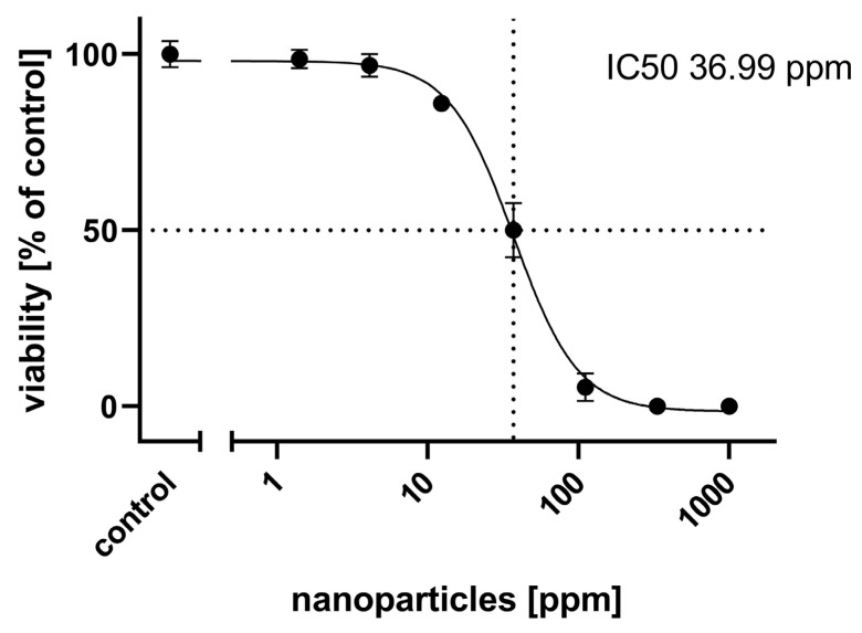
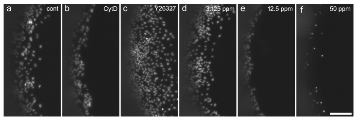
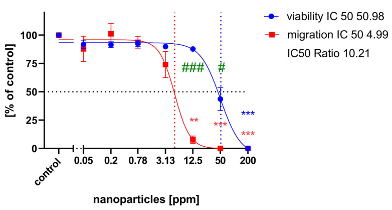
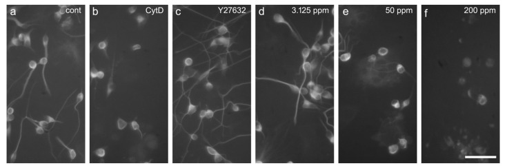
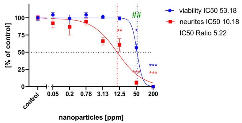
Nanoparticles (NPs) are becoming increasingly important novel materials for many purposes, including basic research, medicine, agriculture, and engineering. Increasing human and environmental exposure to these promising compounds requires assessment of their potential health risks. While the general direct cytotoxicity of NPs is often routinely measured, more indirect possible long-term effects, such as reproductive or developmental neurotoxicity (DNT), have been studied only occasionally and, if so, mostly on non-human animal models, such as zebrafish embryos. In this present study, we employed a well-characterized human neuronal precursor cell line to test the concentration-dependent DNT of green-manufactured copper sulfide (CuS) nanoparticles on crucial early events in human brain development. CuS NPs turned out to be generally cytotoxic in the low ppm range. Using an established prediction model, we found a clear DNT potential of CuS NPs on neuronal precursor cell migration and neurite outgrowth, with IC50 values 10 times and 5 times, respectively, lower for the specific DNT endpoint than for general cytotoxicity. We conclude that, in addition to the opportunities of NPs, their risks to human health should be carefully considered.
Keywords: DNT; NT-2; NTera-2; migration; nanoparticles; neurite outgrowth.
Conflict of interest statement
The authors declare no conflicts of interest.
Figures
Similar articles
-
Evaluation of Zebrafish Toxicology and Biomedical Potential of Aeromonas hydrophila Mediated Copper Sulfide Nanoparticles.Oxid Med Cell Longev. 2022 Jan 28;2022:7969825. doi: 10.1155/2022/7969825. eCollection 2022. Oxid Med Cell Longev. 2022. PMID: 35126821 Free PMC article.
-
Neurite outgrowth in human induced pluripotent stem cell-derived neurons as a high-throughput screen for developmental neurotoxicity or neurotoxicity.Neurotoxicology. 2016 Mar;53:271-281. doi: 10.1016/j.neuro.2016.02.003. Epub 2016 Feb 4. Neurotoxicology. 2016. PMID: 26854185
-
Developmental Neurotoxicity of Fipronil and Rotenone on a Human Neuronal In Vitro Test System.Neurotox Res. 2021 Aug;39(4):1189-1202. doi: 10.1007/s12640-021-00364-8. Epub 2021 Apr 19. Neurotox Res. 2021. PMID: 33871813 Free PMC article.
-
An industry perspective: A streamlined screening strategy using alternative models for chemical assessment of developmental neurotoxicity.Neurotoxicology. 2019 Jul;73:17-30. doi: 10.1016/j.neuro.2019.02.010. Epub 2019 Feb 17. Neurotoxicology. 2019. PMID: 30786249 Review.
-
Current problems of in vivo developmental neurotoxicity tests and a new in vivo approach focusing on each step of the developing central nervous system.Congenit Anom (Kyoto). 2012 Sep;52(3):129-39. doi: 10.1111/j.1741-4520.2012.00376.x. Congenit Anom (Kyoto). 2012. PMID: 22925213 Review.
References
-
- Jadoun S., Arif R., Jangid N.K., Rajesh K.M. Green synthesis of nanoparticles using plant extracts: A review. Environ Chem. Lett. 2021;19:355–374. doi: 10.1007/s10311-020-01074-x. - DOI
-
- Botha N.L., Cloete K.J., Šmit Ž., Isaković K., Akbari M., Morad R., Madiba I., David O.M., Santos L.P.M., Dube A., et al. Ionome mapping and amino acid metabolome profiling of Phaseolus vulgaris L. seeds imbibed with computationally informed phytoengineered copper sulphide nanoparticles. Discov. Nano. 2024;19:8. doi: 10.1186/s11671-023-03953-y. - DOI - PMC - PubMed
MeSH terms
Substances
LinkOut - more resources
Full Text Sources
Research Materials
