. 2024 May 22;25(11):5651.
doi: 10.3390/ijms25115651.

Structural and Interactional Analysis of the Flavonoid Pathway Proteins: Chalcone Synthase, Chalcone Isomerase and Chalcone Isomerase-like Protein

Affiliations

Structural and Interactional Analysis of the Flavonoid Pathway Proteins: Chalcone Synthase, Chalcone Isomerase and Chalcone Isomerase-like Protein

Jacob A Lewis et al. Int J Mol Sci. .

Abstract

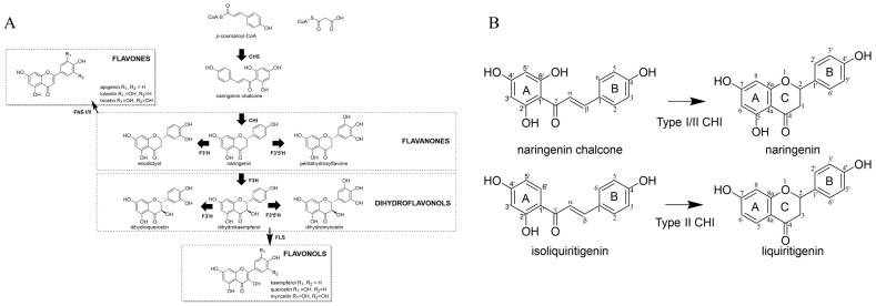
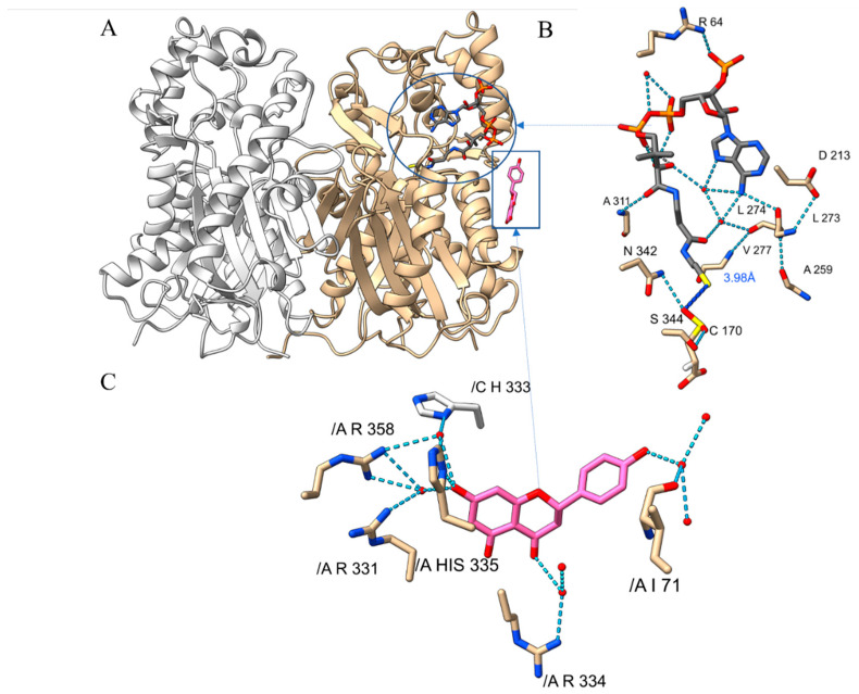
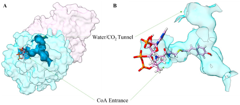
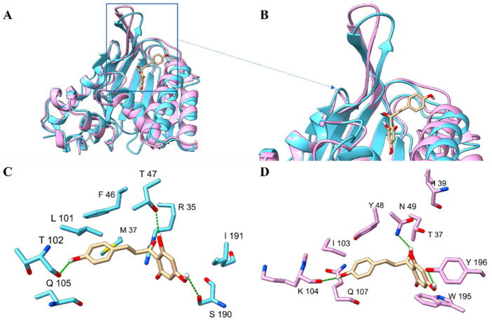
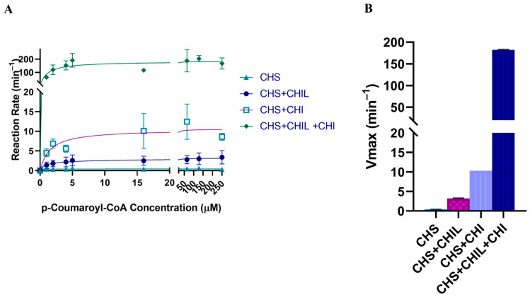
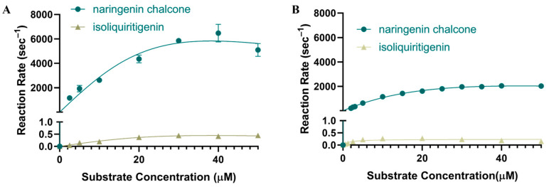
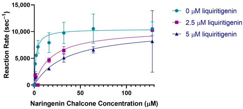
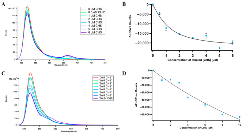
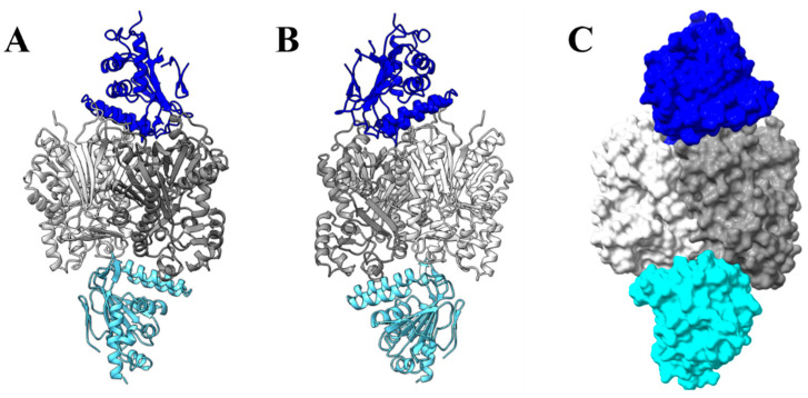
Chalcone synthase (CHS) and chalcone isomerase (CHI) catalyze the first two committed steps of the flavonoid pathway that plays a pivotal role in the growth and reproduction of land plants, including UV protection, pigmentation, symbiotic nitrogen fixation, and pathogen resistance. Based on the obtained X-ray crystal structures of CHS, CHI, and chalcone isomerase-like protein (CHIL) from the same monocotyledon, Panicum virgatum, along with the results of the steady-state kinetics, spectroscopic/thermodynamic analyses, intermolecular interactions, and their effect on each catalytic step are proposed. In addition, PvCHI's unique activity for both naringenin chalcone and isoliquiritigenin was analyzed, and the observed hierarchical activity for those type-I and -II substrates was explained with the intrinsic characteristics of the enzyme and two substrates. The structure of PvCHS complexed with naringenin supports uncompetitive inhibition. PvCHS displays intrinsic catalytic promiscuity, evident from the formation of p-coumaroyltriacetic acid lactone (CTAL) in addition to naringenin chalcone. In the presence of PvCHIL, conversion of p-coumaroyl-CoA to naringenin through PvCHS and PvCHI displayed ~400-fold increased Vmax with reduced formation of CTAL by 70%. Supporting this model, molecular docking, ITC (Isothermal Titration Calorimetry), and FRET (Fluorescence Resonance Energy Transfer) indicated that both PvCHI and PvCHIL interact with PvCHS in a non-competitive manner, indicating the plausible allosteric effect of naringenin on CHS. Significantly, the presence of naringenin increased the affinity between PvCHS and PvCHIL, whereas naringenin chalcone decreased the affinity, indicating a plausible feedback mechanism to minimize spontaneous incorrect stereoisomers. These are the first findings from a three-body system from the same species, indicating the importance of the macromolecular assembly of CHS-CHI-CHIL in determining the amount and type of flavonoids produced in plant cells.

Keywords: Panicum virgatum; Sorghum bicolor; anthocyanidins; anthocyanin; flavones; flavonols; metabolon.


Conflict of interest statement

The authors declare no conflicts of interest.

Figures

Figure 1
Figure 1
Flavonoid pathway. (A) General overview of the flavonoid pathway. (B) Type I (naringenin chalcone) and Type II (isoliquiritigenin) and their corresponding products with IUPAC numbering. Molecular graphics were created utilizing Chemdraw 22.2.0 (Waltham, MA, USA).
Figure 2
Figure 2
The crystal structure of PvCHS. (A) Ribbon diagram representing the overall structure and secondary structures of PvCHS homodimer (tan and white color for each subunit). One naringenin molecule (pink and boxed) at the surface and a CoA molecule (grey and circled) at the active site were depicted by the stick models. The locations of N- and C-terminal structural elements are labeled. (B) A bound CoA (gray) molecule in the active site of PvCHS with the sulfinic acid labeled as CSD, the location where the polyketide is assembled. Hydrogen bonds are shown with dotted lines and crystallographic waters are represented as red spheres. (C) Naringenin (pink) molecule at the surface of PvCHS. Chain identification is listed in C (as /C) to discern A chain (as /A). Molecular graphics images were produced using the ChimeraX package (UCSF).
Figure 3
Figure 3
Alignment of the amino acid sequences of enzymes deposited in PDB that share a high degree of similarity. (A) CHS with PvCHS (PDBID: 8V8M), OsCHS (PDBID: 4YJY), PmCHS (PDBID: 6CQB), AtCHS (PDBID: 6DXB), FhCHS (PDBID: 4WUM), McCHS (PDBID: 1CGK), MsCHS (PDBID: 5UC5), and GmCHS (PDBID: 7BUR); (B) CHI/CHIL with PvCHI (PDBID: 8V8L), PvCHIL (PDBID: 8V8O), AtFAP (PDBID:4DOL), AtCHIL (PDBID:4DOK), MsCHI (PDBID:1EYP), DaCHI (PDBID:5YX3), MtCHI (PDBID:6MS8), AtCHI (PDBID:4DOI). The regions of α-helices are shown in purple, and β-strands are shown in yellow. The alignment was created using BLASTP to search for similar enzymes, aligned using Clustal Omega, and visualized using Jalview (University of Dundee, Dundee, UK).
Figure 4
Figure 4
The binding Pocket of PvCHS. (A) Space-filling representation of a PvCHS homodimer with the substrate binding pocket shown in blue. Different subunits are shown in different colors. Calculations were made using CASTp3.0 [56], and the figure was made using ChimeraX (UCSF). (B) Zoomed-in view of the substrate binding tunnel with p-coumaroyl-CoA. Molecular graphics images were produced using the ChimeraX package (UCSF).
Figure 5
Figure 5
The structure of PvCHI and PvCHIL. (A) Ribbon diagram representing the three-dimensional structures of PvCHI (blue) and PvCHIL (pink). Two disordered loops in the crystal structure PvCHIL are shown as a dotted line. (B) The active site, as identified via molecular docking, is superimposed into PvCHI and PvCHIL. (C) Residues from PvCHI that interact with the superposition naringenin chalcone at the active site. (D) Residues from PvCHIL that are in the active site near the superimposed naringenin chalcone. Different active site residues: PvCHI F46 (Y48 PvCHIL), T47 (N49), R35(T37), M37 (I39), L101 (I103), T104 (K104), Q105 (Q107), S190 (W195), and I191 (Y196). Molecular graphics images were produced using the ChimeraX package (UCSF).
Figure 6
Figure 6
Steady-state kinetics of PvCHS with varying concentrations of p-coumaroyl-CoA and fixed concentration of malonyl-CoA. The product formation is treated as the sum of naringenin and naringenin chalcone. (A) Michaelis-Menten curve comparing PvCHS (turquoise), PvCHS and PvCHIL (dark blue), PvCHS and PvCHI (purple), and PvCHS, PvCHIL and PvCHI (green). The Km remained largely unaffected in all assays at 1 µM. The Vmax for PvCHS by itself was 0.417 μM min−1; for PvCHS with PvCHIL, it was 7.7-fold higher; for PvCHS and PvCHI, it was 24-fold higher, and for the three-protein system, it was 437-fold higher. (B) The Vmax for PvCHS (turquoise), PvCHS and PvCHI (purple), PvCHS and PvCHI (light blue), and PvCHS, PvCHIL, and PvCHI (dark blue) represented in a bar graph with the error in the calculated Vmax by regression shown at the 95% confidence interval. Graphics and calculations were conducted utilizing GraphPad Prism 8.0.2 (San Diego, CA, USA).
Figure 7
Figure 7
Steady-state enzyme kinetics of CHI. (A) PvCHI utilizing both naringenin chalcone (blue) and isoliquiritigenin (gold). The Vmax was 8012 µM s−1 and 0.63 µM s−1, respectively. The Km was 16.04 µM for naringenin chalcone and 17.60 µM for isoliquiritigenin. (B) Steady-state kinetics of SbCHI utilizing both naringenin chalcone (blue) and isoliquiritigenin (gold). The Vmax was 2929 µM s−1 and 0.24 µM s−1, respectively. The Km was 16.98 µM for naringenin chalcone and 1.58 µM for isoliquiritigenin. Graphics and calculations were conducted utilizing GraphPad Prism 8.0.2 (San Diego, CA, USA).
Figure 8
Figure 8
PvCHI inhibition by liquiritigenin. PvCHI enzyme kinetics for naringenin chalcone in the presence of liquiritigenin at 0 (turquoise), 2.5 µM (purple), and 5 µM (blue). Error increased at 150 µM concentration of naringenin chalcone due to saturation of the detector. Graphics and calculations were conducted utilizing GraphPad Prism 8.0.2 (San Diego, CA, USA).
Figure 9
Figure 9
Fluorescence resonance energy transfer (FRET). (A) The concentration of PvCHI was held constant at 1 µM while PvCHS concentration was gradually increased. PvCHI was labeled with ATTO 488 NHS-ester dye, causing a peak at 521 nm. PvCHS was labeled with Atto 647N NHS-ester dye, causing an emission peak at 655 nm. The spectra for each concentration were taken in triplicate (n = 3). (B) The emission of PvCHI labeled with ATTO 488 NHS-ester was titrated with PvCHS labeled with ATTO 647N. The plot was then fitted with the Figure S6, and the Kd resulted in 637 ± 357 nM. (C) The concentration of PvCHIL was held constant at 1 µM while PvCHS concentration was gradually increased. PvCHIL was labeled with ATTO 488 NHS-ester, causing a peak at 521 nm. PvCHS was labeled with ATTO 550 NHS-ester dye, causing a peak at 575 nm. The spectra for each concentration were taken in triplicate (n = 3). (D) The emission of PvCHIL labeled with Atto 488 NHS-ester was titrated with PvCHS labelled with ATTO 550 NHS-ester. The plot was then fitted with the Figure S6, and the Kd resulted in 26.5 ± 21.0 µM.
Figure 10
Figure 10
Energy scan for dihedral angle and the minimal energy conformation for naringenin and isoliquiritigenin. (A) The energy profile for the dihedral angle between the A and B rings of the chalcones was scanned. Optimization was performed using a def2-TZVP basis set in ORCA [64]. A distance between atoms involved in Michael addition is shown in red for (B) naringenin chalcone, (C) deprotonated naringenin chalcone, (D) isoliquiritigenin, (E) deprotonated isoliquiritigenin.
Figure 11
Figure 11
Interaction among PvCHS, PvCHI, and PvCHIL. (A) PvCHIL (dark blue) and PvCHI (light blue) were docked to the dimeric form of PvCHS (grey) using the HDOCK server [66]. The most favorable binding state indicates interaction at opposite sides of PvCHS. (B) Interaction flipped 180 degrees. (C) Space-filling interaction of the complex. Molecular graphics images were produced using the ChimeraX package (UCSF).

Similar articles

Cited by

References

    1. Christensen A.B., Gregersen P.L., Schröder J., Collinge D.B. A chalcone synthase with an unusual substrate preference is expressed in barley leaves in response to UV light and pathogen attack. Plant Mol. Biol. 1998;37:849–857. doi: 10.1023/a:1006031822141. - DOI - PubMed
    1. Guetsky R., Kobiler I., Wang X., Perlman N., Gollop N., Avila-Quezada G., Hadar I., Prusky D. Metabolism of the Flavonoid Epicatechin by Laccase of Colletotrichum gloeosporioides and Its Effect on Pathogenicity on Avocado Fruits. Phytopathology. 2005;95:1341–1348. doi: 10.1094/phyto-95-1341. - DOI - PubMed
    1. Francis G.J.L. Jack Natural Food Colorants: Science and Technology. CRC Press; Boca Raton, FL, USA: 2014.
    1. Sun W., Meng X., Liang L., Jiang W., Huang Y., He J., Hu H., Almqvist J., Gao X., Wang L. Molecular and Biochemical Analysis of Chalcone Synthase from Freesia hybrid in Flavonoid Biosynthetic Pathway. PLoS ONE. 2015;10:e0119054. doi: 10.1371/journal.pone.0119054. - DOI - PMC - PubMed
    1. Weng J., Chapple C. The origin and evolution of lignin biosynthesis. New Phytol. 2010;187:273–285. doi: 10.1111/j.1469-8137.2010.03327.x. - DOI - PubMed

MeSH terms

LinkOut - more resources