. 2024 May 23;25(11):5687.
doi: 10.3390/ijms25115687.

Neuromodulatory Responses Elicited by Intermittent versus Continuous Transcranial Focused Ultrasound Stimulation of the Motor Cortex in Rats

Affiliations

Neuromodulatory Responses Elicited by Intermittent versus Continuous Transcranial Focused Ultrasound Stimulation of the Motor Cortex in Rats

Tsung-Hsun Hsieh et al. Int J Mol Sci. .

Abstract

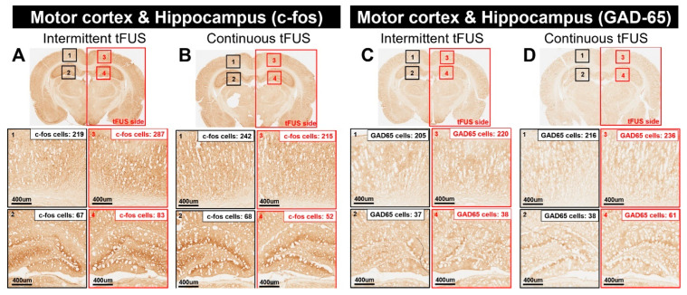
Transcranial focused ultrasound stimulation (tFUS) has emerged as a promising neuromodulation technique that delivers acoustic energy with high spatial resolution for inducing long-term potentiation (LTP)- or depression (LTD)-like plasticity. The variability in the primary effects of tFUS-induced plasticity could be due to different stimulation patterns, such as intermittent versus continuous, and is an aspect that requires further detailed exploration. In this study, we developed a platform to evaluate the neuromodulatory effects of intermittent and continuous tFUS on motor cortical plasticity before and after tFUS application. Three groups of rats were exposed to either intermittent, continuous, or sham tFUS. We analyzed the neuromodulatory effects on motor cortical excitability by examining changes in motor-evoked potentials (MEPs) elicited by transcranial magnetic stimulation (TMS). We also investigated the effects of different stimulation patterns on excitatory and inhibitory neural biomarkers, examining c-Fos and glutamic acid decarboxylase (GAD-65) expression using immunohistochemistry staining. Additionally, we evaluated the safety of tFUS by analyzing glial fibrillary acidic protein (GFAP) expression. The current results indicated that intermittent tFUS produced a facilitation effect on motor excitability, while continuous tFUS significantly inhibited motor excitability. Furthermore, neither tFUS approach caused injury to the stimulation sites in rats. Immunohistochemistry staining revealed increased c-Fos and decreased GAD-65 expression following intermittent tFUS. Conversely, continuous tFUS downregulated c-Fos and upregulated GAD-65 expression. In conclusion, our findings demonstrate that both intermittent and continuous tFUS effectively modulate cortical excitability. The neuromodulatory effects may result from the activation or deactivation of cortical neurons following tFUS intervention. These effects are considered safe and well-tolerated, highlighting the potential for using different patterns of tFUS in future clinical neuromodulatory applications.

Keywords: motor-evoked potentials; neuromodulation; plasticity; rats; transcranial focused ultrasound.


Conflict of interest statement

H.-L.L. served as a technical consultant at NaviFUS Corp., Taipei, Taiwan and currently holds several therapeutic ultrasound-related patents; P.-C.C. concurrently served as a part-time research and development scientist at NaviFUS Corp., Taiwan.

Figures

Figure 1
Figure 1
Representative motor-evoked potential (MEP) responses of a rat before and after receiving either sham, intermittent, or continuous transcranial focused ultrasound stimulation (tFUS) are displayed for each measurement period (A). The average normalized MEP amplitudes across the three intervention protocols (sham, intermittent, or continuous tFUS) are shown (B). The mean cortical excitability responses were calculated for each intervention group within 30 min following the different stimulations (C). Data are presented as mean ± SEM for 7–8 rats per group. ** indicates p < 0.01, denoting significant differences in MEP size post-intervention compared with the baseline measured before tFUS, as determined by a post-hoc Fisher’s LSD test. n.s.: no significance.
Figure 2
Figure 2
Representative immunohistochemically stained sections highlighting the regions of interest (ROI) comprising the motor cortex and hippocampus. The data illustrate the quantity of c-Fos-positive cells within the ROI following intermittent transcranial focused ultrasound stimulation (tFUS) (A) and continuous tFUS (B). Similarly, the number of GAD-65-positive cells in the motor cortex and hippocampus is shown after exposure to intermittent tFUS (C) and continuous tFUS (D).
Figure 3
Figure 3
Representative images of glial fibrillary acidic protein (GFAP) immunostaining in the motor cortex of rats sacrificed 1 day, 3 days, and 7 days after receiving sham, continuous, or intermittent tFUS are presented. Compared to rats that received sham stimulation, no obvious astrogliosis was observed at or near the sonicated sites in the brains of those treated with either continuous or intermittent tFUS.
Figure 4
Figure 4
Representative images showcasing immunostaining for glial fibrillary acidic protein (GFAP) within the intracerebroventricular (ICV) area of rats sacrificed at 1 day, 3 days, and 7 days post-exposure to either sham, continuous, or intermittent tFUS intervention. In comparison to the sham group, no obvious astrogliosis was detected at or around the sonication locations in brains subjected to either continuous or intermittent tFUS treatment.
Figure 5
Figure 5
Experimental setup for measuring motor cortical excitability after transcranial focused ultrasound stimulation (tFUS) in anesthetized rats. The center of the tFUS transducer was positioned and focused on the right motor cortex (A). The characterizations of the tFUS pressure fields. One sagittal (Y–Z plane) and one transverse (X–Y plane) scan of ultrasound pressure distribution using a hydrophone-based US field-mapping system (B). The diameter and length of the half-maximum pressure amplitude of the ultrasound field and transcranial ultrasound field were within 2 and 10 mm, respectively. The placement and assembly of recording electrodes of motor-evoked potentials (MEPs) elicited by the transcranial magnetic stimulation (TMS) coil (C). MEP data were recorded from the brachioradialis muscle and were analyzed to evaluate any changes in cortical excitability resulting from the intervention protocols.
Figure 6
Figure 6
Schematic diagram of the experimental design for testing changes in cortical excitability after tFUS or sham stimulation in anesthetized rats. In this representative experiment, anesthetized rats received intervention protocols for 600 sec. Motor-evoked potentials (MEPs) were measured at baseline and 0, 10, 20, and 30 min after tFUS intervention. Subsequently, the rat brains were removed and subjected to immunohistochemistry for further investigation.

Similar articles

References

    1. Darmani G., Bergmann T.O., Butts Pauly K., Caskey C.F., de Lecea L., Fomenko A., Fouragnan E., Legon W., Murphy K.R., Nandi T., et al. Non-invasive transcranial ultrasound stimulation for neuromodulation. Clin. Neurophysiol. 2022;135:51–73. doi: 10.1016/j.clinph.2021.12.010. - DOI - PubMed
    1. Darrow D.P. Focused Ultrasound for Neuromodulation. Neurotherapeutics. 2019;16:88–99. doi: 10.1007/s13311-018-00691-3. - DOI - PMC - PubMed
    1. Guerra A., Bologna M. Low-Intensity Transcranial Ultrasound Stimulation: Mechanisms of Action and Rationale for Future Applications in Movement Disorders. Brain Sci. 2022;12:611. doi: 10.3390/brainsci12050611. - DOI - PMC - PubMed
    1. Baek H., Pahk K.J., Kim H. A review of low-intensity focused ultrasound for neuromodulation. Biomed. Eng. Lett. 2017;7:135–142. doi: 10.1007/s13534-016-0007-y. - DOI - PMC - PubMed
    1. Tyler W.J., Tufail Y., Finsterwald M., Tauchmann M.L., Olson E.J., Majestic C. Remote excitation of neuronal circuits using low-intensity, low-frequency ultrasound. PLoS ONE. 2008;3:e3511. doi: 10.1371/journal.pone.0003511. - DOI - PMC - PubMed

Substances

LinkOut - more resources