. 2024 May 24;25(11):5716.
doi: 10.3390/ijms25115716.

Reduction in Hippocampal Amyloid-β Peptide (Aβ) Content during Glycine-Proline-Glutamate (Gly-Pro-Glu) Co-Administration Is Associated with Changes in Inflammation and Insulin-like Growth Factor (IGF)-I Signaling

Affiliations

Reduction in Hippocampal Amyloid-β Peptide (Aβ) Content during Glycine-Proline-Glutamate (Gly-Pro-Glu) Co-Administration Is Associated with Changes in Inflammation and Insulin-like Growth Factor (IGF)-I Signaling

Laura M Frago et al. Int J Mol Sci. .

Abstract

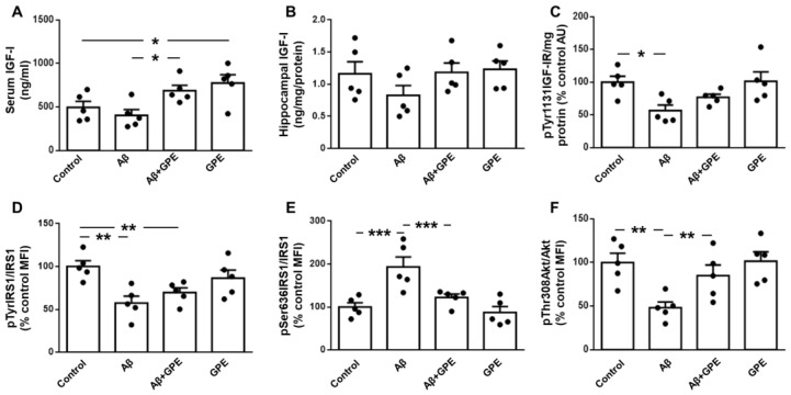
Alzheimer's disease (AD) is characterized by the deposition in the brain of senile plaques composed of amyloid-β peptides (Aβs) that increase inflammation. An endogenous peptide derived from the insulin-like growth factor (IGF)-I, glycine-proline-glutamate (GPE), has IGF-I-sensitizing and neuroprotective actions. Here, we examined the effects of GPE on Aβ levels and hippocampal inflammation generated by the intracerebroventricular infusion of Aβ25-35 for 2 weeks (300 pmol/day) in ovariectomized rats and the signaling-related pathways and levels of Aβ-degrading enzymes associated with these GPE-related effects. GPE prevented the Aβ-induced increase in the phosphorylation of p38 mitogen-activated protein kinase and the reduction in activation of signal transducer and activator of transcription 3, insulin receptor substrate-1, and Akt, as well as on interleukin (IL)-2 and IL-13 levels in the hippocampus. The functionality of somatostatin, measured as the percentage of inhibition of adenylate cyclase activity and the levels of insulin-degrading enzyme, was also preserved by GPE co-treatment. These findings indicate that GPE co-administration may protect from Aβ insult by changing hippocampal cytokine content and somatostatin functionality through regulation of leptin- and IGF-I-signaling pathways that could influence the reduction in Aβ levels through modulation of levels and/or activity of Aβ proteases.

Keywords: Alzheimer’s disease; Gly-Pro-Glu; IGF-I signaling; cytokines; inflammation.


Conflict of interest statement

The authors declare no conflicts of interest. The funders had no role in the design of the study; in the collection, analyses, or interpretation of data; in the writing of the manuscript; or in the decision to publish the results.

Figures

Figure 1
Figure 1
Effects of Aβ25-35 (300 pmol/day) and glycine-proline-glutamate (GPE) co-administration on hippocampal Aβ25-35 levels and phosphorylation of pro-inflammatory and leptin signaling targets. Levels of (A) Aβ25-35, relative protein levels of (B) p38 mitogen-activated protein kinase (pMAPK) phosphorylated (p) at Thr180 and Tyr182 (pThr180Tyr182p38MAPK) and (C) nuclear factor kappa B (NFκB) phosphorylated at Ser536 (pSer536NFkB), (D) serum leptin levels and relative protein levels of (E) signal transducer and activator of transcription 3 (STAT3) phosphorylated at Tyr705 (pTyr705STAT3), and (F) STAT3 phosphorylated at Ser727 (pSer727STAT3) in ovariectomized (Ovx) rats (control), Ovx rats treated with β-amyloid 25-35 peptide (Aβ), Ovx rats treated with Aβ25-35 plus GPE (Aβ + GPE), and Ovx rats treated with GPE (GPE). Data are expressed as mean ± SEM. N = 5. MFI, median fluorescent intensity * p < 0.05, ** p < 0.01.
Figure 2
Figure 2
Effects of Aβ25-35 (300 pmol/day) and GPE co-administration on IGF-I levels and IGF-I-related signaling targets. Serum (A,B) and hippocampal levels of insulin-like growth factor-I (IGF-I) and relative protein levels of (C) IGF-I receptor (IGF-IR) phosphorylated at Tyr1131 (pTyr1131IGF-IR), (D) insulin receptor substrate 1 (IRS1) phosphorylated at Tyr residues (pTyrIRS1), (E) IRS1 phosphorylated at Ser636 (pSer636IRS1), and (F) Akt phosphorylated at Thr308 (pThr308Akt) in ovariectomized (Ovx) rats (control), Ovx rats treated with β-amyloid 25-35 peptide (Aβ), Ovx rats treated with Aβ25-35 plus GPE (Aβ + GPE), and Ovx rats treated with GPE (GPE). Data are expressed as mean ± SEM. N = 5. AU, absorbance units, MFI, median fluorescent intensity * p < 0.05, ** p < 0.01, *** p < 0.001.
Figure 3
Figure 3
Effects of Aβ25-35 (300 pmol/day) and GPE co-administration on serum and hippocampal cytokine levels. Serum levels of interferon (IFN)-γ (A), interleukin (IL)-2 (C), IL-13 (E), and IL-17A (G) and hippocampal concentrations of IFN-γ (B), IL-2 (D), IL-13 (F) and IL-17A (H) in ovariectomized (Ovx) rats (control), Ovx rats treated with β-amyloid 25-35 peptide (Aβ), Ovx rats treated with Aβ25-35 plus GPE (Aβ + GPE), and Ovx rats treated with GPE (GPE). Data are expressed as mean ± SEM. N = 5. * p < 0.05, ** p < 0.01.
Figure 4
Figure 4
Effects of Aβ25-35 (300 pmol/day) and GPE co-administration on basal adenylyl cyclase (AC) activity (pmol/min/mg protein), as well as on somatostatin (SRIF)-mediated inhibition of AC activity in hippocampal membranes ((A) and (B), respectively), percentage of SRIF inhibition of AC activity (C), levels of neprilysin (D) and insulin-degrading enzyme (IDE) (E) in ovariectomized (Ovx) rats (control), Ovx rats treated with β-amyloid 25-35 peptide (Aβ), Ovx rats treated with Aβ25-35 plus GPE (Aβ + GPE), and Ovx rats treated with GPE (GPE). Data are expressed as mean ± SEM. N = 5. * p < 0.05, ** p < 0.01.
Figure 5
Figure 5
Correlation of Aβ25-35 with (A) interleukin (IL)-2 content, (B) percentage of inhibition of adenylate cyclase (AC) activity, (C) neprilysin, and (D) insulin-degrading enzyme (IDE) levels in the hippocampus. Correlation coefficients (r) and p values are represented for each analysis. NS, non-significant.
Figure 6
Figure 6
Effects of Aβ25-35 (1 μM) and GPE co-administration on phosphorylation of signaling targets and levels of insulin-degrading enzyme (IDE) in neuronal and glial cultures. Relative protein levels in neuronal and glial cultures of ((A) and (D), respectively) of the signal transducer and activator of transcription 3 (STAT3) phosphorylated (p) at Ser727 (pSer727STAT3), ((B) and (E), respectively) insulin receptor substrate 1 (IRS1) phosphorylated at Tyr residues (pTyrIRS1) and protein concentrations, and ((C) and (F), respectively) insulin-degrading enzyme (IDE). Data are expressed as mean ± SEM. N = 5. * p < 0.05.
Figure 7
Figure 7
Effects of Aβ25-35 (1 μM) and GPE co-administration on interleukin secretion in glial cultures. Protein levels in culture media of interferon (IFN)-γ (A), interleukin (IL)-2 (B), IL-13 (C), and IL-17A (D). Data are expressed as mean ± SEM. N = 5. * p < 0.05.
Figure 8
Figure 8
Proposed model for reduction in Aβ25-35 levels by activation of leptin- and IGF-related signaling in Aβ-treated rats after co-administration of GPE. Infusion of Aβ25-35 reduces leptin signaling, whereas GPE preserves it. The activation of STAT3 after GPE co-administration increases hippocampal levels of IL-2 and IL-13. Aβ25-35 can also block Akt signaling, and GPE counteracts Aβ effects by increasing mTOR activation and IDE synthesis. The increase in hippocampal IL levels may activate their receptors and augment Akt phosphorylation, which activates CREB and increases SRIF synthesis. These events may augment IDE levels and activity in the hippocampus, thereby reducing Aβ levels. Aβ, beta-amyloid; Akt, protein kinase B; CREB, cAMP responsive element binding protein; GPE, glycine-proline-glutamate; IDE, insulin-degrading-enzyme; IRS1, insulin receptor substrate 1; SRIF, somatostatin; STAT-3, signal transducer and activator of transcription 3. Dark ellipses indicate previously published results.

Similar articles

References

    1. Ludewig S., Korte M. Novel insights into the physiological function of the APP (Gene) family and its proteolytic fragments in synaptic plasticity. Front. Mol. Neurosci. 2017;9:161. doi: 10.3389/fnmol.2016.00161. - DOI - PMC - PubMed
    1. Li R., Li Y., Zuo H., Pei G., Huang S., Hou Y. Alzheimer’s amyloid-beta accelerates cell senescence and suppresses SIRT1 in human neural stem cells. Biomolecules. 2024;14:189. doi: 10.3390/biom14020189. - DOI - PMC - PubMed
    1. Fornari Laurindo L., Aparecido Dias J., Cressoni Araújo A., Torres Pomini K., Machado Galhardi C., Rucco Penteado Detregiachi C., Santos de Argollo Haber L., Donizeti Roque D., Dib Bechara M., Vialogo Marques de Castro M., et al. Immunological dimensions of neuroinflammation and microglial activation: Exploring innovative immunomodulatory approaches to mitigate neuroinflammatory progression. Front. Immunol. 2024;14:1305933. doi: 10.3389/fimmu.2023.1305933. - DOI - PMC - PubMed
    1. Merighi S., Nigro M., Travagli A., Gessi S. Microglia and Alzheimer’s disease. Int. J. Mol. Sci. 2022;23:12990. doi: 10.3390/ijms232112990. - DOI - PMC - PubMed
    1. Wang H., Sun M., Li W., Liu X., Zhu M., Qin H. Biomarkers associated with the pathogenesis of Alzheimer’s disease. Front. Cell. Neurosci. 2023;17:1279046. doi: 10.3389/fncel.2023.1279046. - DOI - PMC - PubMed

MeSH terms

LinkOut - more resources