

GFAP as Astrocyte-Derived Extracellular Vesicle Cargo in Acute Ischemic Stroke Patients-A Pilot Study
- PMID: 38891912
- PMCID: PMC11172178
- DOI: 10.3390/ijms25115726
GFAP as Astrocyte-Derived Extracellular Vesicle Cargo in Acute Ischemic Stroke Patients-A Pilot Study
Abstract
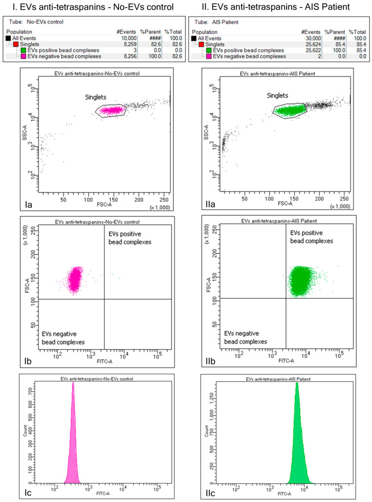
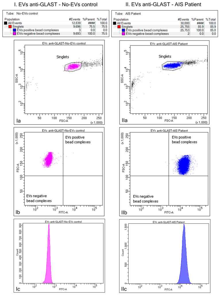
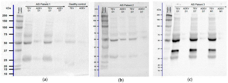
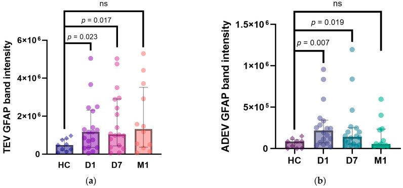
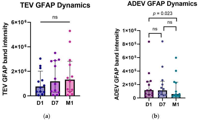
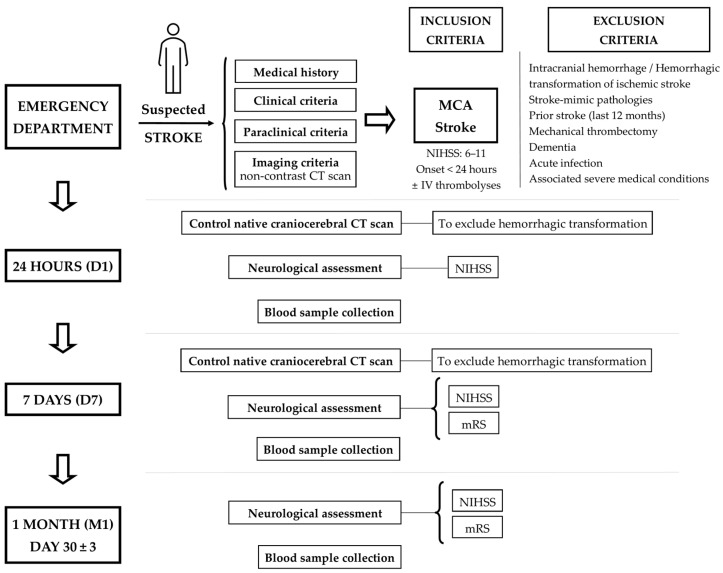
The utility of serum glial fibrillary acidic protein (GFAP) in acute ischemic stroke (AIS) has been extensively studied in recent years. Here, we aimed to assess its potential role as a cargo protein of extracellular vesicles (EVs) secreted by astrocytes (ADEVs) in response to brain ischemia. Plasma samples from eighteen AIS patients at 24 h (D1), 7 days (D7), and one month (M1) post-symptoms onset, and nine age, sex, and cardiovascular risk factor-matched healthy controls were obtained to isolate EVs using the Exoquick ULTRA EV kit. Subsets of presumed ADEVs were identified further by the expression of the glutamate aspartate transporter (GLAST) as a specific marker of astrocytes with the Basic Exo-Flow Capture kit. Western blotting has tested the presence of GFAP in ADEV cargo. Post-stroke ADEV GFAP levels were elevated at D1 and D7 but not M1 compared to controls (p = 0.007, p = 0.019, and p = 0.344, respectively). Significant differences were highlighted in ADEV GFAP content at the three time points studied (n = 12, p = 0.027) and between D1 and M1 (z = 2.65, p = 0.023). A positive correlation was observed between the modified Rankin Scale (mRS) at D7 and ADEV GFAP at D1 (r = 0.58, p = 0.010) and D7 (r = 0.57, p = 0.013), respectively. ADEV GFAP may dynamically reflect changes during the first month post-ischemia. Profiling ADEVs from peripheral blood could provide a new way to assess the central nervous system pathology.
Keywords: acute ischemic stroke; astrocyte-derived extracellular vesicles; glial fibrillary acidic protein; western blotting.
Conflict of interest statement
The authors declare no conflicts of interest.
Figures
Similar articles
-
Astrocyte Dysfunction Reflected in Ischemia-Induced Astrocyte-Derived Extracellular Vesicles: A Pilot Study on Acute Ischemic Stroke Patients.Int J Mol Sci. 2024 Nov 20;25(22):12471. doi: 10.3390/ijms252212471. Int J Mol Sci. 2024. PMID: 39596535 Free PMC article.
-
In vivo evidence of increased vascular endothelial growth factor in patients with major depressive disorder.J Affect Disord. 2025 Jan 1;368:151-159. doi: 10.1016/j.jad.2024.09.073. Epub 2024 Sep 14. J Affect Disord. 2025. PMID: 39278472
-
Plasma Glial Fibrillary Acidic Protein in the Differential Diagnosis of Intracerebral Hemorrhage.Stroke. 2017 Sep;48(9):2586-2588. doi: 10.1161/STROKEAHA.117.018409. Epub 2017 Jul 27. Stroke. 2017. PMID: 28751552
-
Extracellular Vesicles Derived From Neural Stem Cells, Astrocytes, and Microglia as Therapeutics for Easing TBI-Induced Brain Dysfunction.Stem Cells Transl Med. 2023 Mar 17;12(3):140-153. doi: 10.1093/stcltm/szad004. Stem Cells Transl Med. 2023. PMID: 36847078 Free PMC article. Review.
-
Astrocyte-Derived Extracellular Vesicles for Ischemic Stroke: Therapeutic Potential and Prospective.Aging Dis. 2024 May 7;15(3):1227-1254. doi: 10.14336/AD.2023.0823-1. Aging Dis. 2024. PMID: 37728588 Free PMC article. Review.
Cited by
-
Extracellular Vesicle-Associated Angiopoietin-2 and Cell Migration-Inducing Protein in Lung Cancer Progression and Brain Metastases.Cureus. 2025 Mar 7;17(3):e80200. doi: 10.7759/cureus.80200. eCollection 2025 Mar. Cureus. 2025. PMID: 40190907 Free PMC article.
-
Isolating Astrocyte-Derived Extracellular Vesicles From Urine.Int J Nanomedicine. 2025 Feb 26;20:2475-2484. doi: 10.2147/IJN.S492381. eCollection 2025. Int J Nanomedicine. 2025. PMID: 40027875 Free PMC article.
References
-
- Feigin V.L., Stark B.A., Johnson C.O., Roth G.A., Bisignano C., Abady G.G., Abbasifard M., Abbasi-Kangevari M., Abd-Allah F., Abedi V., et al. Global, Regional, and National Burden of Stroke and Its Risk Factors, 1990–2019: A Systematic Analysis for the Global Burden of Disease Study 2019. Lancet Neurol. 2021;20:795–820. doi: 10.1016/S1474-4422(21)00252-0. - DOI - PMC - PubMed
MeSH terms
Substances
Grants and funding
LinkOut - more resources
Full Text Sources
Medical
Miscellaneous
