. 2024 May 26;25(11):5775.
doi: 10.3390/ijms25115775.

Ex Vivo Analysis of Cell Differentiation, Oxidative Stress, Inflammation, and DNA Damage on Cutaneous Field Cancerization

Affiliations

Ex Vivo Analysis of Cell Differentiation, Oxidative Stress, Inflammation, and DNA Damage on Cutaneous Field Cancerization

Lara Camillo et al. Int J Mol Sci. .

Abstract

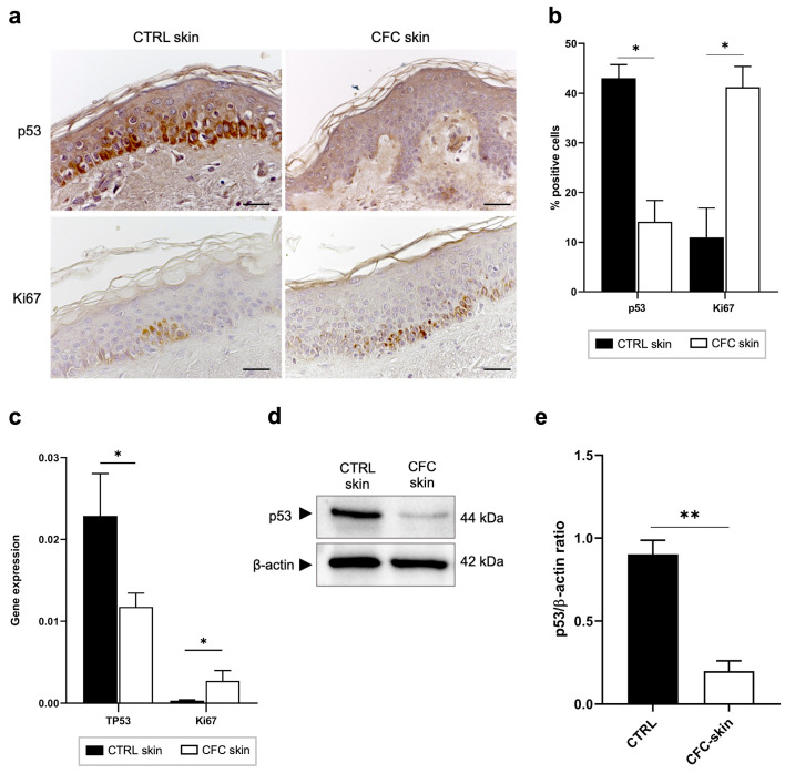
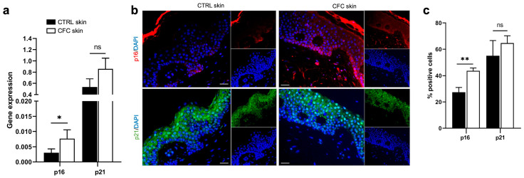
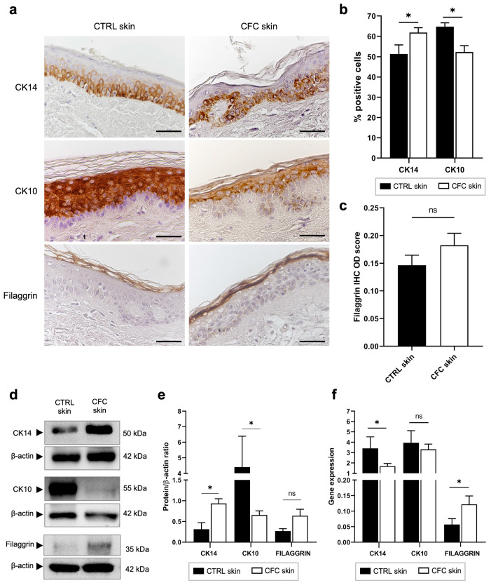
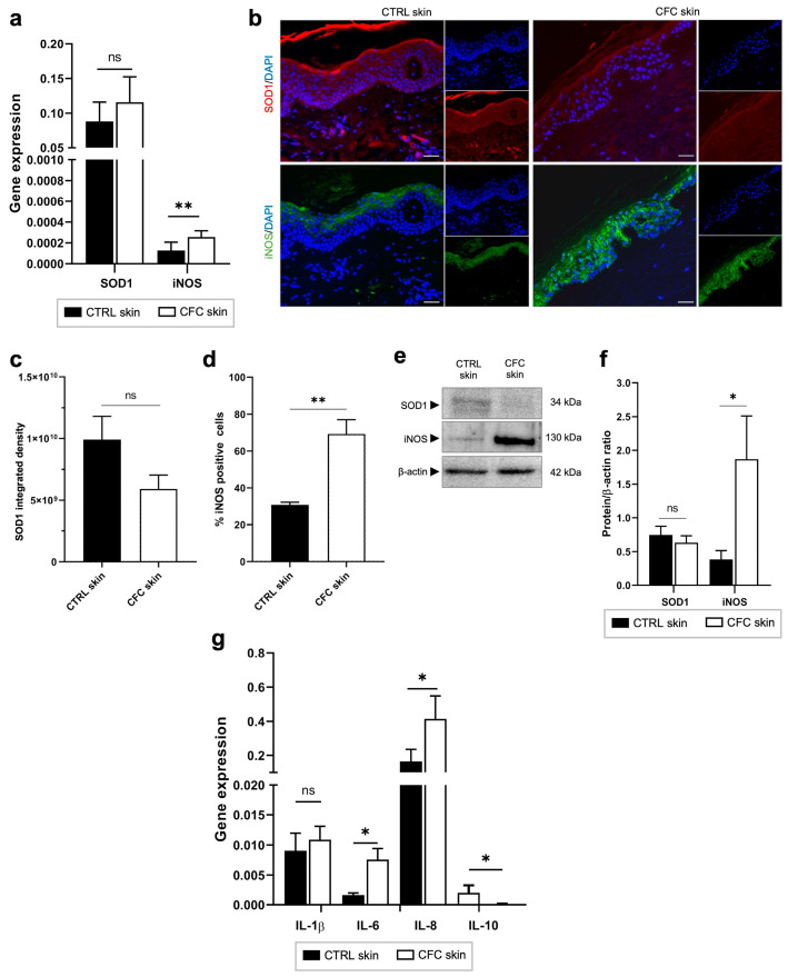
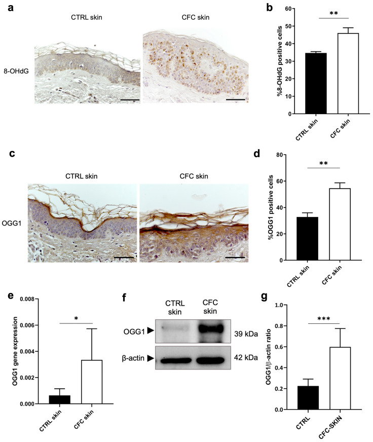
Cutaneous field cancerization (CFC) refers to a skin region containing mutated cells' clones, predominantly arising from chronic exposure to ultraviolet radiation (UVR), which exhibits an elevated risk of developing precancerous and neoplastic lesions. Despite extensive research, many molecular aspects of CFC still need to be better understood. In this study, we conducted ex vivo assessment of cell differentiation, oxidative stress, inflammation, and DNA damage in CFC samples. We collected perilesional skin from 41 patients with skin cancer and non-photoexposed skin from 25 healthy control individuals. These biopsies were either paraffin-embedded for indirect immunofluorescence and immunohistochemistry stain or processed for proteins and mRNA extraction from the epidermidis. Our findings indicate a downregulation of p53 expression and an upregulation of Ki67 and p16 in CFC tissues. Additionally, there were alterations in keratinocyte differentiation markers, disrupted cell differentiation, increased expression of iNOS and proinflammatory cytokines IL-6 and IL-8, along with evidence of oxidative DNA damage. Collectively, our results suggest that despite its outwardly normal appearance, CFC tissue shows early signs of DNA damage, an active inflammatory state, oxidative stress, abnormal cell proliferation and differentiation.

Keywords: DNA damage; field cancerization; oxidative stress; skin cancer; ultraviolet light.


Conflict of interest statement

The authors declare no conflicts of interest.

Figures

Figure 1
Figure 1
p53 and Ki67 expression is modulated on CFC skin samples. (a) Representative IHC and (b) % of p53 and Ki67 positive cells counted in four random areas of CTRL (n = 7) and CFC (n = 10) skin samples expressed as means ± SEM. (c) TP53 and Ki67 gene expression expressed as means ± SEM of CTRL (n = 12) and CFC (n = 21) skin samples. (d) Representative western blotting and (e) densitometric analysis of p53 expression tested on CTRL (n = 6) and CFC (n = 10) skin samples expressed as mean ± SEM. CTRL, control; CFC, cutaneous field cancerization; ns, no significance. Scale bar: 10 μm. * p < 0.05, ** p < 0.01.
Figure 2
Figure 2
p16 expression is increased in CFC skin samples. (a) p16 and p21 gene expression indicated as means ± SEM of CTRL (n = 12) and CFC (n = 21) skin samples. (b) Representative IF and (c) % of p16 and p21 positive cells counted in four random areas of CTRL (n = 7) and CFC (n = 10) skin samples expressed as means ± SEM. CTRL, control; CFC, cutaneous field cancerization; ns, no significance. Scale bar: 10 μm. * p < 0.05, ** p < 0.01.
Figure 3
Figure 3
Keratinocyte differentiation is altered in CFC skin. (a) Representative IHC expression of CK14, CK10 and Filaggrin and (b) % of IHC CK14 and CK10 stain-positive cells counted in four random areas of CTRL (n = 7) and CFC (n = 10) skin samples expressed as means ± SEM. (c) Filaggrin IHC OD score expressed as mean ± SEM of CTRL (n = 7) and CFC (n = 10) samples. (d) Representative Western blot and densitometric analysis (e) of CK14, CK10 and Filaggrin tested on CTRL (n = 6) and CFC (n = 10) skin samples expressed as means ± SEM. (f) CK14, CK10 and Filaggrin gene expression indicated as means ± SEM of CTRL (n = 12) and CFC (n = 21) skin samples. CTRL, control; CFC, cutaneous field cancerization; ns, no significance; OD, optical density. Scale bar: 10 μm. * p < 0.05.
Figure 4
Figure 4
Oxidative stress and inflammatory markers are expressed in CFC skin. (a) SOD1 and iNOS gene expression evaluated on CTRL (n = 12) and CFC (n = 20) samples and expressed as means ± SEM. (b) Representative IF of SOD1 and iNOS and (c) analysis of IF SOD1 integrated density and (d) % of IF iNOS positive cells expressed as means ± SEM of CTRL (n = 7) and CFC (n = 10) skin samples. (e) Representative Western blot and densitometric analysis (f) of SOD1 and iNOS on CTRL (n = 6) and CFC (n = 10) skin samples expressed as mean ± SEM. (g) IL-1β, IL-6, IL-8, IL-10 gene expression expressed as means ± SEM of CTRL (n = 12) and CFC (n = 21) samples. CTRL, control; CFC, cutaneous field cancerization; iNOS, inducible nitric oxide synthase; SOD1, superoxide dismutase; ns, no significance. Scale bar: 10 μm. * p < 0.05, ** p < 0.01.
Figure 5
Figure 5
Evaluation of OGG1 expression as marker of DNA damage. (a) Representative IHC and (b) % of 8-OHdG positive cells of cells counted in four random areas of CTRL (n = 7) and CFC (n = 10) samples expressed as means ± SEM. (c) OGG1 gene expression expressed as means ± SEM of CTRL (n = 12) and CFC (n = 21) skin samples. (d) Representative IHC expression and (e) % of OGG1 positive cells counted in four random areas of CTRL (n = 7) and CFC (n = 10) samples expressed as means ± SEM. (f) Representative Western blot and densitometric analysis (g) of OGG1 performed on CTRL (n = 6) and CFC (n = 10) samples expressed as means ± SEM. CTRL, control; CFC, cutaneous field cancerization; OGG1, 8-oxoguanine DNA glycosylase; 8-OHdG, 8-hydroxy-2-deoxyguanosine. Scale bar: 10 μm. * p < 0.05, ** p < 0.01, *** p < 0.001.

Similar articles

Cited by

References

    1. Curtius K., Wright N.A., Graham T.A. An Evolutionary Perspective on Field Cancerization. Nat. Rev. Cancer. 2018;18:19–32. doi: 10.1038/nrc.2017.102. - DOI - PubMed
    1. Slaughter D.P., Southwick H.W., Smejkal W. Field Cancerization in Oral Stratified Squamous Epithelium; Clinical Implications of Multicentric Origin. Cancer. 1953;6:963–968. doi: 10.1002/1097-0142(195309)6:5<963::aid-cncr2820060515>3.0.co;2-q. - DOI - PubMed
    1. Braakhuis B.J.M., Tabor M.P., Kummer J.A., Leemans C.R., Brakenhoff R.H. A Genetic Explanation of Slaughter’s Concept of Field Cancerization: Evidence and Clinical Implications. Cancer Res. 2003;63:1727–1730. - PubMed
    1. Willenbrink T.J., Ruiz E.S., Cornejo C.M., Schmults C.D., Arron S.T., Jambusaria-Pahlajani A. Field Cancerization: Definition, Epidemiology, Risk Factors, and Outcomes. J. Am. Acad. Dermatol. 2020;83:709–717. doi: 10.1016/j.jaad.2020.03.126. - DOI - PubMed
    1. Fernandez Figueras M.T. From Actinic Keratosis to Squamous Cell Carcinoma: Pathophysiology Revisited. J. Eur. Acad. Dermatol. Venereol. 2017;31((Suppl. S2)):5–7. doi: 10.1111/jdv.14151. - DOI - PubMed

MeSH terms